2006:2273 - TOMFARNEY, CO. WEXFORD, Wexford
County: Wexford
Site name: TOMFARNEY, CO. WEXFORD
Sites and Monuments Record No.: SMR WX030-090
Licence number: E1190
Author: MAEVE SIKORA AND FIONA REILLY
Author/Organisation Address: —
Site type: Early Bronze Age graves
Period/Dating: —
ITM: E 685841m, N 631790m
Latitude, Longitude (decimal degrees): 52.430857, -6.737679
Introduction
In early July 2006 a stone cist containing a cremation was discovered when a sewerage trench was being excavated at Tomfarney, near Clonroche, Co. Wexford. The site was reported to the National Museum and an initial site visit was carried out by Mary Cahill and Maeve Sikora on 10 July. At this time a portion of the cist contents had been removed and a large amount of cremated bone was visible on the ground surface around the cist. The surface bone was collected and brought to the Museum. A rescue excavation was carried out later that week by Maeve Sikora and Fiona Reilly.
Location (Fig. 3.234)
The cist was in the townland of Tomfarney, near Clonroche, south Co. Wexford,368 on elevated ground at an altitude of 100–110m above sea level. No other cists are known from this townland, but according to locals there was a tradition of a burial in the townland. The name Tomfarney is understood to mean ‘the mound or hillock of the alder trees’. An early Bronze Age burial is known from the townland of Misterin, approximately 5km to the south of Tomfarney townland.

Fig. 3.234—Location map, Tomfarney, Co. Wexford.
Description of cist
The site was heavily disturbed at the time of investigation, as the sewerage trench had cut through the centre of the long axis of the cist (Pl. 74; Fig. 3.235). Despite this, it was possible to see that the cist had been roughly rectangular in plan, with its long axis aligned northeast/south-west. Excavation commenced by attempting to find the cut of the pit dug to contain the cist, and the contents of the cist were excavated thereafter. Owing to the nature of the soil it was extremely difficult to discern the line of the cut. The soil also contained a large amount of shale and was extremely compacted. The suboval pit measured approximately 2.1m long by 1.3m wide. It had been dug into the bedrock, a type of shale which occurred close to the ground surface. Overlying the bedrock was a c. 0.1m-thick layer of compact orange clay with very frequent shale inclusions. The topsoil, which lay directly over this layer, was also very compact with many shale inclusions, but was darker brown in colour.
All of the in situ cist slabs were of granite, which contrasted with the shaly bedrock of the site. A number of displaced granite slabs in the vicinity of the site are likely to be part of the cist removed by the digger.
Five upright slabs that formed part of the cist chamber remained in situ but most were slanted inwards from the base, probably partly owing to the recent bulldozing works; three slabs were found at the north-eastern end and two at the south-western end. The northeastern end slab measured 0.61m long by 0.45m high by 0.13m thick. It bore a number of long, linear marks, which were examined and were found to be natural. This end was supported externally by six large granite slabs and a number of other smaller packing stones. The south-western end was also flanked by six granite slabs and a number of other smaller packing stones.

Fig. 3.235—Plans and section of grave, Tomfarney, Co. Wexford. Top left: preexcavation plan. Right and lower left: plan and section showing cist walls in situ.
Owing to the disturbance caused by the digger to the long axis of the cist, it was not possible to determine how many slabs had been originally present on each side. The internal width of the cist chamber was approximately 0.7m, and the interior length (from end slab to end slab) of the cist was 0.95m. The south-western end slab had been badly disturbed by the digger and inclined sharply inwards. One paving slab remained on the floor of the cist at the north-eastern end, measuring 0.65m long by 0.31m wide by 0.11m thick. It is probable that the remainder of the cist had also been paved, but the south-western end had been badly disturbed by the digger and any floor slab in this area would have been removed. The loose soil in this area was cleared and was found to immediately overlie bedrock. The cist had been covered by a large capstone, which had been displaced by the digger. It measured 1.52m long by 1.2m wide by approximately 0.1m thick.
The cist had originally contained a very large cremation deposit, much of which had been removed by the bulldozer. The deposit contained the remains of at least seventeen people, including adults, male and female, children and infants. A substantial portion of the bone was found in situ in the north-eastern end of the cist at the time of excavation. The deposit had probably covered the entire cist, but apart from the deposit at the north-eastern end only a small portion of in situ remains were recovered from the south-western end, where the bone had been set tightly against the end slab. Very large fragments of cremated bone were found packed tightly against the north-eastern end and side slabs, and the deposit measured approximately 0.1m thick. A perforated flat stone was found in the upper layer of the cremation here, and a number of small, rounded pebbles were also recovered from the loose disturbed soil in the cist. A small amount of soil was found covering the cremation deposit at the north-eastern end of the cist. This was compact dark clay with frequent shale inclusions. The cist fill was bulk-sampled, and sieving of this material in the laboratory produced a small biconical clay bead.
Perforated stone, 2006:67 (Pl. 75; Fig. 3.236)
A flat piece of shale with a large central perforation was found mixed in with cremated bone in the cist. It is roughly trapezoidal in shape, with rounded edges. The circular perforation is roughly, but not exactly, central. Some wear is visible around the perforation, particularly on one face, which may indicate that the object was suspended or that something was passed through the perforation. Cracks in the stone are visible on the side of the object. One face bears orange-brown accretions while the other shows the natural mid-grey colour of the rock. The surfaces of the object are not smooth, and rock has laminated on both faces. The rock has been identified as calcareous limestone, and splits such as that visible in the side of this object are typical of this type of rock (Matthew Parkes, pers. comm.). Perforated stones have been found in a number of graves, but nothing comparable to this object is known to the authors.

Fig. 3.236—Artefacts from grave, Tomfarney, Co. Wexford.
Biconical clay bead, 2006:69 (Fig. 3.236)
A bead of fine baked clay, biconical in shape with a small central perforation. The surface is smooth, with a few nicks visible around the widest part of the bead. This object was retrieved after excavation during the processing of the cremation deposit. It is comparable to a number of beads found within a cremation deposit at Burtown Little, Co. Kildare.
Dimensions: D 8.37mm; H 6.3mm; D perforation 1.53mm.
Comment
The burial at Tomfarney is remarkable in terms of both the size of the cremation and the unusual associated artefacts. The cremation of at least seventeen individuals of different ages, perhaps in a single episode, must have been a significant event, and even the construction of a suitably sized pyre or pyres would have required some effort. A sample of bone submitted for radiocarbon dating yielded a date of 3656±27 BP, which calibrates to 2134–1947 BC.369 The number of individuals recorded is by far the greatest known from any Bronze Age cist or pit burial from Ireland. Nine to eleven were recorded in a cremation deposit at Altaghaderry, Co.Donegal (this volume, pp 98–111), and also included a range of individuals based on age and gender. It is not possible to ascertain whether there were multiple episodes of deposition in either case, as both cists had been severely disturbed prior to investigation. If the remains were deposited in a single episode it is possible that the burial occurred as a result of some calamitous event which affected men, women and children in the local community.
Both the clay bead and the perforated stone are unusual in a Bronze Age burial context in Ireland. The bead is very similar to a number of beads found within a cremation deposit at Burtown Little, Co. Kildare, where 25 beads were recovered during the processing of the cremation. The cremation was from a pit which was situated between two ring-barrows. The excavator has suggested that these beads may have been part of a bracelet. It is possible that more beads were originally present in the Tomfarney burial, but these may have been lost when the cist contents were disturbed during discovery. Apart from the Burtown Little parallel, no other examples of this type of ceramic bead are known. A radiocarbon date for the Burtown Little burial centres on the mid-eighteenth century BC, which is much later than the Tomfarney date (Headland Archaeology, pers. comm.). However, biconical beads occur in various media at different stages of the Bronze Age.
HUMAN REMAINS
LAUREEN BUCKLEY
There were eleven different samples from the cremation and, although some attempt had been made to differentiate between them, most were labelled ‘general cremation’ or ‘main cremation’. As the samples were so large they were examined separately, but essentially there was no stratigraphic reason to separate them, so a summary combining all the samples is given.
SUMMARY REPORT
The entire sample consisted of 11,023g of very efficiently cremated bone. Almost all of it was creamy/white in colour, indicating that all the organic content had been burnt off. There was only a tiny amount of blue-coloured bone, where the cremation process had been less complete and the bone still contained an organic portion. These fragments were confined mainly to the backs of the legs, where oxygen may not have been able to circulate. Both air and heat are necessary for efficient combustion.
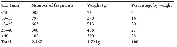
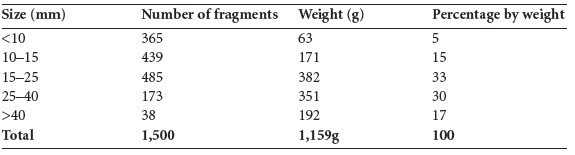
Table 3.140—Fragmentation of bone from the complete sample.

The fragmentation of the bone is shown in Table 3.140, with the largest fragment 120mm in length. It can be seen that although there is a small proportion of very small fragments, most of the sample is divided equally into moderate- and large-sized fragments. Identification is generally easier if the fragments are large or very large in size.
Identification
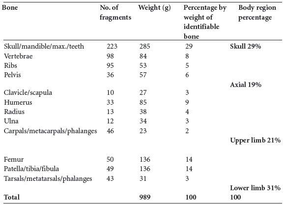
It was possible to identify 5,331g (48%) of the bone. The proportion identified reflects the fragmentation of the sample and compares well to other cremation burials from the same period. Table 3.141 shows the weight of each portion of the skeleton identified, the proportion of the parts in relation to the total identified bone, and a summary of the areas of the skeleton represented (skull, axial, lower limb and upper limb).
Table 3.141—Proportions of identified bone from the complete sample.

The proportions of the various body regions expected in a normal cremation are as follows: skull 18.2%; axial 23.1%; upper limb 20.6%; and lower limb 38.1% (McKinley 1989). These should be the same, no matter how many individuals are present or whether there are adults or juveniles present. In reality, however, it is frequently found that where more than one individual is present the proportion of skull is higher than normal. In this case the proportion of skull was twice what it should be. This is because skull is easier to identify positively, whereas long bone fragments may not be so readily distinguished. Therefore, where multiple bodies are present, the amount of skull identified far exceeds the proportion of long bones. The proportions of other parts of the skeleton in this cremation sample were all slightly reduced, with the lower limb reduced by the highest proportion.
Identification
A full description of the identified elements is given in each of the separate sample reports. The overall number of distinguishing elements is summarised in Table 3.142.
Table 3.142—Summary table of identified adult distinguishing elements.

Table 3.142 (cont.)—Summary table of identified adult distinguishing elements.

Adults
Tooth sockets
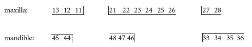
Table 3.143 indicates the number of particular tooth sockets recovered. The numbers in bold refer to socket position number, and the numbers below each socket refer to the numbers of individual sockets identified.
Table 3.143—Summary of recovered permanent tooth sockets.

The most numerous single socket recovered was the lower right first molar, 46, of which there were seven, indicating that at least seven individuals were present.
Teeth
Roots from a number of teeth were recovered. Since the crowns were shattered and it was mostly the teeth that were identified, these are assumed to be erupted permanent teeth. It was not always possible to classify the tooth roots exactly, and occasionally it could only be stated that the tooth was single-rooted, which means that it could have been an incisor, canine or premolar.
If all the possible incisors, canines and premolars are added together, there are 81 of these types of teeth present. With a maximum of twenty of these tooth types per individual, this means that at least five individuals are represented.
If the molar teeth are considered together, there are 62–65 teeth present. With twelve molars per individual, this means that the molar teeth represent at least six individuals.
Juvenile bones
A small amount of juvenile bone was identified. This included infant, juvenile and adolescent bone.
Infant bone
This included an unsided petrous part of a temporal bone, a basilar occipital bone, bodies of three infant cervical vertebrae, and left side, right side and one other partial neural arch from an infant, including one thoracic vertebra.
There were also four infant ribs, left and right infant scapulae, distal hand phalanges, and one unsided infant femur shaft. The bones represent a minimum of one infant.
Juvenile and adolescent bone
Skull bones included a right side of a frontal bone, a left orbit, right orbit, one other incomplete orbit, a left petrous temporal, right petrous temporal and an unsided petrous temporal bone and a fragment of occipital bone. There were also two left and one right zygomatic bones. A fragment of maxilla was also present.
Vertebrae included a left side of a first cervical vertebra, the dens of a C2, the left side of a C2, the right side of a neural arch from a cervical, fragments of thoracic bodies, two othervertebral bodies, one vertebral body with the right side of an arch, and at least two adolescent lumbar bodies. There was one right juvenile rib and two unsided ribs, as well as part of an ilium from a juvenile and an adolescent ilium with unfused iliac crest epiphyses.
Juvenile arm bones present included a coracoid process from a scapula, the proximal half of the shaft of a humerus, the distal third of a humerus, a distal epiphysis of a humerus, the distal part of a shaft including the metaphysis of a right radius, and one proximal and one distal epiphysis from an unsided radius. There was also a proximal ulna shaft and a distal ulna shaft with unfused head. One proximal, one middle and four distal hand phalanges were present.
Only a metaphyseal surface from a distal tibia and one proximal foot phalanx were identified from the leg bones. A minimum of two juveniles were represented by the juvenile bones recovered.
Juvenile teeth
There were a number of teeth from juveniles present. These included erupted deciduous teeth, unerupted deciduous teeth and permanent teeth in the process of development. It was assumed that complete or non-fractured crowns had not been erupted at the time of death and that roots with partial crowns represented teeth that were erupted at the time of death. Table 3.144 shows the juvenile teeth present, their degree of development and the possible age of the individual concerned.
Table 3.144—Summary of recovered juvenile teeth, development and possible age.

From the degree of development of the dentition it seems that there were at least two juveniles present. One was aged 4–6 years. The other could have been aged 2 1⁄2–10 years but was more likely to have been between 2 1⁄2 and 3 1⁄2 years.
In addition, there was an upper premolar found with the tips of the roots not fully closed that could have come from an adolescent aged 11–13 years. There was also evidence among the bones for an adolescent.
There also appears to have been dentition from at least two infants, one around six months of age (6mths ± 2mths) and the other around one year of age (7–15mths).
Minimum number of individuals
There are at least twelve adult individuals present, as represented by the number of petrous portions of the left temporal bone. There was also at least one adolescent present. Two juveniles were identified, one aged 4–6 years and the other aged 2 1⁄2–10 years but probably 2 1⁄2–3 1⁄2 years. At least two infants were also identified, one aged around six months and the other aged around one year. The minimum total number of individuals is therefore seventeen.
Age and sex
The sexing of fragmented cremated remains is difficult, as it depends entirely on certain features of the skull and pelvis surviving in large enough fragments for the characteristics to be discernible. It is further complicated by the fact that a prominent feature is more easily recognisable as being male but a less developed feature cannot with certainty be identified as being female. In addition, sex is best determined by looking at all the morphological features in a skeleton, and this is simply not possible in a cremation where the skeleton is fragmented and mixed with several other individuals.
Table 3.145—Summary table of sexed individuals.

Table 3.145 summarises the features identified in this cist. It can be seen that the most common feature from which sex could be determined was the left orbital rim. Four of these were present, of which three were of the male type and one of the female type. Although there were two female sciatic notches present, one was from a left bone and one was from a right bone, so they could be from the same individual. The results do not point to the number of males being greater than the number of females but merely indicate that it is easier to identify males than females from the skull features.
It was not possible to reliably age the adult skeletons. One female was, however, identified from the auricular surface of the pelvis as being at least 35 years of age but possibly older than 45 years at the time of death.
Various degenerative features present in fragments of the vertebrae suggest that older adults were present, but owing to the severe fragmentation it is not possible to say how many of the adult individuals may have been older adults.
Skeletal pathology
All of the pathology noted on the cremated fragments was degenerative joint disease and osteoarthritis of the joints. Degeneration of the joint surfaces owing to wear and tear leads to bone formation known as marginal lipping at the edges, destruction of the articular surface giving a porotic appearance, and eventually to eburnation or polishing of the joint surface itself. The presence of eburnation is diagnostic of osteoarthritis.
In the remains from this site osteoarthritis was noted on a left temporo-mandibular joint. Both the temporal fossa and the mandibular condyle were affected. The temporal fossa had pitting and eburnation of the articular surface, while the surface of the condyle was severely flattened and porotic with marginal lipping also. Osteoarthritis with eburnation of the articular surfaces was also noted in a first cervical vertebra, lower cervical vertebrae, thoracic vertebrae and in lumbar vertebrae.
The cervical vertebrae seemed to be severely affected. This often happens in older individuals and suggests that there was at least one older adult present. The effects of osteoarthritis can be exacerbated by trauma, however, and without the complete skeleton present it is not possible to say whether there was any complicating factor in the development of the osteoarthritis.
Degenerative joint disease with porosity of the joint surfaces and marginal lipping was also noted in several cervical and thoracic posterior joint surfaces.
The joints between the vertebral bodies are not arthrodial so osteoarthritis cannot occur between them. Nevertheless, similar bone changes, porosity and marginal lipping of the edges of the vertebral bodies can also occur with age and these are known as osteophytosis. Osteophytosis of the vertebral bodies was identified in cervical and thoracic vertebral bodies.
Schmorl’s nodes, which are depressions on the superior and inferior surfaces of the vertebral bodies, were noted in two thoracic vertebrae. These are caused by strenuous manual labour and also become more common with age.
Summary and conclusions
The entire sample from this site consisted of 11,023g of cremated bone. Almost all the bone was creamy/white in colour, indicating that it had been efficiently cremated, with all the organic content burnt off. Small fragments of bone mainly from the back of the leg areas were blue in colour where cremation had not been efficient, probably owing to restriction in oxygen supply. The largest bone fragment was 120mm in length, and most of the sample consisted of moderate to large fragments of bone. There was, however, a relatively small proportion of very large fragments more than 40mm in length. It is generally easier to identify bone if the fragments are large.
In this sample it was possible to identify 48% of the bone. The proportion of skull was twice what would be expected, but as the skull is easier to identify than other bone this is a common occurrence, especially in multiple cremations.
It was estimated that at least seventeen individuals were present in the cremation. These comprised twelve adults, one adolescent, one juvenile aged 4–6 years, another juvenile probably aged 2 1⁄2–3 1⁄2 years, an infant around one year old and another infant aged about six months. At least three of the adults were male and one was female. It was only possible to age the female as being at least 35 years of age. There was considerable evidence that at least one older adult was present in the collection, however, as several fragments of vertebrae were found that had evidence of severe osteoarthritis. Osteoarthritis gets worse with advancing age. There was evidence that at least one individual was engaged in heavy manual labour. Osteoarthritis was also present in at least one temporo-mandibular joint.
This particular cist appeared to be packed full of cremated bone, and it is unfortunate that it was partially destroyed and that there was no chance of distinguishing deposits within the cist. It contained the highest number of individuals of any cist examined during this study, with males, females, adolescents, juveniles and infants interred together.
DESCRIPTION OF SAMPLES
All the samples that were examined separately are described below.
Sample 1, main cremation
This sample consisted of 1,721g of cremated bone. The bone was efficiently cremated and was a creamy brown colour, although there were some small blue fragments from the back of the femur. The fragmentation of the sample is shown in Table 3.146, with the largest fragment being 106mm in length.
Table 3.146—Fragmentation of bone, sample 1, main cremation.
It can be seen that 50% of the sample consisted of large or very large fragments, and most of the remainder consisted of moderate-sized pieces. There was a very low proportion of smaller fragments.
Identification
It was possible to identify 989g (57.5%) of the cremated bone. The proportion of identified bone from the various parts of the skeleton is shown in Table 3.147.
The identifiable features of the various bones are noted below.
Skull
Frontal: a left and a right orbit, a fragment of another unsided orbit, a fragment from around the glabella and the right supraorbital ridge from a male individual.
Parietal: a large section of the posterior part of a left parietal bone with the lambdoid and sagittal sutures visible and with the parietal foramen present.
Occipital: a large fragment of squamous occipital, a piece of the right side of the squamous occipital with the lambdoid suture visible, and a fragment of the left side of the occipital with the lambdoid suture visible.
Table 3.147—Proportion of identified bone, sample 1, main cremation.
Temporal: three adult left petrous temporal bones, another unsided partial petrous temporal, a left temporo-mandibular fossa, another mandibular fossa, possibly from a right temporal bone, a fragment consisting of the left anterior suture and part of the left mandibular fossa, another fragment from a right bone with a pronounced posterior zygomatic arch and one left mastoid process.
Also present was a complete right zygomatic bone and part of another right zygomatic bone, a fragment of sphenoid and part of a right nasal bone.
Juvenile skull: one right petrous temporal bone.
Mandible
Two left and two right mandibular condyles, as well as fragments of the body with tooth sockets.
Maxilla
Three fragments of alveolus with tooth sockets.
Dentition
Various fragments with sockets for the following teeth were present:
Teeth: teeth present included one upper canine root, one upper molar root and some partial molar crowns. There was also part of the root and crown of one mandibular molar.
Vertebrae
These included an axis, C2, consisting of the left and right, superior and inferior articular surfaces and the body. The left side of the arches of three lower cervical vertebrae as well as one partial body were also present. The body of a thoracic vertebra, possibly T1 or T2, was present, as well as the left side of the body of T12. There were five other mid-lower thoracic vertebral bodies, and one body from an adolescent or young adult with the epiphyses just fused. There were several other unidentified small fragments of thoracic vertebral bodies and neural arches. At least five lumbar vertebral bodies were present, as well as the right side of one arch and several disarticulated articular surfaces.
Ribs
There was a minimum of eight left and nine right adult ribs, including a first rib from the left and the right sides. There were several small fragments of shaft.
Sternum
A very small fragment of the body of a sternum was present.
Pelvis
There was one right acetabulum and a fragment from another acetabulum, a fragment of right ilium from around the sciatic notch, one left sciatic notch from a male individual, one auricular surface, several large and small fragments of iliac fossa and fragments of iliac crest including anterior superior and inferior iliac spines.
The body of the first sacral vertebra and the left superior articular surface were identified, as well as a right ala and fragments from three lower sacral bodies.
Scapula
Fragments included the glenoid fossa and part of the lateral border from a left scapula, the lateral border from a right bone, a fragment of a medial border and a coracoid process. There was also a coracoid process from a juvenile scapula.
Clavicle
The lateral half of the shaft of a left clavicle, a fragment from the mid-shaft area and the lateral half of a right clavicle.
Humerus
The distal joint surface from a right bone with most of the trochlea and part of the capitulum present, the medial epicondyle and the trochlea of a left bone, four other fragments of distal joint surface, one proximal joint surface, the distal half of a shaft from a right bone and several fragments from the proximal and middle parts of humerus shaft.
Radius
The proximal third including the head from a right bone, and several fragments from the mid-shaft area. Also present were small fragments of one proximal and one distal articular surface. The distal part of a shaft and metaphysis from the right bone belonging to a juvenile or adolescent was also present.
Ulna
This included a large fragment from the proximal half of a right bone, small fragments from the proximal end near the olecranon of a left and a right bone, the radial articular surface from the proximal end of another bone, fragments from the mid-shaft area and the distal shaft.
Carpals/metacarpals/phalanges
Carpals present included a left scaphoid, another partial scaphoid, a left hamate, left lunate, a right pisiform, one trapezium, ten metacarpal shafts, one proximal joint end and two metacarpal heads. There were a minimum of eleven proximal, one middle and six complete distal phalanges. There was also a juvenile proximal phalanx present.
Femur
Identifiable fragments included the proximal thirds from a left and a right bone, a fragment of distal joint surface and several other fragments of shaft. Two of the fragments from the posterior surface were very blue in colour.
Patella
An almost complete right patella and the medial half of a left patella.
Tibia
Fragments present include a large fragment from the distal half of the shaft of a left bone, fragments from the proximal and middle parts of the shaft of a right bone, one tibial tuberosity and a fragment from a distal joint end. The distal metaphyseal surface of a tibia from a juvenile was also present.
Fibula
The distal end of a right bone and several fragments from the proximal and middle parts of the shaft.
Tarsals/metatarsals/phalanges
There were fragments from at least two tali, one left navicular, two calcanea and three cuneiforms. The head of a first metatarsal was present and there were fragments of the distal ends of three other metatarsals; one left fifth metatarsal shaft and two other metatarsal shafts were also present. A minimum of six proximal foot phalanges, including a first, were present, as well as one middle phalanx.
Pathology
There was evidence of osteoarthritis of the temporo-mandibular joints. One left temporomandibular fossa had pitting and eburnation of the articular surface. A left mandibular condyle had evidence for severe osteoarthritis, with severe flattening, marginal lipping and porosity of the joint surface. Osteoarthritis was also present on the thoracic vertebrae, with lipping, surface porosity and eburnation of at least five posterior articular surfaces.
Sample 2, main cremation
This sample consisted of 1,984g of cremated bone. The bone was efficiently cremated and was a creamy white colour. The fragmentation of the sample is shown in Table 3.148, with the largest fragments being 64mm in length.
Table 3.148—Fragmentation of bone, sample 2, main cremation.

It can be seen that 50% of the sample consisted of small fragments less than 15mm in length and that only a quarter of the sample consisted of large or very large fragments. The high level of fragmentation makes identification more difficult.
Identification
It was possible to identify 541g (28%) of the cremated bone. The proportion of identified bone from the various parts of the skeleton is shown in Table 3.149. The identifiable features of the various bones are noted below.
Skull
Frontal: part of the left frontal with the left orbit and left zygomatic suture visible and the same area from another left frontal bone, both of which were probably from a male individual. There was also a fragment of a supraorbital ridge from a male.
Parietal: several large fragments of parietal bone, including some with the coronal suture visible and some with the sagittal and lambdoid sutures present.
Occipital: several fragments of occipital bone with lambdoid suture visible.
Temporal: one fragment from a left petrous temporal, another unsided petrous temporal, part of a left mastoid process and the posterior zygomatic arch from a male, and the posterior part of a right temporal bone.
Also present was a fragment of the upper part of a right zygomatic bone.
Juvenile skull: a large fragment of the right side of a frontal bone and other small fragments of juvenile calvarium, as well as a right zygomatic bone.
Mandible
An almost complete left mandibular ramus with the mandibular condyle and the coronoid process present, the right inferior angle and a fragment of the anterior part of the body.
Table 3.149—Proportion of identified bone, sample 2, main cremation.

Maxilla
The left nasal part of the maxilla and one fragment of alveolus with tooth sockets.
Dentition
Sockets for the following teeth were present:

From the maxilla there were at least three upper first molars, one second molar and three other upper molars. These consisted of roots only. There were also roots from five premolars and one canine.
Juvenile teeth included the roots of one lower molar with a small part of the crown, probably 85; the complete crown of a lower first deciduous incisor and the partially formed crown of a second deciduous incisor; the crown of an upper deciduous molar; the partially an upper first permanent molar and the crown of a lower permanent incisor.
Vertebrae
These included an atlas cervical vertebra, C1, consisting mainly of the dens articulation area; there was also an axis, C2, consisting of the dens, and another C2 consisting of the dens and the left and right, superior and inferior articular surfaces. The bodies of three lower cervical vertebrae and a number of articular surfaces were also present. The body of a thoracic vertebra, possibly T10, was present; the left side of another thoracic body and also the left posterior articular surfaces were present, as well as several other unidentified small fragments of thoracic vertebral bodies and neural arches. Small fragments of lumbar vertebral bodies and several disarticulated articular surfaces were identified.
Juvenile fragments included the left side of a second cervical vertebra with the neural arch fused to the centrum, indicating that the juvenile had reached at least two years of age. There were also a few fragments of juvenile thoracic bodies and the left half-arch from an infant.
Ribs
There were a minimum of five right adult ribs as well as several other fragments of shaft. At least one right juvenile rib was present.
Pelvis
There was a large fragment of adult left ilium and several smaller fragments of unsided ilium. A few fragments of acetabulum were noted.
Scapula
Fragments included one fragment of left scapula from around the base of the acromion and infra-spinatus fossa, a partial glenoid and base of the acromion from the right bone and part of an acromion from an unsided bone.
Humerus
This consisted mainly of fragments of the shaft from an adult bone, including fragments from the proximal and middle areas. There were also some fragments of the distal half as well as fragments of the trochlea and capitulum.
The distal third of a juvenile humerus was also present.
Radius
Fragments from the distal half of the shaft, as well as one distal end with the ulnar notch and part of the distal joint surface from a right bone.
Ulna
This included a large fragment from the mid-shaft area of an adult, a few fragments from the distal shaft, the olecranon and the superior part of the proximal joint surface from a left bone, a fragment from one other proximal joint surface and one distal joint surface.
Carpals/metacarpals/phalanges
Carpals present included an almost complete left trapezium, an almost complete right lunate, a right scaphoid and one other scaphoid and a triquetral bone. There were two metacarpal shafts, three proximal joint ends and eight metacarpal heads, including one from a first metacarpal. There were a minimum of twelve distal phalanges, including two first distal phalanges, eight middle and eight proximal phalanges, including two first proximal phalanges. There was also an infant distal phalanx.
Femur
Identifiable parts included the distal joint surfaces from at least two adult bones, two proximal ends, the neck of a right bone, several fragments of the middle and distal posterior surface of the shaft and a few fragments from the proximal shaft, all from adult bones. Also present was one unsided shaft of an infant bone.
Patella
Two fragments from the lateral half of the same right patella and one other fragment were present.
Tibia
Fragments present included the medial surface from the proximal half of the shaft, several other fragments from the middle and distal parts of the bone, and one fragment of distal joint surface.
Fibula
One fragment from the proximal shaft was present.
Tarsals/metatarsals/phalanges
There were fragments from at least two tali, one navicular, one calcaneum and one first cuneiform. The head of a first metatarsal was present, and there were fragments of the proximal ends of four other metatarsals and the distal ends of five metatarsals. Two sesamoid bones were also present. A minimum of seven proximal foot phalanges, including a first, were present, as well as four middle and two distal phalanges, also including a first distal.
Pathology
There was mild marginal osteophytosis on the body of a second cervical vertebra and DJD on some of the articular surfaces from the lower cervical vertebrae. The inferior surface of one cervical body was very porotic and reactive.
The tenth thoracic vertebra had a deep Schmorl’s node on the inferior surface of the body, near the posterior edge. It also had mild osteophytosis. There was severe porosity of one other thoracic vertebral body.
Sample 3, main cremation
This sample consisted of 844g of cremated bone. The bone was efficiently cremated and was a creamy white colour, although there were some very small blue fragments from the skull and ribs. The fragmentation of the sample is shown in Table 3.150, with the largest fragment being 120mm in length.
It can be seen that over 70% of the sample consisted of large or very large fragments and most of the remaining sample consisted of moderate-sized pieces. There was a very small proportion of smaller fragments.
Table 3.150—Fragmentation of bone, sample 3, main cremation.

Identification
It was possible to identify 577g (68%) of the cremated bone. The proportion of identified bone from the various parts of the skeleton is shown in Table 3.151.
Table 3.151—Proportion of identified bone, sample 3, main cremation.

The identifiable features of the various bones are noted below.
Skull
Frontal: fragments of squamous frontal bone.
Parietal: a large fragment of parietal bone and several small fragments, the posterior part of a right parietal bone with the lambdoid and sagittal sutures visible, and several other small fragments of lambdoid and sagittal sutures.
Occipital: fragments of squamous occipital.
Temporal: large piece of left temporal bone with most of the mastoid area present, although the mastoid process was not complete; a fragment of squamous temporal from just above the external auditory meatus; the anterior part of a left temporal bone with the anterior suture and mandibular fossa present; one left and two right petrous parts of the temporal bone.
Also present were a left zygomatic bone and a fragment of sphenoid.
Mandible
A left ramus was almost complete, including the condyle and most of the coronoid; there was a fragment of the anterior part of the body, split through the alveolus but with the genial tubercles and some sockets present. Another small fragment with tooth sockets was from the same mandible.
Maxilla
A large piece of the left side of the maxilla and a small piece of the right side were present; there were also two pieces of the palate roof.
Dentition
Sockets for the following teeth were present:

Vertebrae
These included two atlas vertebrae (C1), both consisting of the left sides of the arches and the right side of an arch of a lower cervical vertebra. The thoracic vertebrae consisted of a body of T1, the left side of the body and part of the arch of another upper thoracic vertebra, the complete body and part of an arch of a middle thoracic vertebra, the left side of another thoracic body, the left side of the body and part of the arch of T11, other small fragments of arches and vertebral bodies, and the body of a T11 from an adolescent. The left side of a lumbar body was present; there was an almost complete arch and the right side of a body and superior part of the arch from an adolescent.
Ribs
There were at least five left and four right adult ribs, including a first rib from the left side and a second rib from the left side and also one from the right side. There were several other fragments of shaft. One rib was very small and may have been from a juvenile. One of the rib fragments was very blue in colour.
Pelvis
The posterior part of the right ilium with an almost complete auricular surface and posterior iliac crest was present; the superior part of another auricular surface and part of the iliac fossa from another right ilium was also present, as well as other fragments of ilia including two right sciatic notches, a fragment of the superior part of an acetabulum and the anterior iliac spine from a right ilium. Also present were a right ischium, a fragment of the body of a pubic bone and two fragments of iliac crest with the epiphysis unfused. The right ala from a first sacral vertebra and a fragment of the posterior surface survived.
Scapula
There was a fragment from a right bone that included part of the glenoid fossa, the base of the acromial spine and part of the lateral border. A fragment from a left scapula from near the glenoid area with the base of the acromion was also present; there was also another fragment of a lateral border, a right acromion, another fragment of acromion, possibly a left, and a small fragment of another acromion.
Clavicle
Part of the lateral end of a left clavicle was present.
Humerus
There was one fragment of a humerus head and several fragments of shaft, including a large fragment from the proximal half of the bone and one fragment from the deltoid tuberosity.
Radius
There were fragments of two radial heads, a large fragment of proximal shaft, other fragments of shaft including the medial and lateral borders, and the distal third of a left radius with the joint surface visible.
Ulna
This included a fragment from the proximal end of a bone with the inferior part of the joint surface visible, as well as fragments from the middle and distal shaft areas.
Carpals/metacarpals/phalanges
There was one almost complete second metacarpal shaft with the proximal end missing, the distal end of a first metacarpal, two other metacarpal shafts and one proximal phalanx.
Femur
There was one large fragment from the distal joint surface of a right bone, several fragments of shaft, some from the anterior surface, and one fragment of linea aspera, as well as fragments from the popliteal fossa from the posterior surface of the distal third of the bone.
Patella
Two fragments of incomplete bones were present.
Tibia
There were large fragments of shaft, including the anterior border and medial and lateral surfaces; most of the distal shaft of a left tibia was present and there were some fragments of the posterior surface, including a nutrient foramen. Two fragments of the proximal joint surface were present.
Fibula
A fragment from the distal joint surface from an unsided bone was present, as well as two large fragments of shaft.
Tarsals/metatarsals/phalanges
There was an almost complete right talus, the posterior parts of two calcanea, a fragment of cuboid, two fragments of cuneiform bones, the proximal ends of three metatarsals including a left fifth and part of the shaft of a fifth metatarsal, the distal end of a first metatarsal and the proximal end of one other metatarsal. Two proximal and one middle foot phalanges were present.
Pathology
Moderate degenerative joint disease was present on a posterior articular surface of one thoracic vertebra.
Sample 4, main cremation
This sample consisted of 244g of cremated bone. The bone was efficiently cremated and was a creamy white colour.
Table 3.152—Fragmentation of bone, sample 4, main cremation.

The fragmentation of the sample is shown in Table 3.152, with the largest fragments being 55mm in length. Although there was a small proportion of small fragments in this sample, there was also a low proportion of larger fragments. Most of the fragments could be classed as medium-sized, 15–40mm in length.
Identification
It was possible to identify 68g (28%) of the cremated bone. The proportion of identified bone from the various parts of the skeleton is shown in Table 3.153.
The identifiable features of the various bones are noted below.
Skull
Parietal: one large and several small fragments of adult parietal bone.
Temporal: the mandibular fossa from an adult left temporal bone.
Table 3.153—Proportion of identified bone, sample 4, main cremation.

Teeth
Only the root of a lower incisor, possibly 42, was identified.
Vertebrae
The body of a lower cervical vertebra and the left side of an arch with superior and inferior articular surfaces were present. Two complete neural arches from the thoracic vertebrae were present, along with some fragments of articular surfaces from other neural arches. One fragment of a lumbar vertebral body survived.
Sacrum
Most of the body of the first sacral vertebra was present.
Ribs
There was a minimum of one right adult rib, but several other fragments of shaft were also found.
Pelvis
Only the lesser sciatic notch from an adult ilium remained.
Humerus
This consisted of a fragment of a trochlea from a distal joint surface.
Radius
Fragments from the mid-shaft area were identified.
Ulna
A fragment from near the proximal end of a right bone, including the radial articulation area from a proximal joint surface; fragments from the mid-shaft area and a fragment of the distal joint surface.
Carpals/metacarpals/phalanges
There were two metacarpal shafts, the proximal joint surface of a first metacarpal and one distal joint surface. There were three middle hand phalanges; one was complete and the other two consisted of the proximal halves only.
Femur
Three shaft fragments from an adult bone remained.
Tibia
There was a fragment from the posterior surface with the soleal line present, as well as fragments from the medial and anterior surface.
Fibula
One fragment from the proximal shaft was present.
Juvenile bones
Also identified were a petrous temporal bone from an infant, the left and right scapulae from an infant, a fragment of juvenile occipital bone and a fragment of maxilla.
Sample 5, main cremation
This sample consisted of 537g of cremated bone. The bone was efficiently cremated and was a creamy white colour. There were a few small areas of brown staining and some encrustation of mineral deposits. There were numerous longitudinal and horizontal fissures on the bone.
Table 3.154—Fragmentation of bone, sample 5, main cremation.

The fragmentation of the sample is shown in Table 3.154, with the largest fragment being 63mm long. Most of this sample consisted of larger fragments more than 25mm in length and there were no very small fragments present.
Identification
It was possible to identify 363g (68%) of the cremated bone. The proportion of identified bone from the various parts of the skeleton is shown in Table 3.155.
Table 3.155—Proportion of identified bone, sample 5, main cremation.

The identifiable features of the various bones are noted below.
Skull
Frontal: most of a left orbit with the rim of the female type, and some squamous frontal bone; another left orbit with orbital foramen visible as well as some of the frontal sinus—the rim was thicker and may be of the male type. There was also a large fragment of the left squamous part from the same skull and a fragment with the frontal crest visible.
Parietal: several fragments of adult parietal bone with some lambdoid and sagittal sutures visible.
Temporal: the mandibular fossa from an adult right temporal bone and some squamous temporal bone.
Occipital: several fragments of squamous occipital bone; on some the lambdoid suture was visible and it was partially fused. There was also a fragment of basal occipital bone.
Sphenoid: fragments from the bodies of two sphenoid bones were present.
Mandible
Part of the right ramus with the coronoid process and mylohyoid groove visible was present, as well as one mandibular condyle and part of the anterior body with the genial tubercles visible. There were also two fragments from the left side of the mandible.
Dentition
Sockets for the following teeth were present:
Vertebrae
The left side of a first cervical vertebra with superior and inferior articular surfaces was present. There was a body from a lower cervical vertebra, three other incomplete bodies and the left side of two neural arches and one right side of an arch. There were at least five thoracic arches present, including one with the right side of the body attached. Two inferior articular surfaces remained from the arches of two lumbar vertebrae.
Ribs
There was a left first rib, another unsided first rib, two other left ribs and several fragments of
shaft.
Pelvis
There were several fragments of ilium, including the posterior part near the iliac crest, two fragments of auricular surface, a fragment from near the sciatic notch, the lateral edge of an acetabulum and part of an acetabular surface, as well as the right ischium. There was one fragment of iliac crest with an unfused epiphysis.
Clavicle
The lateral third of a bone was present.
Humerus
There were large sections of shaft, including the distal third of a left humerus, several fragments of middle and proximal shaft, the distal joint surface of a right humerus, the incomplete distal joint surface of a left humerus and one other fragment of trochlea.
Radius
One radial head, possibly from a female, was present; there were two fragments from the proximal shaft, including one with the radial tuberosity present, as well as other fragments from the mid-shaft and distal shaft areas.
Ulna
A large fragment and other smaller fragments from the proximal half of the shaft, fragments from the mid-shaft area and a fragment from the distal shaft with the head unfused were present.
Carpals/metacarpals/phalanges
There was a partial scaphoid and one metacarpal shaft present, as well as three almost complete proximal hand phalanges.
Femur
There was one fragment of neck and several fragments of shaft from the middle and distal parts, including some of the popliteal surface.
Patella
One small fragment from the anterior surface was present.
Tibia
There was a large fragment from a left bone with the anterior border, medial surface and a small part of the lateral surface from the mid-shaft region present. There was also a fragment from the posterior surface near the proximal end and other shaft fragments from near the distal end. A fragment from the medial condyle of the proximal joint surface was also present.
Fibula
Fragments from the proximal and mid-shaft areas were present.
Tarsals/metatarsals/phalanges
A partial navicular bone and the proximal joint surface of a fifth metatarsal were identified.
Juvenile bones
Also identified were the left side of a juvenile first cervical vertebra, the dens of a juvenile second cervical vertebra and a fragment from the proximal end of an ulna.
Pathology
One of the adult cervical vertebral bodies had slight osteophytic lipping and porosity of the inferior surface. There was also marginal lipping of one of the superior articular surfaces from a lower cervical neural arch.
Sample 6, south-east corner
This sample consisted of 1,300g of cremated bone. The bone was efficiently cremated and was a creamy brown colour, with only a few blue fragments, including one from the back of the femur.
Table 3.156—Fragmentation of bone, sample 6, south-east corner

The fragmentation of the sample is shown in Table 3.156, with the largest fragment being 70mm long. It can be seen that 50% of the sample consisted of moderate to large fragments 15–40mm in length, with the remainder divided almost equally into small and very large fragments.
Identification
It was possible to identify 626g (48%) of the cremated bone. The proportion of identified bone from the various parts of the skeleton is shown in Table 3.157.
Table 3.157—Proportion of identified bone, sample 6, south-east corner.

The identifiable features of the various bones are noted below.
Skull
Frontal: a large fragment of squamous frontal, split through the diploe, a fragment of the right squamous frontal near the coronal suture and an unsided fragment of orbit.
Parietal: a large fragment of the posterior part of a right parietal bone with most of the lambdoid suture and a small amount of occipital bone; one fragment with the sagittal suture almost completely fused and the surrounding left and right parietal bones; a fragment of the anterior part of a left parietal bone with the coronal and squamous sutures visible; a fragment of parietal bone with the sagittal suture visible; a fragment of parietal bone with the left squamous suture visible; a fragment of the posterior part of a left parietal bone; and several other parietal fragments.
Occipital: a large fragment of squamous occipital.
Temporal: three adult left petrous temporal bones, the anterior part of a right temporal bone with the anterior suture and part of the mandibular fossa.
Also present were most of a right zygomatic bone, the superior part of a left zygomatic bone and the right greater wing of sphenoid.
Mandible
There were five fragments of the body of the mandible with sockets, some of which belonged to one mandible; there were also fragments from two left rami.
Maxilla
A fragment of alveolus with unidentifiable tooth sockets and the nasal part of the right maxilla.
Dentition
Various fragments with sockets for the following teeth were present:

Teeth: adult teeth present included the roots of the right mandibular first molar, 46, and the roots of the right mandibular third molar, 48, both of which fitted into the sockets. There were also roots from another lower molar, possibly 36, and from another lower third molar, as well as a root from a mandibular premolar, 34, the root and partial crown of a mandibular canine and the root of another canine, and two incisor roots. Maxillary teeth included the roots of the left third molar, 28, the roots of another molar, possibly 27, the roots of the right third molar, 18, the roots of another upper third molar, the root of an upper canine, premolar roots, possibly 24 or 25, one other upper premolar, and the root and partial crown of the left lateral incisor, 22. In addition, there were seven other single-rooted teeth present, ten partial molar roots and several shattered crown fragments.
Juvenile teeth consisted of the unerupted but almost completely formed crowns of the deciduous lower second molars, 75 and 85, and two unidentified lower molar roots with the tips not closed and the crown shattered.
Vertebrae
These included the left and the right side of an arch of C1, the left side of the arch and body of an axis, C2, the body of C3, five other lower cervical bodies, the left side of the arches of three lower cervical vertebrae and one right side of an arch. The left side of a body of an upper thoracic vertebra with the attached superior articular surface, one complete and one partial upper thoracic bodies, an almost complete middle thoracic body, the left side of a middle thoracic body and one other thoracic body were present, as well as one left and three right sides of the neural arches. One neural arch and a partial neural arch from upper lumbar vertebrae were present, as well as the right side of the neural arch of the fifth lumbar vertebra. There were several disarticulated articular surfaces.
Ribs
There were at least three left and ten right adult ribs, including two first ribs. There were several small fragments of shaft.
Sternum
A very small fragment of the body of a sternum was present.
Pelvis
The posterior part of a left ilium including most of the auricular surface, a pre-auricular sulcus and part of the sciatic notch, probably from a late middle adult or older female, the inferior part of a right acetabulum and most of the ischial tuberosity, the left lesser sciatic notch and fragments of the iliac fossa. The body of the first sacral vertebra and part of another sacral body was present.
Scapula
Fragments included a glenoid fossa, one coracoid process and most of another, part of the lateral border from a right scapula and fragments of two acromial spines.
Humerus
Fragments included the proximal, middle and distal parts of the shaft from a left and a right bone, as well as small fragments from the distal joint surface.
Radius
A fragment from the proximal part of the shaft, fragments from the middle and distal shaft areas and the distal end of a right radius, including the joint surface.
Ulna
This included a fragment from the proximal half of a right bone with the inferior part of the proximal articular surface, fragments from the proximal shaft of another bone, fragments from a proximal joint end, sections of the mid-shaft, the distal third of a right bone and the distal shaft of two other bones.
Carpals/metacarpals/phalanges
Carpals present included a fragment of scaphoid, a hook of a left hamate, one lunate, an almost complete left trapezoid, a minimum of twelve metacarpal shafts (including two firsts and one second metacarpal), seven proximal phalanges (including a first), seven middle and nine complete distal phalanges.
Femur
There was a large fragment of the head and most of the neck of a right bone, probably from a female; several fragments of shaft, some from the anterior surface and some from the posterior surface with the linea aspera present; several fragments from the posterior surface near the distal end and three fragments of distal joint surface. One large fragment from the posterior surface was a blue/grey colour and was poorly cremated.
Patella
The inferior surface of a left patella was present.
Tibia
All the fragments were shaft fragments from the medial, anterior and lateral surfaces and from left and right bones.
Fibula
Fragments from the proximal and middle parts of the shaft and one fragment of a distal joint end were present.
Tarsals/metatarsals/phalanges
There was one partial talus, one partial navicular, one partial cuboid and fragments of cuneiforms. The distal ends of two first metatarsals, one fifth metatarsal and eight other metatarsals were present, as well as ten proximal foot phalanges, including a first, and one first distal phalanx. There were also three sesamoid bones present.
Pathology
Severe osteophytic lipping, porosity and eburnation of the joint surfaces were present in three of the lumbar posterior articular surfaces.
Sample 7, south-east corner
This sample consisted of 1,547g of cremated bone. The bone was efficiently cremated and was a creamy white colour with numerous horizontal fissures.
Table 3.158—Fragmentation of bone, sample 7, south-east corner.

The fragmentation of the sample is shown in Table 3.158, with the largest fragment being 76mm in length. It can be seen that although there is a good proportion of larger fragments (35%), nearly half of the sample consisted of relatively small fragments, less than 15mm in length.
Identification
It was possible to identify 529g (34%) of the cremated bone. The proportion of identified bone from the various parts of the skeleton is shown in Table 3.159.
Table 3.159—Proportion of identified bone, sample 7, south-east corner.

The identifiable features of the various bones are noted below.
Skull
Frontal: small fragments of squamous frontal.
Parietal: a fragment of the posterior part of a right parietal bone, near lambda, with the lambdoid and sagittal sutures visible and with the parietal foramen present, as well as numerous small fragments of parietal bone.
Temporal: one left petrous temporal bone, one right petrous temporal, and the middle part of another right petrous temporal, the anterior suture and mandibular fossa of a left temporal bone, a right mastoid area with the posterior zygomatic arch of the male type, and another right mastoid area.
Also present were two left and one right zygomatic bones, part of another right zygomatic bone, a fragment of the greater wing of sphenoid and part of an ethmoid bone.
Juvenile skull: two left partial zygomatic bones.
Mandible
One left and two unsided mandibular condyles; the left side of the mandible near the gonial
angle, as well as small fragments of the body with tooth sockets.
Maxilla
Nasal part of the left maxilla and a few unidentified sockets.
Dentition
Various fragments with sockets for the following teeth were present:

premolar roots including two upper premolars, six lower and two upper molar roots and
some partial molar crowns.
Juvenile teeth: the crown of the upper deciduous molar, 55, was intact and complete but
was probably not erupted. The two deciduous canines, 53 and 63, were present with the
crowns just formed and intact, and the occlusal surface of a permanent first molar, 16, was
complete and intact.
Vertebrae
These included the dens articulation areas from three first cervical vertebrae and the right
articular surfaces from one C1, the dens from one axis, C2, two lower cervical vertebral bodies
and the right side of an arch. The body of a thoracic vertebra and the left sides of two other
thoracic bodies were present, as well as the left side of a thoracic arch and one thoracic spine.
There were several other unidentified small fragments of thoracic vertebral bodies and
disarticulated joint surfaces. At least three lumbar vertebral bodies were present, as well as the
left side of one arch.
Ribs
There were at least six left and three right adult ribs, as well as several fragments of shaft.
Pelvis
There was a large fragment from a left iliac fossa with an unfused iliac crest, a large fragment
from the right ilium with a sciatic notch of the female type and one fragment of acetabulum.
One sacral body and fragments of the left and right ala were present.
Scapula
One fragment from the base of an acromion was present.
Humerus
One humeral head and several fragments from the mid shaft and distal parts of the bone were present.
Radius
The proximal third including part of a joint surface, fragments from the mid-shaft and distal shaft areas and two distal articular surfaces were identified.
Ulna
This consisted of shaft fragments, including the distal shafts from a minimum of three bones.
Carpals/metacarpals/phalanges
Carpals present included a partial scaphoid, partial hamate and partial lunate; there were five metacarpal heads and three metacarpal shafts, eight proximal, three middle and four distal phalanges.
Femur
Identifiable fragments included one femoral head, three fragments of distal joint surfaces probably from two bones, and fragments of shaft from the proximal and distal halves.
Tibia
Fragments from the mid-shaft and distal shaft areas were present.
Fibula
This consisted of small fragments from the proximal and mid-shaft areas.
Tarsals/metatarsals/phalanges
A medial cuneiform, a partial navicular, six metatarsal shafts, one sesamoid bone and two first proximal phalanges were present.
Juvenile bones
The right side of an arch from a juvenile cervical vertebra, the right side of a neural arch of a thoracic vertebra from an infant, four infant ribs, the proximal half of the shaft of a humerus and the distal epiphysis of a humerus.
Pathology
One of the dens articulation areas on an axis vertebra had severe osteoarthritis.
Sample 8, south-east corner
This sample consisted of 612g of cremated bone. The bone was efficiently cremated and was a creamy white colour, although some fragments were stained brown from mineral deposits. The fragmentation of the sample is shown in Table 3.160, with the largest fragment being 75mm in length. It can be seen that there was a very low proportion of very small fragments but that the percentage of very large fragments was also low. Most of the fragments were medium-sized, with nearly two thirds of the sample consisting of fragments less than 25mm in length.
Identification
It was possible to identify 264g (43%) of the cremated bone. The proportion of identified bone from the various parts of the skeleton is shown in Table 3.161.
Table 3.160—Fragmentation of bone, sample 8, south-east corner.

Table 3.161—Proportion of identified bone, sample 8, south-east corner.

The identifiable features of the various bones are noted below.
Skull
Frontal: glabella and part of the right orbit.
Parietal: a large fragment of the posterior part of a left and of a right parietal bone, with
the lambdoid and sagittal sutures visible in the left bone, and two other large fragments of parietal bone.
Occipital: fragments of squamous occipital.
Temporal: the mastoid area of a left temporal bone with the posterior zygomatic arch of the male type, the anterior portion of a left temporal with the anterior suture and mandibular fossa, a fragment of the right anterior suture and temporal fossa, a fragment of a mastoid process, a piece of squamous temporal with squamosial suture and a small fragment of petrous temporal.
Also present was a right zygomatic bone, a left and a right nasal bone and the body of a
hyoid bone.
Mandible
Fragments of the body with unidentifiable tooth sockets.
Maxilla
The left side of a maxilla in two pieces and another complete left side of a maxilla.
Dentition
Various fragments with sockets for the following teeth were present:

Juvenile teeth: a fragment of the right side of a juvenile maxilla was present with the following tooth sockets:
partial crown of a deciduous second molar, 75, and the crown of a lower first molar, 36, not
fully formed and unerupted.
Vertebrae
These included the arch of an axis vertebra, C2, one complete body and part of another body
of lower cervical vertebrae, and the left side of an arch of a lower cervical vertebra. The body
of a thoracic vertebra was present, as well as the right side of an arch and several articular
surfaces. One lumbar vertebral body and a few disarticulated articular surfaces were present.
Ribs
There was a minimum of two left and two right adult ribs and several small fragments of shaft.
Pelvis
There were two fragments of ilium and one fragment of acetabulum.
Scapula
A fragment of a glenoid fossa, part of the lateral border and one right acromion were present.
Clavicle
One fragment from the lateral end of a right clavicle was present.
Humerus
This consisted of one fragment from the proximal shaft.
Radius
A partial radial head and a few fragments of mid-shaft and distal shaft were identified.
Ulna
This included a large fragment from the proximal half of a left bone but the upper part of the olecranon was missing; a fragment from the proximal shaft of another left bone was present and there was one fragment of distal shaft.
Carpals/metacarpals/phalanges
Carpals present included a left scaphoid, a hook of a hamate, left lunate, the distal halves of two metacarpal shafts, and the proximal halves of a first and a second metacarpal. There were at least three proximal, two middle and five distal phalanges.
Femur
Fragments from the mid-shaft area and one fragment of distal joint surface.
Patella
Two fragments from two bones were present.
Tibia
A large fragment from the mid-shaft area from a right bone and other fragments of distal shaft were present.
Fibula
One fragment of shaft was identified.
Tarsals/metatarsals/phalanges
There were fragments from at least two navicular bones, one cuboid and one cuneiform; the shafts and distal ends of six metatarsals were present, as well as the distal ends of five metatarsals. There were also two proximal and two middle foot phalanges.
Juvenile bones
A right orbit of a juvenile and a fragment of one other juvenile orbit, one juvenile rib, one
middle and four distal juvenile hand phalanges and one proximal foot phalanx were present.
Pathology
Severe DJD was present on the posterior articular surfaces of one lower thoracic vertebra and moderate DJD was present on another articular surface.
Sample 9, northern corner, upper cist
This sample consisted of 919g of cremated bone. The bone was efficiently cremated and was a creamy white colour, although there were a small number of blue fragments.
Table 3.162—Fragmentation of bone, sample 9, northern corner, upper level.Table 3.162—Fragmentation of bone, sample 9, northern corner, upper level.
Table 3.162—Fragmentation of bone, sample 9, northern corner, upper level.
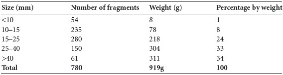
The fragmentation of the sample is shown in Table 3.162, with the largest fragment being 95mm in length. It can be seen that two thirds of the sample consisted of large or very large fragments and most of the remainder consisted of moderate-sized pieces. There was only a very low proportion of smaller fragments.
Identification
It was possible to identify 634g (69%) of the cremated bone. The proportion of identified bone from the various parts of the skeleton is shown in Table 3.163.
The identifiable features of the various bones are noted below.
Skull
Frontal: one unsided orbit.
Parietal: a large section of the posterior part of a parietal bone with the lambdoid sutures visible and with part of the occipital bone present, the posterior parts of left and right parietal bones with the sagittal suture almost obliterated and the left parietal foramen visible, and several other fragments of parietal bone.
Occipital: a large fragment of squamous occipital with the external occipital protuberance, strongly of the male type, and a right occipital condyle.
Temporal: fragments of the left and right temporal bones from around the mastoid and asterion areas, a right anterior suture and mandibular fossa.
Also present was a complete left and a complete right zygomatic bone and part of another right zygomatic bone, as well as a greater wing of sphenoid.
Mandible
A right mandibular condyle, coronoid process and part of the ramus, a fragment of the left side of the body split through the alveolus, the left angle and part of the ramus with one molar socket.
Table 3.163—Proportion of identified bone, sample 9, northern corner, upper level.

Maxilla
The orbital and nasal parts of the left bone; the right side of the maxilla with tooth sockets.
Dentition
Sockets for the following teeth were present:
Juvenile dentition: sockets for the following teeth were present:
Vertebrae
These included the dens articulation area of a first cervical vertebra, the dens from an axis, C2, and two left sides and one right side of the arches of lower cervical vertebrae, as well as two partial bodies of lower cervical vertebrae. The body, left side of the arch and right superior articular surface of a thoracic vertebra was present. There was also a body and the left and right superior articular surfaces from another thoracic vertebra, and the right side of the body and right superior articular surface from a twelfth thoracic vertebra. One other partial body and two other incomplete arches were present. In addition, there were a few transverse processes and several articular surfaces. A body and right superior surface from a lumbar vertebra was present, along with two other right sides of the body and fragments of posterior articular surfaces.
Ribs
There were at least three left and two right adult ribs present.
Pelvis
There was one right superior ramus from the pubic bone, part of an acetabulum, and fragments of ilium from around the acetabulum and from the iliac fossa. There was also a fragment of iliac crest from a juvenile.
Scapula
Fragments included the inferior part of the lateral border from a left bone, one coracoid process, the base and part of the acromion from a right bone, one fragment of glenoid fossa and a fragment of the blade.
Humerus
The proximal end and part of the shaft of a right humerus with the greater and lesser tubercles was present. There were fragments of proximal shaft from a right bone, and another fragment of proximal shaft probably from a left bone was also present. There were fragments of middle and distal shaft, including a fragment with the olecranon fossa visible. Two fragments of distal joint ends were also present.
Radius
The head and part of the neck from an adult bone survived, as well as another partial head and neck from another bone. There were fragments of at least two distal joint ends, one from a right bone, as well as fragments from proximal and middle shaft areas.
Ulna
This included most of the proximal half of the shaft of a left bone, the mid-shaft area of a right bone, other mid-shaft fragments, one distal shaft fragment, three fragments of the proximal joint surface and a small fragment of a distal joint surface.
Carpals/metacarpals/phalanges
A right lunate was present and there was a first metacarpal with the proximal end missing, the distal half of a second metacarpal, most of a third metacarpal, the proximal end and styloid process of a third metacarpal and one other metacarpal shaft. There were at least four proximal and two distal hand phalanges.
Femur
The posterior surface of the distal third of the shaft of a left femur was present. There were also mid-shaft fragments from the posterior surface of a left femur with the linea aspera present. These were from the same bone as the distal shaft. The proximal part of the shaft of a right femur was present and there was a neck of a left femur. Fragments from the anterior surface of a distal shaft area were also present.
Patella
An almost complete right patella and a smaller, almost complete left patella were present.
Tibia
Fragments from two proximal joint surfaces were present and there were large fragments from the mid-shaft area of a left tibia with the lateral surface present, as well as the anterior and part of the medial surfaces. The upper shaft of a right tibia with medial surface and part of the lateral surface visible was identified. There was also part of the posterior surface from the proximal end with the nutrient foramen visible. There were other fragments from the mid-shaft and the distal shaft areas.
Fibula
The distal end of a right bone, part of the distal end from another bone, a large fragment from the proximal shaft and a few fragments from the middle part of the shaft were present.
Tarsals/metatarsals/phalanges
The superior articular surface from one talus was identified, as well as the anterior half of a left calcaneum, one cuneiform, one navicular and a fragment of cuboid bone. The right fifth metatarsal was present, as well as the head of one other metatarsal and the proximal ends of two metatarsals. There was one proximal foot phalanx.
Pathology
There was a Schmorl’s node on the superior surface of a thoracic body. Osteoarthritis with eburnation of the joint surface was present in one of the thoracic posterior articular surfaces.
Sample 10, upcast
This sample consisted of 1,159g of cremated bone. The bone was efficiently cremated and was a creamy brown colour, with only one or two white or blue/white fragments.
Table 3.164—Fragmentation of bone, sample 10, upcast.
The fragmentation of the sample is shown in Table 3.164, with the largest fragment being 110mm in length. It can be seen that nearly 50% of the sample consisted of large or very large fragments, and most of the remainder consisted of moderate-sized pieces. There was a very low proportion of smaller fragments.
Identification
It was possible to identify 662g (57%) of the cremated bone. The proportion of identified bone from the various parts of the skeleton is shown in Table 3.165.
Table 3.165—Proportion of identified bone, sample 10, upcast.

The identifiable features of the various bones are noted below.
Skull
Frontal: several large fragments of squamous frontal bone, the glabella and part of the left
orbit and a small section of another orbit.
Parietal: there were several large fragments of parietal bone.
Occipital: a large fragment of squamous occipital with a very pronounced external occipital protuberance, indicating that it was probably from a male individual. There were also several other fragments of squamous occipital.
Temporal: one fragment of mastoid process and another mastoid process that was split vertically, as well as the area from just above the mastoid in the left temporal bone. There were three adult left petrous temporal bones and another unsided partial petrous temporal. Also present was a complete left zygomatic bone and an almost complete right zygomatic bone.
Juvenile skull: a left orbit from a juvenile skull and a left petrous temporal from a juvenile were present, as well as an unsided juvenile petrous temporal bone. A basilar occipital bone from an infant was also present (L 14.77mm), probably from an infant in the first year of life (Scheuer and Black 2000, 62).
Mandible
Two left and one other mandibular condyles as well as several fragments of the body from the same bone with tooth sockets.
Sockets for the following teeth were present:
In addition, there was another piece of mandible near the internal angle, with a socket possibly for another left third molar.
Maxilla
One fragment of the left side with sockets, a fragment from just below the zygomatic bone as well as a nasal part.
Sockets for the following teeth were present:
Teeth: the roots of the lower left second premolar, 35, were present, as well as two upper premolars and one other root of an anterior tooth. There were also two molar roots present.
Vertebrae
These included an axis, C2, consisting of the left and right superior articular surfaces, the body and the odontoid process (dens). There was a fragment from a superior articular surface of a lower cervical vertebra. The bodies of two juvenile vertebral bodies as well as another juvenile body with the right side of the arch were present. There were also bodies from three infant cervical vertebrae and part of a neural arch. The bodies of two thoracic vertebrae with the costal facets from the left side visible were present, as well as several articular facets from the posterior arch. There was a fragment of a lumbar body and another body with unfused epiphyses. There were a few articular surfaces present.
Ribs
Several small fragments of shaft were present but there were a minimum of four left ribs.
Sternum
A sterno-clavicular notch of the manubrium was present.
Pelvis
There were fragments of ilium, including a large fragment with most of the auricular surface from a right bone, as well as one fragment of acetabulum. There was also a fragment from the ala of a sacrum.
Scapula
The base of an acromion was present.
Clavicle
A fragment of clavicle shaft remained.
Humerus
The anterior surface of the distal third of a probable left bone was present, as well as part of the olecranon fossa from the posterior surface. There was also an olecranon fossa and trochlea from another bone and one other trochlea. Several fragments of shaft were present, including fragments from the anterior surface of the distal third of a right bone as well as fragments from the proximal end with muscle attachment areas visible.
Radius
Fragments from the proximal, middle and distal areas of shaft were identified, including a radial tuberosity. There was one fragment of radial head and a fragment of distal joint surface from a right bone.
Ulna
The olecranon and superior articular surface from a left bone and a similar fragment from a right bone were present. A partial fragment of the superior articular surface and olecranon from a left bone and the same fragment from a right bone were also present. There were also a few fragments from the proximal and distal areas of the shaft.
Carpals/metacarpals/phalanges
Bones present included a right trapezoid and a hook of hamate. There were five metacarpal shafts, two metacarpal heads and also the head of a first metacarpal. There were nine proximal phalanges including a first phalanx, all with their distal ends present, as well as two almost complete distal phalanges.
Femur
There were several fragments of shaft, including fragments from the anterior and posterior surfaces of the proximal, middle and distal areas, including a large fragment from the posterior surface at the distal end. A few fragments of well-developed linea aspera were present. A lesser trochanter was identified and there were fragments of proximal and distal joint ends, with a minimum of two distal ends and one proximal end present. There were also two proximal epiphyses from a juvenile.
Patella
The superior half of a right bone with strong muscle insertions was present.
Tibia
A large fragment of the right shaft, including almost all the distal half of the bone. There was another fragment of mid-shaft from a right bone, possibly the same bone as the distal half. A large fragment of shaft from a left bone was also present. There was a fragment with the interosseous border present, as well as several other fragments of shaft from the proximal end and a fragment of distal joint surface.
Fibula
The proximal third of a shaft and another fragment from mid-shaft were identified.
Tarsals/metatarsals/phalanges
There was a minimum of one calcaneum, two tali, one almost complete right first cuneiform, one middle cuneiform, one cuboid, a proximal end of the left fifth metatarsal, one other metatarsal, distal ends of two first metatarsals and distal ends of three other metatarsals. There were also three proximal phalanges and one distal phalanx.
Pathology
A superior articular surface from a cervical vertebra had evidence of severe osteoarthritis, with gross lipping and eburnation of the surface.
Sample 11, north-eastern corner
This sample consisted of 192g of cremated bone. The bone was efficiently cremated and was creamy white in colour.
Table 3.166—Fragmentation of bone, sample 11, north-eastern corner.

The fragmentation of the sample is shown in Table 3.166, with the largest fragment being 65mm in length. It can be seen that over 50% of the sample consisted of small or moderatesized pieces, and there was a low proportion of very large fragments.
Identification
It was possible to identify 50g (26%) of the cremated bone. The proportion of identified bone from the various parts of the skeleton is shown in Table 3.167.
The identifiable features of the various bones are noted below.
Skull
Frontal: fragments of squamous frontal and a fragment of the orbital part.
Parietal: fragments of parietal bone.
Temporal: a fragment of squamous temporal bone was present, as well as the anterior part of the right temporal bone.
Also present was part of an ethmoid bone and a root of an upper premolar tooth.
Vertebrae
These included a complete vertebral body from a first thoracic vertebra, some isolated posterior articular surfaces and one thoracic spinous process.
Table 3.167—Proportion of identified bone, sample 11, north-eastern corner.

Ribs
There were a few fragments of shaft.
Pelvis
Two fragments of ilium remained.
Scapula
One fragment of acromion was present.
Humerus
There was one fragment of shaft and a fragment of the distal end from a right humerus.
Ulna
This included the top of an olecranon and two fragments from the distal area of the shaft.
Carpals/metacarpals/phalanges
There were at least four proximal hand phalanges, one middle and one complete distal phalanx.
Femur
There were small fragments from the middle and distal areas of the shaft.
Tarsals/metatarsals/phalanges
The head of a first metatarsal and the head of one other metatarsal were identified.
368. Parish of Chapel, barony of Bantry. SMR WX030-090——. IGR 285910 131742.
369. UBA-9625. Calculated to 2 sigma by 14CHRONO Centre, Queen’s University, Belfast, using CALIB REV5.0.2.