2001:1398 - SLIGUFF, CO. CARLOW, Carlow
County: Carlow
Site name: SLIGUFF, CO. CARLOW
Sites and Monuments Record No.: SMR CW019-104
Licence number: E1036
Author: MARY CAHILL
Author/Organisation Address: —
Site type: Graves of indeterminate date
Period/Dating: —
ITM: E 669019m, N 656659m
Latitude, Longitude (decimal degrees): 52.656726, -6.979838
Introduction
In June 2001 human remains were discovered during quarrying operations at Sliguff, Co. Carlow. Workers noticed a skeleton as it tumbled from the top of the quarry. The site was first reported to the Garda Síochána, who removed all visible bone and sent the remains to the State Pathologist’s office for examination. The NMI was subsequently informed and the site was visited by Mary Cahill. Owing to the dangerous condition of the quarry face it was not possible to excavate the burial pit, but the in situ bones were removed. The bones previously collected by the Gardaí were acquired from the Office of the State Pathologist. The human remains were examined by Laureen Buckley.
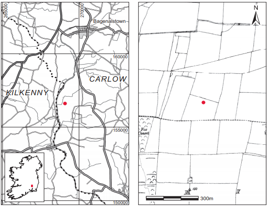
Location (Fig. 6.3)
The site was in the townland of Sliguff, south-west Co. Carlow.4 It was located on a gravel ridge between c. 30 and 60m above sea level. An early Bronze Age cist burial was excavated by Michael Ryan at Sliguff in 1974, approximately 1km from the present discovery (Ryan 1974a).
Description of site
The grave consisted of a simple pit, the outline of which was clearly visible in the quarry face. The outer edge of the pit was also visible from the quarry surface and the outline tapered towards the edge of the quarry, suggesting that the pit was oval in shape. It was aligned east/west and measured 2.3m by 0.4m by 0.6m deep. There was no evidence for stone lining. The grave originally contained an extended inhumation burial (2001:16), which was aligned west/east. The remains were those of a young adult male aged under 25 years at the time of death. By the time of the investigation only the left upper arm remained in situ so the layout of the body is not known, although the length of the pit would suggest that it was extended. The fill of the pit is described as a loose dark mixture of clay and sandy gravel. Some juvenile bones were also found with the adult skeleton, which would suggest that there may have been a number of other burials in the area.
Comment
There was nothing present on site to indicate a date for this burial, but local tradition records that men involved in the 1798 Rising had been buried in the locality. The presence of juvenile remains suggests, however, that other burials were present and that a mixed population was buried here.
HUMAN REMAINS
LAUREEN BUCKLEY
Introduction
The bones (2001:16) were complete and in quite good condition, although there was some decay on the joint ends of the left humerus and femur and also some decay of the outer cortex of the radius and ulna, as well as the femora and tibiae.
Description of burial
The skeleton was almost complete but the right arm and some of the vertebrae were missing. The skull was complete, with the cranium in one piece. Only the third cervical vertebra survived, and five thoracic and the upper three lumbar vertebrae were present. Four ribs from the left side and one from the right remained.
The left scapula was virtually complete and the shaft of the left clavicle was present. Only fragments remained from the right scapula. The bones of the left arm, humerus, radius and ulna, were complete but the right arm was missing. Only one proximal phalanx remained from the hand bones.
The pelvis was almost complete, with both ilia and ischia present, but the pubic bones were missing. The sacrum was also complete. Both femora and both tibiae were present from the leg bones and both were complete. There were no feet bones remaining.
Age and sex
All the features of the skull, the supraorbital ridges, orbital rims, mastoid processes and external occipital protuberance, were of the male type. The mandible also had typical male features. The epiphyseal line was still clearly visible on the iliac crest and the upper three sacral vertebrae were not completely fused together, suggesting that the individual was less than 25 years of age. The epiphyses at the heads of the ribs were just fused. The auricular surface of the ilium still had the ridge development typical of a young adult aged 19–24 years.
Skeletal pathology
Mild cribra orbitalia was present in both orbits. There were two small patches of periostitis on the posterior surface of the left scapula. The new fibre bone was in a very thin layer and the patches were only 1cm in diameter. There was a slight extension of the inferior right posterior articular surface of the first thoracic vertebra, which suggests that there was some compression of the joint between T1 and T2 that may have been caused by trauma. There was a moderate degree of osteophytic lipping around the margins of the articular surface. Mild marginal lipping also occurred between the joints of the third and fourth thoracic vertebrae. Schmorl’s nodes were present on the inferior surface of the body of one thoracic vertebra and on the upper three lumbar vertebrae, with the inferior surface of L1, the superior and inferior surfaces of L2 and the superior surface of L3 affected.
Dental pathology

Attrition: there was light wear on the incisors, canines and first molars but no wear on the second and third molars.
Calculus deposits: in the maxilla there were slight deposits on the buccal surfaces of the canine, right first premolar and second right molar. The left first molar had light deposits on the lingual surface. In the mandible there were slight deposits on the buccal surfaces of the right lateral incisor and canine and the left first premolar. The lingual surfaces of the central incisors, canines, left second premolar, the first molars and left second molar all had moderate calculus deposits. The distal surface of the left third molar had light deposits.
Periodontal disease: there was a very slight degree of alveolar recession around the left second premolar and first molar.
Additional bone
Additional decayed fragments that were probably from a parietal bone of a juvenile skull were present. This suggests that there were other graves in the same location.
Summary and conclusions
The remains represent one young adult male individual, aged 17–25 years at the time of death. The individual had a living stature of 169cm. Cribra orbitalia, which may be indicative of irondeficiency anaemia, was present in both orbits. The presence of Schmorl’s nodes in the vertebral column suggests that he had carried out heavy manual labour. The fact that some compression of joint spaces in the spine had already occurred and that some slight degeneration was already present further indicates that this individual had a strenuous lifestyle. Although the cause of death could not be determined, the presence of a slight amount of active periostitis on the left scapula suggests that the young man may have been weakened by an infection before death. There was only a small amount of ribs left so it was not possible to determine whether there was evidence for infection in the lungs. Lung infections such as pneumonia or tuberculosis can lead to death in a short time. Apart from the main skeleton, some fragments of juvenile skull were found, which suggests that other graves were present in the locality.
4. Parish of Lorum, barony of Idrone East. SMR CW019-104——. IGR 269084 156616.