1993:232 - CLONMEEN SOUTH, CO. LAOIS, Laois
County: Laois
Site name: CLONMEEN SOUTH, CO. LAOIS
Sites and Monuments Record No.: SMR LA033-010
Licence number: E1105
Author: NESSA OCONNOR
Author/Organisation Address: —
Site type: Graves of indeterminate date
Period/Dating: —
ITM: E 622811m, N 673795m
Latitude, Longitude (decimal degrees): 52.814655, -7.661611
Introduction
In June 1993 human remains were discovered during agricultural works near Rathdowney, Co. Laois. An area around a silage pit was being levelled when the bones were discovered against the wall of a ruined church. The burial had been disturbed by the digger and was thought by the landowner to be animal bone until skull fragments were noticed. By this time most of the bone had been disturbed. The NMI was informed of the find and a rescue excavation was undertaken by Nessa O’Connor.
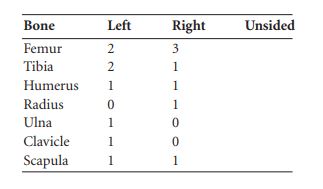
Location (Fig. 6.26)
The site was in the townland of Clonmeen South in south-west County Laois, close to the County Tipperary border.44 It lay against the south wall of a ruined Roman Catholic church, some 200m south-west of Clonmeen Abbey. The burial lay at an altitude of approximately 102m above sea level. The church is marked as being in ruins on the first edition OS 6in. Sheet. It is not possible to estimate the date of the burial other than to say that it is later in date than the church, as it abutted the church wall.
Description of site
The grave was located against the wall of a ruined church. It had been completely disturbed but there was no evidence for stone lining.45 The grave was aligned approximately west/east, with the head to the west. The maximum length recorded was 1.13m and the maximum width
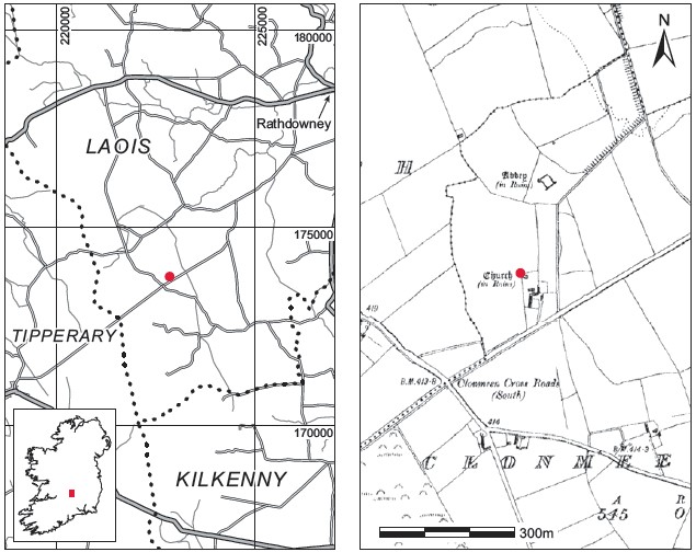
Fig. 6.26—Locationmap, ClonmeenSouth, Co. Laois.
0.48m. The site was completely disturbed, and only a few vertebrae (1999:89) remained in situ. The burial appears to have abutted the church wall, suggesting that it was deposited after the church was built. Other disarticulated bone in the vicinity was retrieved and brought to the Museum for analysis. A minimum number of five individuals were present, comprising three adults, a juvenile and an infant (see below)
.
Comment In the absence of associated finds or other dating evidence these burials must be regarded as undated, though the in situ burial probably post-dates the church.
HUMAN REMAINS
LAUREEN BUCKLEY
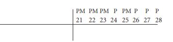
Adult bones
Left femur
(1) The proximal half of a left femur shaft.
(2) The proximal half of a left femur shaft.
(3) The distal half of a left femur.
Right femur
(1) The distal half of a right femur.
(2) The distal half of the shaft of a right femur.
(3) A section from the middle of the shaft of a right femur.
Left tibia
(1) The proximal third of a left tibia.
(2) The shaft of a left tibia.
(3) The distal half of a left tibia.
Right tibia
Only one right tibia was present and this consisted of the distal half of the bone.
Left humerus
One complete left humerus was present and the proximal epiphysis was just fused. This usually fuses by the age of 19–20 years. It was not possible to tell from the size of the head whether the bone was of a male or a female. (HuL1 304mm; HuHd 44.3mm, HuE1 56.6mm.)
Right humerus
One right humerus remained and consisted of the distal third of the shaft.
Radius
One radius only was present, the proximal third of a right radius.
Ulna
Only the proximal third of a left ulna remained.
Clavicle
The medial third of a left clavicle was present and the sternal epiphysis was unfused.
Scapulae
Fragments of a left and a right acromion from the scapulae were present.
Vertebrae
These included the arch of a first cervical vertebra, the centrum of a lower cervical vertebra, one thoracic vertebra and the right side of the neural arch of another thoracic vertebra. There was also a fragment of a sacral vertebra.
Ribs
There was a minimum of one left and three right ribs present, as well as a fragment of the body of the sternum.
Hand bones
Only a right capitate and the left fourth metacarpal remained from the hand bones.
Foot bones
These consisted of a right calcaneum, an unidentified metatarsal shaft and a distal foot Phalanx.
The minimum number of adults was three, based on the minimum number of right femora.
Adult skull bones
(1) Most of the squamous occipital bone and the posterior halves of a left and a right parietal bone from the one skull. The sutures were open so it was probably from a young individual.
(2) The left side of the frontal bone and the left anterior parietal bone from the one skull. It could not be determined with certainty whether or not this was the same skull as (1) above.
(3) Part of the left side of a frontal bone from another individual.
Also present from the skull bones were a left zygomatic bone, a left maxilla, the left side of a mandible and the right side and anterior part of another mandible.
The minimum number of adult skulls present was two, based on the left frontal bone and the mandibles.
In addition, there was a right lateral incisor and a right third molar, which may have belonged to the same individual.
Attrition: attrition was very light, with no wear on the second and third molars and only slight polishing of the first molar. These teeth probably came from a relatively young individual.
Calculus: there were slight deposits on the lingual surfaces of the loose incisor and third molar and moderate deposits on the buccal surface of the first molar. Deposits were moderate on the distal surface of the loose right third molar and this tooth also had considerable deposits on the buccal surface.
Hypoplasia: linear enamel hypoplasia was noted on the left second molar and the right lateral incisor. There were pits of hypoplasia on the left third molar.
Abrasion: there was a chip out of the bucco-occlusal edge of the crown of the first molar in the distal half. The canine had a vertical groove on the buccal surface near the occlusal edge.
Calculus: there were slight deposits of calculus on the buccal surfaces of the incisors and canine and on the lingual surfaces of the central incisor and canine. Deposits were moderate on the lingual surfaces of the premolars and the mesial and distal surface of the central incisor.
Hypoplasia: linear enamel hypoplasia was noted on the central incisor and canine.
There were no teeth remaining for examination. The roots for the second molar were very shallow, however, indicating severe periodontal disease.
Juvenile bones
The juvenile bones included the proximal third of a right ulna with unfused epiphysis, the distal epiphysis of a right tibia, part of a right talus, a fragment of scapula and a right calcaneum. Also present were four rib fragments, an arch of a thoracic vertebra and a hand phalanx. There was also a fragment of parietal bone and a right zygomatic bone from a skull. One tooth, a deciduous upper left first molar, was present and there was a cavity on the distal side of the crown.
Infant bones
The infant bones present consisted of the proximal half of a right femur, a left orbit and a skull Fragment.
Summary and conclusions
There were a minimum of five individuals recovered from this site: three adults, one of which was a young adult, an unaged juvenile and an infant. The right side of a maxilla and a mandible, probably from the young adult, were present and both showed little wear on the teeth and moderate calculus deposits. This individual had also suffered from acute illness or nutritional deficiency during early childhood and adolescence. Another adult mandible was present and although no teeth remained there was evidence of severe periodontal disease. A tooth from the juvenile was present and it had a caries cavity. Caries is rarely seen in juvenile dentition in prehistoric burials; its presence here suggests a late medieval or post-medieval date for these burials.
44. Parish of Rathdowney, barony of Clandonagh. SMR LA033-010. IGR 222866 173756.
45. A large slab found just north of the burial was thought to be connected with the ruined church.