1991:2008 - ARDMORE, CO. GALWAY, Galway
County: Galway
Site name: ARDMORE, CO. GALWAY
Sites and Monuments Record No.: N/A
Licence number: E1069
Author: ERIN GIBBONS
Author/Organisation Address: —
Site type: Late medieval graves, c. AD 12001600, and post-medieval graves, AD 16001800
Period/Dating: —
ITM: E 459520m, N 752300m
Latitude, Longitude (decimal degrees): 53.501920, -10.117571
Introduction
In January 1991 human remains were discovered exposed in a cliff face near Clifden, Co. Galway. A severe storm had revealed a portion of an unburnt skeleton approximately 1m below ground level. The site was reported to the NMI by archaeologist Erin Gibbons and permission was granted to her to investigate the site and to remove any loose material that had been exposed. The human remains were examined by Laureen Buckley and the bone was radiocarbon-dated. This report is based on Erin Gibbons’s account of the site.
Location (Fig. 5.5)
The site was located in the townland of Ardmore in west County Galway, approximately 8km west of Clifden,9 and was known locally as ‘the burial site of the sailor’. Description of site This site had been disturbed and was not completely excavated, so it is not possible to describe the form of the grave in detail. Its most notable feature was a slab (2000:39), 1m in length, which covered the grave. The slab originally lay in an east/west direction but had moved from its original location owing to erosion. A possible stone-lined pit feature was noted in the section to the north of the skeleton. The relationship between this and the burial was not established.
The burial consisted of an extended inhumation (1991:140), aligned east/west. Some
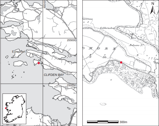
Fig. 5.5—Location map, Ardmore, Co. Galway.
fragments of shell material were recovered from the area around the skull, and a fragment of iron (1991:141) was recovered approximately 0.5m above the skull.
Comment
Radiocarbon dating of a portion of the skull yielded a date of 200±50 BP,10 which is calibrated as 1530–1955. This would indicate an early modern date for the burial and supports the local tradition of its being a sailor’s grave.
HUMAN REMAINS
LAUREEN BUCKLEY
Introduction
The human remains (1991:140) from this coastal site consisted of a well-preserved single skull and some vertebrae. As Viking burials had previously been found in the general area, it was thought that these might be from the same period. Radiocarbon dating results put them in the post-medieval period, however. According to local tradition, two Negroid sailors were washed up and buried here in the seventeenth century. The skull was therefore examined for physical traits that might help to identify ethnic origin.
Description
The cranium was partially fragmented but was virtually complete, with the occipital, both parietal, both temporal and frontal bones present. The base of the skull was very fragmented but the sphenoid bone was present. The maxillae and both zygomatic bones were present from the facial bones and the mandible was complete. All the cervical vertebrae and the first thoracic vertebra were present and complete.
Facial characteristics
Despite the fragmentation of the skull, most of the facial bones were in one piece and it was possible to assess ethnic origin. The observed presence of a nasal sill, retreating zygomatic bones and the flat orthognathous face are all Caucasoid features (Bass 1987). There were no Negroid or Mongoloid features present. The skull is therefore definitely that of a Caucasian.
Sex
The mastoid processes, external occipital protuberance and supraorbital ridges were all of the male type.
Age
Although the base of the skull was fragmented, the basio-sphenoid symphysis appeared to be fused, so the individual was aged at least 21 years at the time of death. Aging of individuals by the degree of suture closure of the skull is very unreliable, but the lack of significant closure indicates that the individual was not old. There was no degeneration of the cervical vertebrae, and in fact the epiphyseal line from the fusion of the epiphyses of the vertebral bodies was still visible in a few vertebrae. Since this epiphysis fuses at 25 years, it is likely that the individual was not much older than 25 years at the time of death. There was not a great deal of wear on the teeth, but post-medieval populations have generally less wear than in earlier periods and in any case it is not possible to assess age from one isolated set of dentition. In view of the lack of other aging features, such as the pubic and auricular surfaces in the pelvis and the sternal ends of the ribs, it is only possible to place this skeleton in the broad age range of early middle adult, 26–35 years, with the proviso that it is probably in the younger half of this range.
Anomalies
The metopic suture was retained. This suture, which separates the frontal bone into two halves, is usually fused by the seventh year of life. Occasionally it is retained as a non-metric trait into adult life but causes no problems for the individual.
Skeletal pathology
Cribra orbitalia was present in both orbits and porotic hyperostosis was present in both parietal bones. Porotic hyperostosis is a sign of iron-deficiency anaemia, which can also cause cribra orbitalia. To counteract the effects of anaemia, the body makes up in quantity what it cannot produce in quality. Therefore the bone marrow, where most blood cells are made, expands at the expense of compact bone, making the outer surfaces thin and porous. The anaemia can be caused not just by inadequate intake of iron in the diet but also by inadequate absorption of iron owing to nutritional imbalance, disease or parasitic infection.
Dental pathology

Anomalies: the lower left second deciduous molar, 75, was retained and the permanent second premolar had not developed. The upper canines were rotated by 45o mesially.
Attrition: there was light wear on the incisors but virtually no wear on the molars.
Calculus: there were light calculus deposits on the lingual surfaces of the upper teeth and most of the buccal surfaces of the lower teeth. Deposits were moderate on the buccal surfaces of the upper canines and first molars as well as the lingual surfaces of the lower premolars and first molars. There were heavy deposits on the lingual surfaces of the lower incisor and canine.
Periodontal disease
There was a slight degree of recession around the sockets of the premolars and molars of the right maxilla and right side of the mandible.
Enamel hypoplasia
Linear enamel hypoplasia was present in the upper incisor, canines, premolars and molars. There were also pits of hypoplasia in the left incisor, canine and second molar and the right premolars and second molars. Hypoplasia is a defect in the enamel caused by nutritional deficiency or acute infection during childhood when the tooth is developing. The age at which the episodes of hypoplasia occurred can be determined by the position of the line on the tooth. In this individual the episodes occurred between the ages of two and six years.
Summary
These remains were the skull and upper vertebrae of an adult Caucasoid male, aged between infection during early childhood and iron-deficiency anaemia in later life, there were no major pathological conditions or injuries on the skeleton. There were a few minor anomalies, including a retained metopic suture, a retained deciduous molar and rotation of canine teeth, which may have had a genetic component.
9. Parish of Omey, barony of Ballynahinch. OS 6in. sheet 35. IGR 059540 252277.
10. GrA-17122.