1990:148 - RATHCONRATH, CO. WESTMEATH, Westmeath
County: Westmeath
Site name: RATHCONRATH, CO. WESTMEATH
Sites and Monuments Record No.: SMR WM018-150
Licence number: E1176
Author: RAGHNALL Ó FLOINN
Author/Organisation Address: —
Site type: Early Bronze Age graves
Period/Dating: —
ITM: E 631555m, N 753053m
Latitude, Longitude (decimal degrees): 53.526495, -7.524083
Introduction
In March 1990 a short cist containing a vessel and a cremation was discovered during ploughing near Mullingar, Co. Westmeath. The large capstone was displaced by the plough, revealing the chamber. The vessel was removed from the cist by the landowner for safekeeping and the find was reported to the NMI. A rescue excavation was carried out by Raghnall Ó Floinn and Stella Cherry. During the excavation, a second large stone, also displaced by ploughing, was noticed. It transpired that this was the capstone of a second cist, which was excavated the following week.

Location (Fig. 3.194)
The cist was located on a low rise in a field known as ‘Racepark’ in the townland of Rathconrath, mid-Co. Westmeath.337 According to the landowners, the Gibson-Brabazon family, the field had not been ploughed in living memory. The surrounding area is rich in early Bronze Age burials. Just over 1km to the south-west, in the townland of Glascarn, an early Bronze Age cist burial containing a cremation, an inhumation and a vase was excavated by Fionnbarr Moore in 1986. The cemetery at Sonnagh Demesne, excavated by Breandán Ó Ríordáin (this volume, pp 552–9), is just 5km to the north-east, and some 2.5km north-west of the site, in the townland of Davidstown, a number of inhumation burials were excavated by Eamonn P. Kelly in 1976 (Vol. 2, pp 201–3).
Grave 1
The cist was trapezoidal in plan and its long axis was aligned south-east/north-west, with the wider end at the south-east (Fig. 3.195). Internally it measured 0.94m long by 0.56m wide by 0.41m high. It was constructed of four slabs set on edge. The trapezoidal plan was exaggerated by the fact that the western side stone had been displaced by the action of the plough, as had its accompanying padstone. Padstones composed of smaller slabs were laid flat along each side, either overlapping or abutting the side stones. Two padstones were found laid diagonally across the southern and northern corners of the cist. The floor of the cist consisted of a single large slab, which had subsequently split in two, dipping towards the centre of the cist. This appears to have been placed in position before the side stones were put in place as it ran under the eastern side stone. The cist had originally been sealed by a tight-fitting capstone large enough to prevent spoil from slipping into the cist chamber.
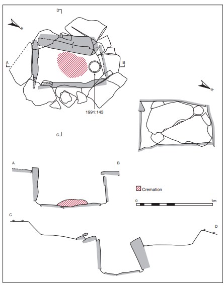
The capstone was suboval in shape, measuring 1.68m long by 1.14m wide. It bore two sets of grooves running roughly at right angles on its upper surface, probably made by ploughshares on previous occasions. At the time of excavation, the capstone lay to the south of the cist, partly covering the padstones on the southern side of the cist. There was no indication of the edge of the pit dug to receive the cist, but it appears that the pit walls lay under the padstones, which were not removed in the course of the excavation. The pit had been dug into a fine, dark grey sandy gravel (visible in the floor of the pit), and this, mixed with boulder clay, was found behind the side stones and under the padstones, where excavated.
The cist contained the cremated remains of two adults, one male, and cremated animal bone (1991:142),338 accompanied by a tripartite bowl. According to the finders, the vessel was placed mouth upwards on the floor of the cist midway along the northern end. The cremation was untouched and no spoil had spilled into the chamber. The cremation consisted of an oval spread of burnt bone, placed centrally, measuring about 0.4m by 0.3m by 0.1m in maximum height. The larger bones lay on top while the smaller were underneath, being concentrated in the eastern side of the spread. Some of the bone had fallen into the central crack in the floor stone. The remains were examined by Laureen Buckley and found to represent a minimum of two individuals, both apparently adult, at least one being an older adult male.
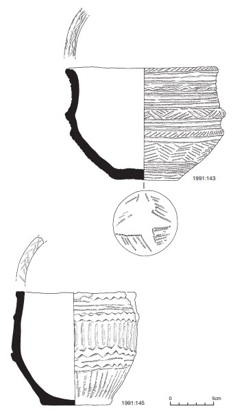
Tripartite bowl, 1991:143 (Fig. 3.196)
This is a buff-coloured bowl, probably of tripartite type, as classified by Ó Ríordáin and Waddell (1993). The vessel is complete, with medium to large grits (maximum 8mm long) visible, particularly on the interior. It is decorated all over with impressed decoration, and the external surface bears two horizontal ribs that give the impression of a shoulder groove. The rim of the vessel is slightly bevelled and bears decoration in the form of a bar chevron in false relief delimited on either side by a single incised line. The outer part of the rim is decorated with obliquely disposed short comb impressions, and the base (of the rim) is delimited by a single horizontal row of comb impressions. The neck/upper section of the vessel is decorated by a series of alternating incised horizontal lines and horizontal bands of comb impressions, which delimit a central band of bar chevron executed in false relief. The upper rib is decorated with a bar chevron, again in false relief, and the blank areas are infilled with comb-impressed decoration. The narrow area between the first and second ribs appears as a shoulder groove and was decorated with three horizontal incised lines. The lower rib is decorated with a series of short, oblique comb impressions. The lowest portion of decoration on the vessel consists of comb impressions forming a chevron motif around the circumference of the vessel. This pattern is repeated six times and the blank areas are filled with comb-impressed decoration. This zone of chevrons is bordered above by impressed horizontal lines and below by impressed lines and lines of comb impressions. The base is decorated with an incised starshaped motif with the angles defined by irregular comb impressions.
Dimensions: H 12.4cm; ext. D rim 17.5cm; int. D rim 15.5cm; D base 7.52cm; T rim 1.1cm.
Grave 2
About 5m north-east of grave 1 a flat stone, less massive in size, had been displaced. This too had a series of ploughmarks on its upper surface. According to the landowner, Mr GibsonBrabazon, this was aligned roughly east/west, i.e. at right angles to the first cist. Investigation of an area measuring c. 1.8m by 1.9m around its original position revealed a second stone, 3m east of grave 1, with roughly the same orientation (Fig. 3.197). This was revealed to be a second capstone of the cist, which had been undisturbed prior to excavation.
The cist was rectangular in plan, with its long axis aligned east-north-east/west-southwest. Internally it measured 0.84m long by 0.51m wide by 0.48m high. It was constructed of four main edge-set slabs, one forming each wall. The end stones inclined inwards from the base and were fitted neatly between the side stones. All the main slabs appeared to be of sandstone. The western end stone did not extend the full width of the cist, and three smaller stones, one above the other, filled the gap at its southern end. The floor was paved with a number of slabs, which had cracked and were sloped downwards towards the centre of the cist.
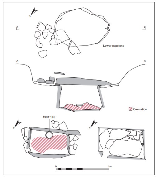
There was a notable slope in the floor of the cist from north to south. The cist was apparently covered with two capstones, one above the other. The uppermost indicated the presence of the cist. This was suboval in shape, measuring 1.46m long by 1.08m wide. Below this was the second capstone, which measured 1m long by 0.7m wide. Fragments of a large padstone were discovered resting on its upper surface at the north-eastern end, but these may have been disturbed on discovery.
The cist contained the cremated remains of a young adult male and a juvenile (1991:144) accompanied by a bowl. As in the case of grave 1, no soil had infiltrated the chamber. The burial deposit consisted of two piles of cremated bone placed along the central axis, forming a figure of eight spread 0.7m long by 0.3m wide. Some of the cremated bone had fallen into the cracks in the paved floor of the cist. Some large pieces of charcoal were visible resting on top of the eastern concentration of bones. Further smaller pieces of burnt bone and dust had been washed through to the base of the deposit. Some of the bones in the western concentration were resting in a vertical position, others at an acute angle as if poured onto the floor of the cist from a container. The fragments of bone were much larger than those found in grave 1 and seemed to represent an adult. Each pile of bones was removed and bagged separately. The westernmost deposit contained the complete cremation of an adult male, while the eastern area consisted of the remains of an older juvenile or adolescent.
Tripartite bowl, 1991:145 (Fig. 3.196)
This vessel is a tripartite bowl decorated with impressed decoration; a portion of the rim and body is missing, but the vessel has been reconstructed. The ware is red to red-brown in colour, with grits visible on the inside and fibrous root-like material visible in parts of the outer surface (possibly as a result of roots in the cist). It bears two horizontal ribs or mouldings, which delimit the widest parts of the vessel. The mouldings or ribs are executed in false relief by the use of impressed triangles on either side, and thus appear as bands of bar chevrons. The decoration is thus in three parts, the narrowest band at the neck, and the two wider bands at either side of the lower rib. The internal profile of the vessel appears almost bipartite, as the short neck is almost vertical (see inner profile).
The rim is flat and bears some decoration, which is extremely faint. The decoration appears to be in the form of either impressed triangles or short impressed strokes.
The rim is delimited externally by a horizontal band of impressed chevrons. Below this is a series of three to four thin horizontal lines formed of cord impressions. This horizontal decoration is bordered by the first rib. Below the first rib is a wide band of decoration (approximately 3.35cm wide) consisting of vertical bands of comb impressions. These bands are very neatly executed and evenly spaced across the circumference of the vessel. This is delimited by the second rib on the vessel. A single line of cord impression occurs directly underneath the second moulding. This is poorly executed; the line is slanting and often non-contiguous. Below this is a single horizontal band of triangular impressions. These, along with the lower line of impressed triangles in the rib, give the impression of a second smaller and flatter moulding. Delimiting this decoration underneath is another horizontal line of cord impression. The final band of ornamentation on the vessel consists of vertical bands of comb impressions, mirrored in the upper band between the two ribs. The upper part of each vertical line is executed using a thin combed instrument, which is followed directly by the impression of a thicker instrument. It is possible that this decoration was executed using a single tool with different-sized teeth, as it is extremely neat and regular. The base bears a very faint diamond-shaped impression.
Dimensions: H 10.8cm; ext. D rim 13.75cm; int. D rim 11.93cm; D base 7.59cm; T rim 1cm.
Comment
The human remains from this site have not been dated, but the bowl from grave 1, on the basis of form and decoration, would be considered as a tripartite (variant) bowl. This unusual vessel is very similar to an unlocalised vessel illustrated by Ó Ríordáin and Waddell (1993, 212, no. 268). The bowl from grave 2 is a very typical tripartite bowl of similar style to those which Brindley (2007, 172–3) places in stage 1 of the development of bowl tradition pottery. This phase is dated to 2160–1980 BC. The bowl from grave 1 is most similar to vessels of Brindley’s stage 3 series (2007, 174–5, 248–9) such as Lug, Co. Offaly (Fig. 3.152), and Baggotstown, Co. Limerick (Fig. 3.96). Stage 3 vessels are dated to 1980–1930/20 BC. A small quantity of animal bone was found in the cremation deposit from cist 1. This may represent the remains of the last meal of the interred or a symbolic offering to accompany the spirit on its final journey.
HUMAN REMAINS
LAUREEN BUCKLEY
Grave 1 (1991:142)
The sample consisted of 5,304 fragments of cremated bone, weighing a total of 2,885g. This, however, included seven fragments (31g) of animal bone. The bone was multicoloured, with practically every bone a different colour. Some fragments were creamy white and some were grey. Several fragments were an orangey brown colour, and several were stained a blue-green colour. In addition, several fragments were encrusted in limestone deposits. It is presumed that the orange colour came from iron deposits and the blue-green colour from copper deposits in the groundwater. Nevertheless, most of the bone seemed to be efficiently cremated, with numerous horizontal fissures on the bone surface, although some of the vertebral bodies had barely changed at all.
Table 3.117—Fragmentation of bone, grave 1, 1991:142.
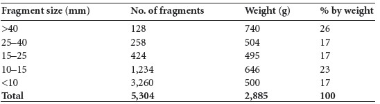
The fragmentation of the sample is shown in Table 3.117, with the largest fragment being 99mm long. It can be seen that there is only a relatively small proportion of very large fragments but that most of the sample is made up of large fragments more than 15mm in length. There is, however, a significant quantity of fragments less than 15mm in length. This probably reflects the thorough collection of all the bone from the cist. In some of the very early excavations of cists only the larger fragments were collected and the smaller fragments seem to have been ignored. In addition, some crushing of smaller fragments may have occurred after deposition, since it was discovered during excavation that the larger fragments of bone had been placed on top of the smaller fragments.
Identifiable bone
A total of 1,249g (44% of the human bone) was identified (Table 3.118). This is a reasonable percentage of identifiable bone, considering that there was a high proportion of large fragments but a relatively small amount of very large fragments. It is similar to other Bronze Age cremations where the proportion of very large fragments was also quite low.
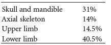
Table 3.119 summarises the proportions of various body parts identified. It can be seen that the skull is overrepresented in this sample, as the proportion is nearly twice what it should be. The axial skeleton is just over half of what it should be. The amount of upper limb bone was also lower than expected and the amount of lower limb was only slightly higher than is normally present in a full cremation.
Since all of the bone was collected from the cist, it is highly likely that when the bone was originally collected from the funeral pyre the skull was favoured over the smaller bones of the vertebrae and ribs, and the larger leg bones were favoured over the arm bones.
Skull
There was a large fragment of the occipital bone with a very pronounced external occipital protuberance. The bone was 12mm thick. Another large section of occipital bone was present with part of the parietal bone attached through a fused lambdoid suture. Two left temporal bones were present, one with the superior part of the temporal fossa and the superior part of the external auditory meatus present. The posterior part of the zygomatic arch was of the male type. Part of the mastoid process was also present and it was very large, also of the male type. The second left temporal bone had most of the mandibular fossa and the petrous part of the temporal bone present. There were another two complete petrous temporal bones. The right orbit and the left supraorbital area were present. It was also of the male type. The right zygomatic bone was present. There were several large fragments of parietal bone and squamous frontal present.
Table 3.118—Proportion of identified bone, grave 1, 1991:142.
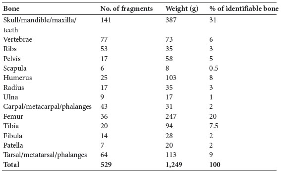
Table 3.119—Summary of identified bone, grave 1, 1991:142.
Mandible and maxilla
Most of the right side of the body of the mandible was present and there were a few small fragments of the mandibular body, including a small fragment with the genial tubercle visible. There was also a left mandibular condyle. A fragment of the middle of the maxilla but with no sockets remaining was present.
Dentition
The following tooth sockets were present:
Mandibular tori were present.
The roots and most of the crown of the third molar, 48, were present. There were two other molar roots and two incisor roots. It was possible to deduce from the shape of the socket for 48 that the third molar had impacted. It was lying on its side in the alveolus. Since most of the crown had not erupted and was protected by the jaw, it had not shattered, as erupted adult teeth normally do during cremation.
Vertebrae
The dens articulation area and most of the left and right articular surfaces of the first cervical vertebra were present, as well as the dens of the second cervical vertebra. A few fragments of the body and some arches from the lower cervical vertebrae were present. Part of the bodies and arches of a few thoracic vertebrae were present, and there were several fragments of body and several fragments of arches of the lumbar vertebrae. There was osteophytic lipping of the cervical bodies and severe osteophytosis on the lumbar vertebrae. There was also some marginal lipping on the posterior articular surfaces of the lumbar vertebrae.
Ribs
Several rib fragments were present; most were from the shaft and some were split through the middle of the bone.
Pelvis
Fragments of acetabulum and other small fragments of ilium were present, as well as one large right ischium.
Scapulae
A fragment of lateral border and the left glenoid and acromion were present.
Humerus
Most of the fragments were shaft fragments, including fragments from the distal end of a left humerus with the capitulum and part of the trochlea present. The distal end of the right humerus was also present and there were a few fragments of a proximal end.
Radius
Fragments from the proximal and distal halves of the shaft were present. There were also two proximal joint ends and fragments of distal joint surfaces.
The heads of the radii were two different sizes (RaHd 21.2mm; RaHd 18.6mm). While owing to the shrinkage of the bone during the cremation process this would not be accurate enough to determine the sex of the individuals, it does indicate that two different individuals were present.
Ulna
There were several fragments from the middle and distal parts of shaft.
Carpals, metacarpals and phalanges
A left and right lunate, left and right trapezium, left and right capitate and left trapezoid were present from the carpal bones, as well as one scaphoid bone. Two first metacarpals, one second, two third metacarpals, one fourth and one fifth metacarpals were present, and there were ten proximal, four middle and seven distal hand phalanges.
Femur
Most of the femur fragments were stained orange, although some had a blue/green coating as well. The proximal third of a right bone was present and the lesser trochanter was very well developed. There were also a few fragments of the proximal articular surface. Most of the fragments were shaft fragments, some with the linea aspera present, and it was very well developed. A large fragment of the distal end of the shaft was present and there was a fragmented distal joint end. One of the fragments was eggshell blue.
Tibia
All the fragments were of the shaft, including a large fragment of the posterior surface with the nutrient foramen present. There was also a fragment with the anterior tubercle visible, as well as fragments from the medial and lateral surfaces.
Fibula
There were fragments of shaft and the distal end of a right fibula, which was a very bright turquoise blue colour.
Patella
One left and one right patella were present.
Tarsals/metatarsals
Fragments from two tali, two naviculars, a fragment of calcaneum, a fragment of cuneiform and a fragment of cuboid bone remained from the tarsal bones. The heads of two first metatarsals, the proximal end of a fifth metatarsal and part of the shaft of one other metatarsal were present, as well as two first proximal phalanges, one distal phalanx and two sesamoid bones.
Minimum number of individuals
There appear to be two individuals in this sample, based on the number of left temporal bones present. Both seem to be adult and at least one is an older adult male with severe osteophytosis in the vertebral column.
Grave 2, west end (1991:144)
This was a short cist in which two cremations had been placed in two piles in the centre, to form a figure of eight. The bone was collected as two samples, one pile from the western side of the cist and one pile from the eastern end. There were also two vessels in this cist. The sample from the western end consisted of 2,722 fragments of cremated bone, weighing a total of 2,074g. The bone was very white and clean and well calcined, with numerous horizontal fissures and distortion of the bone. Some fragments were heavily stained an orange colour, probably caused by iron deposits leaching out of the groundwater. There were deposits of a calcium compound on the long bones. A few small fragments were blue/black in colour, indicating less efficient cremation.
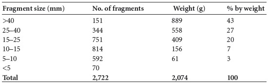
The fragmentation of the sample is shown in Table 3.120, with the largest fragment measuring 145mm. It can be seen that almost half the sample consists of very large fragments and that almost all the sample is made up of large fragments more than 15mm in length. Most of the smaller fragments probably came from post-excavation fragmentation of the bone. It seems reasonable to assume that the bone was not deliberately crushed after collection from the funeral pyre.
Table 3.120—Fragmentation of bone, grave 2, west end, 1991:144.
Identifiable bone
A total of 1,432g (69% of the total bone) was identified (Table 3.121). This is a high percentage of identifiable bone and is due to the number of very large fragments.
Table 3.121—Proportion of identified bone, grave 2, west end, 1991:144.
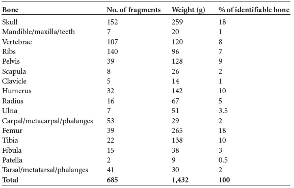
Table 3.122—Summary of identified bone, grave 2, west end, 1991:144.
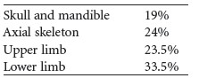
Table 3.122 summarises the main parts of the skeleton identified from this sample. The proportion of skeletal parts is almost exactly what would be expected from a normal cremation. There is only a slightly higher amount of the upper limb at the expense of the lower limb. Since all of the bone was collected from the cist, it seems that all the bone from the skeleton was carefully collected from the funeral pyre, including the fragile bones of the vertebral column and the small bones of the hands and feet.
Skull
Bones present included the left orbital part of the frontal bone and the middle and left parts of the squamous frontal bone, which was stained with orange. Part of the outer cortex of the frontal bone was missing but the maximum thickness of the bone was 6mm. The internal surface with the frontal sinus and crista frontalis was intact.
There was a large fragment of the left side of the occipital bone with a very pronounced external occipital protuberance. The internal occipital protuberance was also visible. The left and right petrous temporal bones were present, as well as the right temporal fossa and anterior suture. The left zygomatic bone and left and right nasal bones were also present, as well as the left and right greater wings of sphenoid.
There were several large fragments of parietal bone present. The sutures did not appear to be well fused and there was a lambdoid ossicle present.
Mandible and maxilla
There was a large section of the left side of the mandible near the angle with some sockets visible. Part of the body, including the anterior part with the genial tubercles on the internal surface, was present.
There was a large fragment of the left side of the maxilla but with no sockets intact.
Dentition
The following tooth sockets were present:

There were two fragments of molar roots, one with a shattered crown, and the roots of two anterior teeth. The roots of a maxillary molar, probably the third left, were also present.
Vertebrae
The dens articulation area and most of the arch of the first cervical vertebra were present, as well as the dens of the second cervical vertebra. At least five bodies and disarticulated arches from the lower cervical vertebrae were present. Parts of the bodies of twelve thoracic vertebrae were present, as well as some fragments of neural arches. Five complete lumbar bodies survived, as well as a few articular surfaces from the neural arches. The fifth lumbar vertebra was very blue.
Ribs
There were at least seven left and nine right ribs present. The sternal ends of five ribs were also present. There were eight tubercles and two heads remaining. The epiphyses at the heads had just fused. The other fragments were fragments of shaft. There was some pale blue staining on the front of the ribs. The sternal ends of the ribs appeared quite young, with some scalloping of their edges.
Pelvis
Fragments of ilium with the iliac crest fused, from at least two ilia, were present. There was also one complete left acetabulum and another fragmented acetabulum. There was a large fragment of the right ilium with the auricular surface present. The right ischium was complete and there was one left and one right pubic bone from a male. The right pubic bone was complete and seemed to be from a young adult. The body of the first sacral vertebra and the ala were also present.
Clavicle
The shafts of one left and one right clavicle were present, as well as a fragment of medial end.
Scapulae
The right glenoid fossa and most of the acromial spine were present. There was also a left acromial spine and a right coracoid process.
Humerus
The proximal ends of two bones, including the proximal ends of the shaft, were present, as well as other shaft fragments and a fragment of trochlea from the distal end.
Radius
The proximal end and head of a right radius was present (RaHd 22.8mm), and there was also a radial tuberosity from a left and a right radius. The distal third of a left radius with fused distal joint end was present, as well as a distal end and fused distal joint end from a right radius. There were also large fragments of shaft from a left and a right radius.
Ulna
There were several large fragments of shaft from left and right ulnae, including the proximal shaft and the distal thirds of shaft from two bones. There was one proximal and one distal joint end.
Carpals, metacarpals and phalanges
A left and right lunate, left and right scaphoid, left triquetral and right capitate were present from the carpal bones. Two first metacarpals and eight metacarpal shafts were present, as well as two fragments of metacarpal heads. There were six proximal, two middle and three distal hand phalanges present.
Femur
There was one very large piece of a left femur, with the head and neck and proximal shaft visible. The epiphyseal line of the head was still vaguely visible. The head and neck of a right bone was present but fragmented. Most of the fragments were shaft fragments and some were very large and thick. Two fragments of distal joint end were present.
Tibia
All the fragments were of the shaft, including some very large fragments. There were fragments of the posterior and lateral surfaces and the interosseous border was visible. There was also a large fragment with the anterior border and medial surface of the right bone visible. A fragment from the proximal posterior shaft with nutrient foramina from two bones, a left and a right, were present; there were also two anterior tubercles visible, as well as fragments from the distal shaft and one distal joint surface.
Fibula
Fragments of shaft and the distal ends of two fibulae were present.
Patella
One left and one right patella were present. They were almost complete and both had a vastus notch.
Tarsals/metatarsals
Fragments from one talus, one navicular, one calcaneum, a fragment of cuneiform and a fragment of another navicular remained from the tarsal bones. There were six metatarsal shafts, including a left fifth metatarsal and a few metatarsal heads. Two first proximal phalanges and one other phalanx were also present.
Minimum number of individuals
There appears to be one individual present, as there is no repetition of skeletal elements. It seems to be the remains of one young adult male, with all his remains carefully collected from the funeral pyre and placed carefully in the cist without unnecessary crushing of the bone.
Grave 2, east end (1991:144)
The sample from the eastern end of the cist consisted of 4,900 fragments of cremated bone, weighing a total of 1,972g. The bone was creamy white with a chalky texture. A few of the metatarsals were a blue colour where the cremation had not been so efficient. Some of the fragments were stained orange and encrusted with deposits, but not as much as those on the western side of the cist.
Table 3.123—Fragmentation of bone, grave 2, east end, 1991:144.
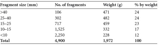
The fragmentation of the sample is shown in Table 3.123, with the largest fragment measuring 110mm. It can be seen that there is only a relatively small proportion of very large fragments but that almost three quarters of the sample is made up of large fragments more than 15mm in length. There is, however, a significant quantity of fragments less than 15mm in length. With half the sample consisting of fragments over 25mm in length, it is unlikely that deliberate crushing of the bone took place.
Identifiable bone
A total of 938g (47% of the bone) was identified (Table 3.124). This is a reasonable percentage of identifiable bone and compares well to the 44% of identifiable bone from cist 1, where there was a similar pattern of fragmentation
Table 3.124—Proportion of identified bone, grave 2, east end, 1991:144.
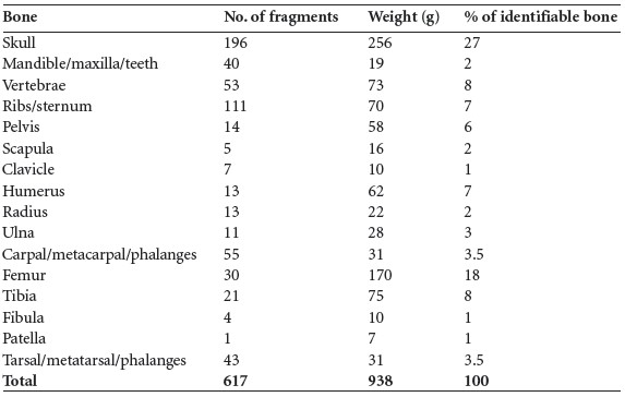
Table 3.125—Proportion of identified bone, grave 2, east end, 1991:144.
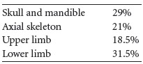
Table 3.125 summarises the main parts of the skeleton identified from this sample. It can be seen that the skull is overrepresented, although not as much as in some Bronze Age cremations, such as that from cist 1. The axial skeleton is as might be expected. The amount of upper limb bone was also similar to that expected and the amount of lower limb was only slightly smaller than is normally present in a full cremation.
Skull
The left and right petrous parts of the temporal bones were present, as well as the mandibular fossa of the right temporal bone. There were large fragments of the frontal bone, with the crista frontalis on the internal surface visible on one fragment. There were several large fragments of parietal bone and squamous frontal present, as well as some fragments of occipital bone.
Mandible and maxilla
Most of the left side of the ramus and two fragments of the body of the mandible were present, making the mandible almost complete. A fragment of the right side and part of the left side of the maxilla were present, along with a fragment of right side of the nasal border from a second maxilla.
Dentition
The following tooth sockets were present:
The upper right first molar, 16, and the lower left first molar, 36, were still in situ in the jaw. The upper first left molar was present and there were two deciduous molars, as well as the crown of an adult upper canine and the crown of a premolar. There were also roots from various incisors and molars.
Vertebrae
The right side of the first and the dens and right side of the second cervical vertebrae were present, as well as five lower cervical bodies and fragments of posterior articular surfaces. Parts of the bodies of ten thoracic vertebrae were present, but there were only a few fragments of vertebral arches. Two of the bodies of lumbar vertebrae were present and there were several fragments of articular surfaces.
Ribs/sternum
Several rib fragments were present; most were from the shaft, although at least four left and two right tubercles were present. The almost complete manubrium of the sternum was present.
Pelvis
A fragment of right ilium with the acetabulum and a fragment of another acetabulum were present, as well as other parts of the left and right ilium. There was also one right ischium and one left ischium with unfused epiphyses. The first sacral body, part of the second sacral body and a fragment of the third were present.
Scapulae
The left lateral border and inferior border were present, as well as the right glenoid and left and right acromions.
Clavicles
The left and right shafts and two sternal ends of clavicle were present.
Humerus
Most of the fragments were shaft fragments, including fragments from the left and the right proximal thirds of the bone, the distal end of a left and a right humerus with the epiphyses fused and the left and right proximal epiphyses.
Radius
The proximal third of a right bone was present and the epiphysis was unfused. The proximal diaphysis and metaphysis from another bone were present, as well as some fragments of shaft. The distal ends of left and right radii were also present. Two proximal epiphyses and a fragment of one other were present.
Ulna
There were several fragments from the middle and distal parts of shaft, the proximal end of a right bone and a distal epiphysis, as well as a distal metaphysis.
Carpals, metacarpals and phalanges
A right lunate, first and second metacarpal shafts and six other metacarpals were present, and there were ten proximal, thirteen middle and eleven distal hand phalanges present.
Femur
The neck areas of the left and right bones were present with the left epiphysis unfused. The right metaphysis had a flat articular surface fused to it. This could be a flattened femoral head, such as is seen in a slipped epiphysis or Perthes disease. As there is no sign of infection this is unlikely to be Perthes disease, but a slipped femoral head epiphysis must be considered (Pl. 108).
Also present was the greater trochanter of one shaft and part of another. There was also the distal third of a shaft, an incomplete distal end and a proximal epiphysis, as well as various fragments of shaft. Most of the femur fragments were large and thick and probably belonged to an adult. The greater trochanter from one shaft and part of a greater trochanter from another bone were present.
Tibia
Fragments of distal and proximal metaphysis and diaphysis were present, as well as one
proximal and two distal epiphyses and fragments of shaft.
Fibula
Fragments of shaft only were present.
Patella
One right patella was present and virtually complete.
Tarsals/metatarsals
Fragments from a talus, two naviculars, fragments from the anterior parts of two calcanea and the posterior part of one calcaneum, a fragment of middle cuneiform and a fragment of cuboid bone remained from the tarsal bones. The heads of two first metatarsals, the left fifth metatarsal and part of the shaft of four other metatarsals were present, as well as two middle and two distal phalanges.
Minimum number of individuals
There appear to be two individuals in this sample, based on the number of phalanges and the fact that some adult femur shaft was present along with all the juvenile bone. Since so little of the adult bone remained, however, and since the amount of adult femur in the western side of the cist was slightly lower than expected, it is probable that the adult bone ‘strayed’ into the eastern side of the cist but really belonged to the western end. The main cremation in the eastern side of the cist seems to be that of an adolescent at least 10–12 years old at the time of death but probably 15–20 years. The individual may have suffered from a slipped femoral epiphysis.
Summary and conclusions
The cremated remains from two cists were examined. The bone from all the cists was efficiently cremated but had been subsequently stained by minerals in the groundwater. A large quantity of bone was collected for each cremation and there was very little crushing of the bone. From the identified bone it could be determined that cist 1 contained the remains of at least two adults, one of whom was an older male who suffered from degenerative joint disease of the spine. One of the individuals had an impacted third molar.
Cist 2 was divided into two areas. The western area contained the complete cremation of a young adult male. Virtually all the burial was collected from the funeral pyre and the bone was not crushed but placed carefully in the cist, with all skeletal elements collected equally. The eastern area consisted of the cremated remains of an older juvenile or adolescent, with some stray adult bone from the western end. It is possible that the juvenile had suffered a slipped femoral epiphysis.
337. Parish of Rathconrath, barony of Rathconrath. SMR WM018-150——. IGR 231612 253031.
338. The animal bone was identified when the human remains were examined and was thus registered with the human remains.