1990:146 - GREENMOUNT, CORK CITY, CO. CORK,, Cork
County: Cork
Site name: GREENMOUNT, CORK CITY, CO. CORK,
Sites and Monuments Record No.: SMR CO074-075
Licence number: E1045
Author: STELLA CHERRY
Author/Organisation Address: —
Site type: Graves of indeterminate date
Period/Dating: —
ITM: E 566838m, N 571045m
Latitude, Longitude (decimal degrees): 51.890585, -8.481770
Introduction
In July 1990 human remains were discovered during construction work at Greenmount in Cork City. The site was being levelled in advance of the construction of a nursing home. The back garden of the property was being levelled to a depth of 1.5m, to bring it in line with the front of the house. It was during this levelling that the human remains were recovered at a depth of 1m. The site was investigated by Stella Cherry and the human remains were examined by Laureen Buckley.
Location (Fig. 6.7)
The site was in Greenmount, close to St Finbar’s Cathedral, in south Cork City. Prior to 1850 the area was known as Gallows Green.8 In 1825 the area was acquired by the Presentation Brothers and a school complex was built on the site of the gallows.
Description of site
After cleaning of the exposed surfaces, two separate deposits were visible. The upper layer consisted of a dark band of refuse material, including pottery, tin cans, glass, metal and shells, and varied in depth from 0.11m to 0.48m. Underneath this was a layer of mid-brown silt that extended to ground level. It was in this layer that the human remains were found, as well as a number of discrete deposits.
A compact surface 0.1m deep was found, which may have been the pathway that crossed the triangle prior to the construction of the houses. Below this was a further 0.16–0.18m of mid-
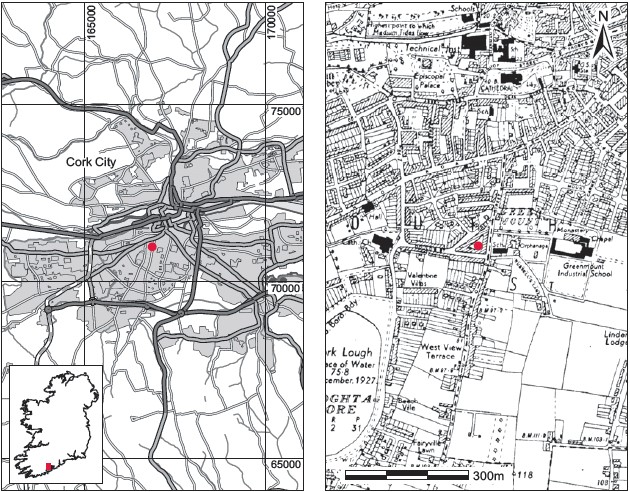
Fig. 6.7—Location map, Greenmount, Co. Cork.
brown silt, which overlay a thin layer (0.02–0.04m thick) of red brick fragments. Beneath this was another 0.16m of mid-brown silt, overlying a layer 0.04m deep of broken slates. The skeletal remains occurred 0.17m below the broken slate layer. While fourteen separate deposits of human remains were excavated, the remains were found to represent at least twelve individuals (1990:30–38).9 There was no evidence of any formal burial and all of the bones were disturbed and placed so that individuals were indistinguishable from each other. No trace of any burial structure or pit was evident within the restricted area examined. The human remains appear to be part of a mass grave, the limits of which have yet to be defined but which probably extends well beyond the area investigated and covers much of the triangular area of land delimited by the houses. The human remains could not be divided into separate skeletons or even burials, so the osteoarchaeologist’s term ‘human remains no. 1–12’ is used here.
The first deposit to be removed was human remains no. 2, protruding in section at a depth of 1.4m. The remains consisted of pelvis fragments, several long bones and some smaller bones. These were arranged in a pile as if they had been deliberately stacked. A quartz pebble was recovered 0.06m above the bones but no other finds were present in the soil that was removed. Human remains no. 6 was excavated immediately after human remains no. 2. It consisted of a fragment of skull bone and possibly a tibia fragment protruding through the earth. The bands of brick and mortar rubble and broken slate were also present over these remains. The skull was exposed but was found to be incomplete, with only the upper portion present. The skull fragment rested on a number of long bones, the remains of more than one individual. In exposing this area, another skull (human remains no. 9) was exposed. Long bones close to this skull were uncovered, and in the process a further three incomplete skulls (human remains nos 10, 11 and 12) were revealed. It was not possible to differentiate between the long bones associated with these skulls. Approximately 0.15m south of these burials was an indiscriminate mass of long bones and skull fragments (human remains no. 8). Further investigation in this area revealed two more skulls (human remains nos 13 and 14) but, as with the other remains, it was impossible to tell which long bones are associated with which individual. Human remains no. 7 lay 1.2m south of these remains and appeared as two femora protruding through the earth. In removing the earth above the bones, only the rubble red brick and mortar layer similar to that overlying skeletons 2, 6 and 8–14 was found to be present. No traces of the compact layer (present over human remains no. 2 only) or the broken slate layer were found here. On exposing the bones it was obvious that this skeleton was also incomplete. Only the pelvis and lower long bones were present; the upper part of the body, including the lower vertebrae, was absent.
The area in which human remains nos 1, 3, 4 and 5 were located consisted of a dark humic refuse layer, which varied in depth from 0.11m to 0.32m and which overlay the mid-brown silt layer where the human remains were visible. Human remains no. 1 consisted of a mandible and some vertebrae and arm bones, all of which were disturbed. Skull fragments were also present but these were badly broken. There were no associated finds. All that remained of human remains nos 3 and 5 were a number of long bones, while no. 4 was only represented by rib bones. Once the surrounding soil was removed from human remains nos 3 and 5 it was obvious that what existed here was a spread of human bones. These could not be identified as individual burials but at least five persons were represented.
Comment
A number of finds were retained, including nine pottery sherds (1990:39.1–9), one tile fragment (1990:39.10), one clay pipe fragment (1990:39.11) and one musket ball (1990:39.12). In the absence of any associated finds or other evidence it is not possible to suggest a date for these burials.
HUMAN REMAINS
LAUREEN BUCKLEY
Introduction, 1990:30–38
The bones found in this mound appear to represent the fragmented and jumbled remains of several individuals. Although the archaeologists have tried to label separate skeletons according to where they were found, the bones had been disturbed so much in the past that the bags pertaining to ‘one’ number always contained elements from more than one individual. Each bag has been examined and its contents detailed in the inventory. Because of the disturbance and fragmentation of the bones, however, it is impossible to separate individual skeletons. As one skeleton may have been distributed throughout several bags, it was decided to consider the bones collectively to discern the minimum number of individuals present.
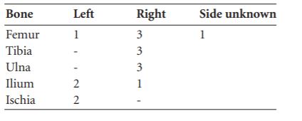
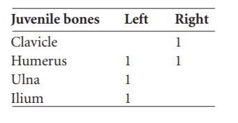
Tables 6.1–3 list the skeletal elements and the total number observed. The number of observations represents the minimum number of each bone.

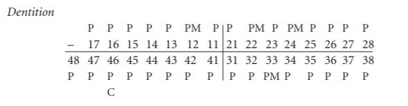
Dentition
The dentition indicates the presence of at least three juveniles.
Summary and conclusions
These bones appear to represent the remains of at least twelve individuals. Because of the fragmentary condition of the skulls it is not always possible to determine whether they belonged to an adult or a juvenile. The minimum number of skulls present was twelve, as indicated by the occurrence of the left temporal bone. There were at least seven adults present and three juveniles. One juvenile, as indicated by the presence of first and second deciduous molars with incompletely formed roots, was aged 11 ⁄2–21 ⁄2 years. Another juvenile was aged between six and nine years. There was at least one juvenile aged between fourteen and sixteen years at time of death. Since the maxillae and mandibles were often incomplete, it was not always possible to determine the state of eruption of the third molar, and so the number of juveniles present may actually be higher.
There were a number of young adults present, as indicated by the presence of epiphyses just fused or starting to fuse. The state of attrition of the teeth also indicates that these individuals were relatively young. There were no degenerative diseases observable in any of the joints remaining. The overall impression is of a young population. Only two adults were able to be sexed and these were male.
The dentition was in very good condition, with very few pathological changes noted. Only nine teeth (7% of the total examined) were affected by caries, which is a very low percentage. Only one tooth had been lost ante-mortem. This was a lower incisor, which had evidence of an abscess cavity. Hypoplastic defects were noted on 56 teeth (42%). Hypoplastic defects arise during a period of dietary deficiency or acute illness during childhood. They take the form of lines or grooves in the enamel. It is not possible to say how many individuals were affected as there were a large number of loose teeth from this site. Nevertheless, 42% of all teeth seems quite high and probably indicates a high degree of nutritional stress or acute infection among this population. The only pathology noted on the bones was on the left orbit of the adult skull from bag no. 8. This orbit had evidence of cribra orbitalia, a condition characterised by pitting of the bone on the orbit. It comes about as a result of proliferation of the bone marrow at the expense of the outer compact bone, which becomes porous. The bone marrow enlarges during iron-deficiency anaemia in an attempt to make up by quantity what the blood lacks in quality. Iron-deficiency anaemia can arise from lack of iron in the diet or excessive loss of iron. It does not appear to have been a significant problem here, however, as only one instance was noted.
INVENTORY
Human remains no. 1
There were three bags of bones from this skeleton; one contained a skull and the others contained post-cranial bones.
Skull
The occipital bone and both parietal bones were complete. Part of the frontal bone was present but the orbital part was missing. Both temporal bones were present. The mandible was complete and part of the maxilla was present.
Dentition:
Attrition: very slight.
Calculus: slight.
Caries: small, buccal crown.
Linear enamel hypoplasia: on all incisors except upper right lateral, all canines except lower left, all premolars except upper left premolars, and on all the second molars.
Age of occurrence: 2–3 years, 3–4 years, 4–5 years, 5–6 years.
Vertebrae
The atlas, axis and the lower five cervical vertebrae were present as well as the first thoracic vertebra.
Long bones
The right humerus was complete and most of the shaft of the left was present. The proximal ends of the right radius and ulna were present. Other bones present included the right clavicle and scapulae and at least four ribs from the right side. The state of fusion of the basiosphenoid symphysis was not observable, but the lack of attrition on the second and third molars indicates that this is probably a young adult individual. All observable epiphyses are fused but the line of fusion can be seen at the proximal humerus and proximal radius. The epiphyses of the clavicle are not observable.
Human remains no. 2
This consisted of a pelvis and some long bones, nearly all from an adolescent individual. The pelvis consisted of a right ilium and ischium. The epiphyses at the ischial tuberosity had just fused but the iliac crest epiphysis was unfused. The long bones present included a proximal half of a left ulna, the proximal half of the shaft of a left radius and a distal radius epiphysis. The distal epiphyses of a left and right femur, the proximal half of a left femur with unfused epiphyses and the proximal epiphysis of a right tibia were also present. The distal ends of the shafts with unfused epiphyses of a left and right tibiae, most of the shaft of a fibula and a distal end of another fibula with unfused epiphysis were also present.
Three immature lumbar vertebrae and the first three sacral vertebrae were present.
There were also a few foot bones and a fragment of femur shaft from an adult individual and a piece of occipital and fragments of parietal bone from a younger child.
Human remains no. 3
This bag contained a fragmented and much-decayed portion of frontal bone, one tooth, the left upper third molar, a rib fragment and the head of a metacarpal.
Human remains no. 4
This contained the proximal half of a child’s right ulna, the proximal half of a child’s radius, fragments of four ribs and the decayed arch of a thoracic vertebra.
Human remains no. 5
This consisted mainly of bone from the upper half of a skeleton. There were a few scapula fragments, the lateral end of a right clavicle, the distal half of an adult right humerus, the proximal half of an adult right ulna, three rib fragments and fragmented arches of three thoracic vertebrae. There was also a part of a vertebral body, a fragment of squamous temporal bone and a much-decayed but almost complete right tibia shaft from an adult.
Human remains no. 3 and no. 5
There were a few bags labelled nos 3 and 5.
(1) This bag contained decayed portions of humerus, radius and ulna shafts, fragments of ilium, the body of one lumbar and arch of another lumbar vertebra, three sacral vertebrae and fragments of frontal, parietal and a left temporal bone. A portion of a maxilla with some teeth in situ was also present.
Dentition:
Molar wear: slight.
Calculus: very slight.
Linear enamel hypoplasia: present on the first premolar and first molar.
Age of occurrence: 2–3 years, 3–4 years.
There was also one upper central incisor and one canine in poor condition, as well as the right third upper molar.
(2) This bag contained several small fragments of bone. The identifiable fragments consisted of a femur head and distal end of a right femur, the proximal end of a left ulna, a fragment of radius shaft and the distal end of a right and left radius. There were also a few fragments of ilium and scapula, some lumbar and sacral vertebrae fragments, some rib fragments and one carpal, metacarpal and two phalanges. There were several small fragments of parietal and frontal bone from the skull.
Long bones
This bag contained several fragmented bones. The minimum number of bones present is given in Table 6.4. In addition to these, there was an almost complete sacrum, a right calcaneum, a right patella, one thoracic vertebra and the fifth right metacarpal.
Table 6.4—Human remains nos 3 and 5: minimum number of bones present.
Human remains no. 6
This consisted of a virtually complete occipital bone, left parietal bone and most of a right parietal and a complete frontal bone. The basilar occipital bone was present but was much decayed. Also present were a few rib fragments and part of the proximal articular surface of a Tibia.
Human remains no. 7
This consisted mainly of the pelvis and legs from more than one individual. A left ilium and ischium were present as well as a fragment of right ilium and two right ischia. The proximal two-thirds of a left and a right femur were present as well as another femur head. Also present were six sacral vertebrae, three incomplete metacarpals, two phalanges and a fragment of the distal end of a right humerus.
Human remains no. 8
(1) Skull—this consisted of a complete skullcap from a sub-adult skeleton. The frontal bone, both parietal bones, occipital and both temporal bones were complete. The sphenoid was present but broken. The basio-sphenoid symphysis was not observable but the size of the skull and lack of suture fusion indicates that it is probably not an adult skull.
(2) This bag contained part of a skullcap from an adult. Most of the frontal and part of both parietal bones were present, as well as part of the left temporal. Of the long bones present a right clavicle, the shaft of a right ulna, a radius shaft and a humerus shaft probably belonged to a juvenile. The head and neck of a left femur with epiphyses just fused was present. The heads of two other femora were present, as well as part of a femur shaft and the distal end of an adult right femur. A left humerus shaft and most of the shaft and lower end of a right humerus were present, as well as a proximal right ulna shaft and mid-shaft sections of two tibiae. Other fragments present included fragments of scapula, fragments of ilium and a right pubis, rib fragments and a few metacarpals. The right side of a mandible with no teeth was present.
(3) This bag contained several small fragments of skull and long bone. The identifiable fragments consisted of one piece of radius shaft, the olecranon of one ulna, a fragment of acetabulum, one rib and a few carpals and metacarpals.
Teeth
There was a bag containing a number of teeth. The following adult teeth were identified.
Incisors: 12 Slight calculus.
21 Moderate calculus, linear enamel hypoplasia 1–2 years, 2–3 years.
32 Moderate calculus.
Premolars: 24 Slight calculus.
34 Grooves of hypoplasia, 3–4 years, 4–5 years.
35 Slight calculus.
25 Slight calculus; linear hypoplasia, 4–5 years, 5–6 years.
Molars: 46 Calculus slight, attrition slight 2+.
37 Calculus moderate, attrition slight 2.
27 Calculus slight, attrition slight 1.
Juvenile teeth
First mandibular molar 46 Roots not fully formed, 6–9 years.
Maxillary first molar 54 Roots not fully formed, 1 1⁄2 – 2 1⁄2 years.
Maxillary second molar 55 Roots not fully formed.
Human remains no. 9
This skull consisted of an almost complete occipital bone, several large fragments of parietal bone, both temporal bones, the orbital part of the frontal bone and fragments of sphenoid
Bone.
Dentition

Calculus: very slight.
Molar attrition: slight.
Human remains no. 10
This skull consisted of a complete frontal bone and a portion of the mandible. Also present were part of the axis and two rib fragments.
Dentition
Attrition: none.
Calculus: slight.
Linear enamel hypoplasia: on canine, 43, age of occurrence 2–3 years. The third molars are probably not erupted.
Human remains no. 11
The occipital and parietal bones of this skull were virtually complete. Both temporal bones were present but the mastoid processes were decayed. Some fragments of frontal bone and sphenoid bone were present.
Human remains no. 12
The occipital bone of this skull was almost complete. The right parietal was complete and most of the left was present. The frontal bone was complete and the left temporal was almost complete. The supercilliary arches and mastoid processes indicate a male skull.
Human remains nos 6, 9, 10, 11, 12
This contained a number of mandibles and maxillae and some loose teeth.
Mandibles
(A)
Calculus: moderate.
Molar attrition: slight.
(B)
Calculus: none.
Attrition: very slight.
Linear enamel hypoplasia: canine and right premolars.
Age of occurrence: 2–3 years, 3–4 years.
(C)
Heavy wear on incisors.
Calculus: moderate.
Linear enamel hypoplasia: on canine.
(D)
Calculus: slight.
Attrition: slight.
Caries: buccal side at cervical margin.
(E)
Calculus: moderate.
Molar attrition: 2.
Maxillae
(A)
Calculus: slight.
Attrition: slight.
Linear enamel hypoplasia: on all teeth except lateral incisor.
Age of occurrence: 1–2 years, 2–3 years, 3–4 years, 4–5 years.
(B)

Calculus: moderate.
Attrition: heavy on incisors, slight on premolars.
Linear enamel hypoplasia: central incisor and canine.
Age of occurrence: 2–3 years.
(C)

Calculus: moderate.
Attrition: slight.
Linear enamel hypoplasia: first molar.
Age of occurrence: 1–2 years, 2–3 years.
(D)
Calculus: slight.
Attrition: slight.
Caries: distal, cervical.
Linear enamel hypoplasia: first molar.
Age of occurrence: 1–2 years, 2–3 years.
(E)

Calculus: moderate.
Attrition: slight.
Linear enamel hypoplasia: all teeth.
Age of occurrence: 2–3 years, 4–5 years, 5–6 years, 6–7 years.
In addition, the following teeth were present, which could not be identified as belonging to any particular mandible or maxillae:
Molars,
Mandibular
47 46 Attrition slight, calculus moderate.
Linear enamel hypoplasia, 5–6 years.
46 Calculus slight, attrition slight.
47 Calculus slight, attrition slight.
Caries: cervical buccal.
37 Calculus moderate, attrition slight.
48 Calculus slight, attrition slight.
48 Calculus slight, attrition slight.
Caries: occlusal.
Molars,
maxillary
16 Calculus slight, attrition slight.
Linear enamel hypoplasia, 5–6 years.
17 Calculus slight, attrition slight.
Caries: distal cervical, large.
27 Calculus slight, attrition slight.
Caries: occlusal, small.
27 Calculus slight, attrition slight.
Premolars
45, 44 Calculus slight; linear hypoplasia, 4–5 years.
34 Hypoplasia, 4–5 years.
25 Hypoplasia, 4–5 years.
Canines
13 Hypoplasia, 2–3 years, 3–4 years.
13
Incisors
12 Linear hypoplasia, 2–3 years, 3–4 years.
22
Long bones nos 6, 9, 10, 11, 12
Besides long bones, this bag contained fragments from at least four skulls as well as three atlas vertebrae, two axes, two lumbar vertebrae and one sacrum. There were a few metacarpals, metatarsals and phalanges present. The long bone and skull fragments are summarised in Tables 6.5 and 6.6.
Table 6.5—Summary of identified adult bone, nos 6, 9, 10, 11, 12.
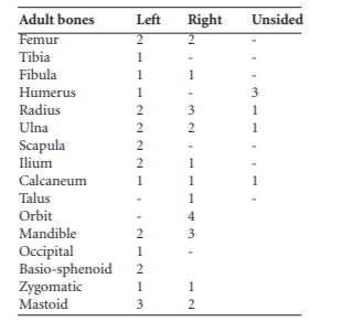
Table 6.6—Summary of identified juvenile bone, nos 6, 9, 10, 11, 12.
Human remains no. 13
This consisted of the left side of a skull with male characteristics. The frontal bone was almost complete and the left parietal and temporal bones were complete. The sagittal suture was fused.
Human remains no. 14
The frontal bone of this skull was almost complete, although the right orbit was missing. The left parietal bone was complete and there was a piece of the right parietal. There was one complete occipital bone and one piece of another frontal bone. A right temporal and the petrous portion of the left temporal were present, as well as several fragments of skullcap.
Dentition
Calculus: slight, some wear on incisors but little on premolars.
Linear enamel hypoplasia: all teeth.
Age of occurrence: 2–3 years, 3–4 years, 4–5 years.
Other bones
Fragments of a right and a left ilium were present. Some loose teeth present were incisors 2 and 1 and molar 8, which had a caries on the distal cervical margin.
Miscellaneous fragments
This bag contained a left humerus from an adult, one radius shaft, one left ilium and some skull fragments.
8. SMR CO074-075——. IGR 166881 070983.
9. The burials are described not in numerical order but in the order in which they were excavated, according to the excavator’s report.