1989:128 - TONAFARNA, CO. LAOIS, Laois
County: Laois
Site name: TONAFARNA, CO. LAOIS
Sites and Monuments Record No.: SMR LA014-086002SMR LA014-013
Licence number: —
Author: MARY CAHILL
Author/Organisation Address: —
Site type: Iron Age and early medieval graves, c. 300 BCc. AD 1200
Period/Dating: —
ITM: E 657839m, N 701307m
Latitude, Longitude (decimal degrees): 53.059273, -7.137135
Introduction
In November 1989 human bone was exposed during sand-digging near Stradbally, Co. Laois. According to the report on file, three or four skeletons had been discovered and work was suspended on the site. The discovery was reported to the Garda Síochána at Stradbally, who informed the NMI. Mary Cahill visited the site on 24 November 1989. The human remains were examined by Laureen Buckley.
Location (Fig. 4.31)
The site was in the townland of Tonafarna in east County Laois, close to the border with County Kildare,69 at an altitude of 70–80m above sea level.
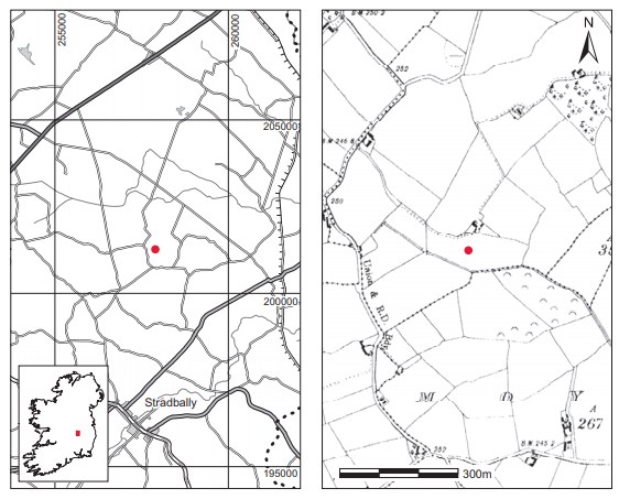
The burials seem to have been inserted into the side of an enclosure, which is not marked on the OS 6in. map. This enclosure survived as a very low, approximately subrectangular banked area with an outer ditch, which had been silted up. The burials were placed on the south side at a depth of 0.3–0.4m, just above the deposit of sand and gravel. No associated sites are known from the vicinity; the closest sites shown on the SMR are three enclosures in the adjacent townland of Killeen.70
Description of site
There was no evidence of grave structures and the graves appear to have been later intrusions into the side of the enclosure. All of the burials were inhumations (1989:54) and no accompanying artefacts were found. As they were not excavated in situ, the disposition of the skeletons is not known. The remains were analysed by Buckley and found to represent a minimum of five adults and four juveniles. According to the landowner, the previous owner had used a metal-detector and had found a cross and rosary beads some years ago on the land, but the find-spot is not known. A sample of bone was submitted for radiocarbon dating and yielded a result of 1265±40 BP, which calibrates to 665–869.71
Comment
This site was dated solely on the basis of the radiocarbon date obtained from some of the bone. From Buckley’s analysis it appears that the cemetery contained a normal mixed population, consisting of both adults and juveniles. As is the case with many of the sites, this may be compared to other early medieval enclosed cemeteries that are not associated with a church, and may be ancestral burial grounds that remained in use in the first few centuries after Christianity was introduced into Ireland (see O’Brien 2003; Stout and Stout 2008; Ó Carragáin 2009).
HUMAN REMAINS
LAUREEN BUCKLEY
Description of remains
Since the remains (1989:54) had been so completely disturbed and appear to represent more than one individual, all that could be done was to describe each bone and count the minimum number of individuals present.
Adult bones
Left femurs
1. A virtually complete left femur. An accurate measurement of the diameter of the head could not be taken but the epiphyseal line was still visible, indicating that it was probably from a young adult (FeL2 459mm; FeE1 75.1mm).
2. The proximal third of a left femur with complete head and neck.
3. The proximal thirds of two left femur shafts only.
4. The distal ends of two left femurs only.
The minimum number of left femurs, based on the proximal thirds of the shafts, is four.
Right femurs
1. One complete right femur. It was not possible to determine from the bone measurements whether the individual could be male or female (FeL1 463mm; FeD1 23.7mm; FeD2 28.0mm; FeE1 75.3mm).
2. The proximal two-thirds of a right femur. From the size of the diameter of the head it is probably from a male individual (FeHd 55.0mm).
3. The proximal two-thirds of a right femur. The diameter of the head indicates that this could be from a female individual and the epiphyseal line is still visible so it is from a young adult individual (FeHd 41.2mm).
4. The proximal halves of the shafts of two right femurs.
5. The proximal joint end only of a right femur.
6. The distal third of the shaft of a right femur.
7. The distal half of a right femur. From the size of the epicondylar width it is very likely that this is from a male individual.
The minimum number of right femurs present, based on the number of proximal halves of the shaft present, is five.
Left tibiae
1. The proximal third of the shaft only.
2. The distal half of a left tibia.
3. The distal third of a left tibia.
The minimum number of left tibiae present, based on the distal thirds, is two.
Right tibiae
1. The proximal third of a right tibia only
2. The distal third of a right tibia.
3. A fragmented shaft only.
The minimum number of right tibiae present is one.
Fibulae
Only three fragments of shaft from fibulae were present but they could not be sided.
Left humerus
1. One almost complete left humerus with only the distal end missing was present. From the size of the bone and the diameter of the humerus head, it is probable that it came from a female individual (HuHd 43.4mm).
2. The proximal half of a left humerus only. The size of the head indicates that it could be from a male individual (HuHd 47.3mm).
3. The distal half of a left humerus.
4. The distal third of a left humerus.
The minimum number of left humeri is three.
Right humerus
1. A complete right humerus, which from its appearance and measurements is probably a match for the almost complete left humerus described above (HuL1 322mm; HuHd 43.0mm).
2. The proximal halves of the shafts of two right humeri.
3. A complete shaft of a right humerus.
4. The distal third of a right humerus.
The minimum number of right humeri present is four.
Left radius
1. The proximal third of the shaft of a left radius.
2. The distal half of the shaft of a left radius.
The minimum number of left radii present is one. There were no right radii present.
Left ulnae
1. The proximal two-thirds of the shafts of two left ulnae were present.
Right ulnae
1. The proximal third of a right ulna.
2. The proximal half of the shaft of a right ulna.
The minimum number of right ulnae present is two.
Left clavicle
1. A complete left clavicle, probably belonging to a male as it was 154mm in length.
Right clavicles
1. The lateral halves of three right clavicles.
2. The medial half of a right clavicle.
The minimum number of right clavicles present is three.
Left scapulae
1. One almost complete left scapula (glenoid cavity breadth 27.1mm).
2. The lateral border of a left scapula.
The minimum number of left scapulae present is two.
Right scapulae
1. One almost complete right scapula (glenoid length 36.3mm; glenoid breadth 26.2mm).
2. Another almost complete right scapula, but glenoid fossa damaged.
3. The acromion of a right scapula.
The minimum number of right scapulae present is three.
Pelvis
There were two left ilia and two right ilia present. Both had wide sciatic notches. Two left and two right ischia were also present.
Sacrum
1. One sacrum consisting of the first three sacral vertebrae was present.
2. Another sacrum consisting of the first sacral vertebra and the body of the second sacral vertebra was also present.
Vertebrae
There were two first cervical vertebrae present, one second cervical and two other lower cervical vertebrae. Seven thoracic vertebrae were also present, and there were three complete lumbar vertebrae along with the neural arch of one other lumbar vertebra.
Ribs
Two ribs from the left and ten from the right were present.
Tarsals and metatarsals
There were two left tali, one right talus and a left and a right calcaneum present from the tarsal bones. Two first left metatarsals, a second, third and fifth left metatarsal and also one right first and fourth metatarsal were present. There were also two foot phalanges.
Pathology
Schmorl’s nodes were present on five thoracic and two lumbar vertebrae. Osteochondritis dessicans was noted on the left and right calcanea. Slight porosity indicative of degenerative joint disease was present on the lateral end of the left clavicle. Degeneration of the hip joint was also present, with marginal lipping and porosity of the inferior surface of the acetabulum in one right pelvis. One of the left ilia had a bar on the bone. This is thought to be caused by trauma.
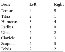
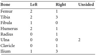
Summary of the adult infracranial bones
Below are the minimum numbers of adult bones present in this collection. From the minimum number of right femurs present it can be ascertained that the minimum number of individuals present in this collection is five. There is at least one male and one female present, although there could be two females. The sternal end of a right clavicle was unfused and a left ischial tuberosity had just fused, indicating the presence of at least one young adult.
Table 4.8—Adult infracranial bones.
Adult skull bones
The following minimum numbers of skull bones were present.
Table 4.9—Skull bones present.
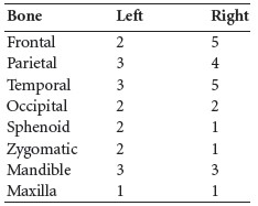
From the minimum number of the right side of the frontal bone and right temporal bones it can be determined that at least five skulls were present in this collection, which is consistent with the findings from the long bones. At least four of the right temporal bones had mastoid areas that were of the male type. One occipital bone was also from a male, as was one of the frontal bones.
Pathology
A very small flat osteoma was present on a left parietal bone. It measured 1.2cm in diameter and was smooth and circular in appearance.
Mild cribra orbitalia was present in a left and a right orbit.
Dentition
Mandible 1:
Attrition: there was very slight wear on the remaining teeth, with no wear on the third molar.
Calculus: there were slight deposits on the buccal surfaces of all the remaining teeth apart from the canine, where deposits were moderate. There were also slight deposits on the lingual surfaces of the canine, premolar and first molar.
Caries: a very small cavity was present on the distal side of the crown, near the occlusal edge of the first molar.
Mandible 2:
[insert EBS-122-3.jpg here]
Attrition: there was moderate wear on the remaining molars except for the left third molar, which had no wear. The premolars also had moderate wear but there was heavy wear on the incisor and canine.
Calculus: there were light deposits on the lingual surfaces of the incisors, canines and first right premolar, as well as on the buccal surface of the first left premolar. Deposits were moderate on the buccal surfaces of the right canine and first premolar and on the lingual surfaces of the right third molar and left first premolar. There were considerable deposits of calculus on the buccal surfaces of the right incisors, on the buccal and lingual surfaces of the left second molar and on the buccal, lingual and distal surfaces of the left third molar.
Periodontal disease: a slight degree of alveolar recession was present around the roots of most teeth and a moderate degree was present around the roots of the first left molar.
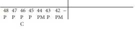
Abscess: two external abscesses were present in the right side of the mandible around the first and second molars. The sockets for the mesial root of the second molar, 47, and the distal root of the first molar, 46, were almost destroyed and beginning to meet.
Mandible 3:
No teeth were present in the mandible but the molars had been lost during life and the gaps in the sockets were almost but not completely closed.
The mandibular foramen on the right side was very enlarged.
The sockets for the anterior teeth were partially broken so it is not possible to tell whether or not alveolar recession was present.
Two additional teeth, a lower left central incisor, 31, with slight wear and moderate calculus and an upper molar, probably 17, with heavy wear and a large cavity on the mesial half of the crown and root were also present.
Maxilla 1:
Attrition: there was very slight wear on the anterior teeth, with moderate wear on the first molar and no wear on the second and third molars.
Calculus: there were slight deposits on the buccal surfaces of all the remaining teeth apart from the right canine and the first premolars, where deposits were moderate. There were also slight deposits on the lingual surfaces of the right molars and first left molar.
Juvenile bones
A number of juvenile long bones and skull bones were present and are described below.
Left femurs
1. A complete left femur with all the epiphyses unfused was present; length of diaphysis 288mm, diameter 17.3mm.
2. The proximal half of another left femur, smaller than the first, was also present; diameter 13.2mm.
Right femurs
1. The distal part of the shaft of a right femur.
2. Part of the proximal end of the shaft of a right femur.
Left tibiae
1. The proximal end of a left tibia from an adolescent. The bone is fragmented but from its size it is probably from an adolescent. The epiphysis is unfused.
2. Most of the shaft of a left tibia from a smaller juvenile.
Right tibiae
1. The distal half of a right tibia from an adolescent.
2. The mid-shaft area of a right tibia from a juvenile, the same size as left tibia (2).
3. The proximal two-thirds of a right tibia from a smaller juvenile than (2) above; mid-shaft diameter 13.2mm, width 36.2mm.
Fibula
1. The proximal half of a left fibula from an adolescent.
Left humerus
1. Distal half of a shaft from a small juvenile.
2. Proximal third of a shaft from a smaller juvenile.
Right humerus
1. A complete right humerus from an adolescent. The proximal epiphysis was unfused but the distal epiphysis was fused, with the medial epicondyle just fused; HuL1 310mm.
Radius
1. The distal half of a right radius from an adolescent.
Ulnae
1. Two ulnae from a small infant were present. It was not possible to side them, as they were so decayed.
Ilium
1. One almost complete left ilium from a juvenile with unfused acetabulum; breadth 94.7mm.
2. One right ilium from a juvenile; breadth 93.6mm, height 84.4mm.
3. Fragments of a right ilium from another juvenile.
4. An almost complete right ilium from an adolescent. The acetabulum was probably fused but the iliac crest was unfused.
Other juvenile bones
The lateral half of a right clavicle from a juvenile was present. There was one thoracic vertebra with the neural arch fused to the body and two ribs from a small juvenile. One unidentified metatarsal from a juvenile was present and there was also a left first metatarsal from an adolescent with the distal epiphysis unfused.
Summary of juvenile bones
The following minimum numbers of juvenile infracranial bones were present.
Table 4.10—Minimum numbers of juvenile infracranial bones.
It appears from the above table that the minimum number of juveniles present was three; as the ulnae were from an infant, however, this must also be added to the number, giving a minimum number of juveniles of four. At least one was an adolescent and two were juveniles, with one juvenile being older than the other.
Juvenile skull bones
1. One occipital, left and right parietals and part of the left side of the frontal bone was present in one piece and is probably from the adolescent skull. There was also a left temporal bone present.
2. Matching left and right temporal bones.
3. Fragments of two mandibles.
4. A basilar occipital bone from an infant; length 14.8mm, breadth 20.2mm.
The fact that the breadth of this bone is greater than the length indicates that the infant had reached a viable state of gestation and was at least eight and a half months in utero. In fact, using the equations given by Kosa (1991, 36) it is possible to work out an estimate of body length from the length of the occipital bone and hence to approximate the age of the infant. The estimate of body length determines that this was over the length of an average neonate and therefore was probably from an infant of around three months of age.
Dentition
Mandible 1:

Development: the lower right permanent central incisor, 41, was present and the root was one-quarter formed. The socket for 36 indicates that the roots were one-third formed, and the crown for the second molar, 37, had just started to form. This stage of development and of dental eruption is consistent with the juvenile being aged between five and seven years at the time of death.
Attrition: there was moderate wear on the incisors only.
Caries: there was a moderate cavity on the occlusal surface of the second left deciduous molar, 75, and a very small cavity on the occlusal surface of the first deciduous molar, 74. In both cases the cavities were on the mesial side of the tooth.
Mandible 2:

The crown of the first permanent molar, 36, was present but the root had not formed. This is consistent with the juvenile being aged 3–5 years at the time of death.
Summary
These disarticulated remains consisted of a minimum of five adults and four juveniles. The adults consisted of at least one female and four males, one of whom was a young adult. The juveniles consisted of one adolescent and two younger juveniles, one aged 5–7 years at the time of death and the other aged 3–5 years. There was also an infant present. A small amount of pathology was noted on the bone. One of the adult skulls had a small osteoma of a left parietal bone. This is a benign bone tumour that is quite common and does not cause adverse effects. At least one individual had evidence of cribra orbitalia. This is characterised by pitting of the orbital bone and can be caused by iron-deficiency anaemia.
There was some evidence of trauma, with a left and a right calcaneum, probably a matching pair, having osteochondritis dessicans. In this condition a small area of bone on a joint surface becomes necrotic and leaves a small, generally circular lytic lesion on the bone surface. It is thought to be caused by trauma. A more unusual lesion also possibly caused by trauma was present on a left ilium. There was a build-up of bone on the internal surface to form what is known as a bar on the bone. This lesion has been noted before in archaeological specimens although the exact cause is uncertain and again it is thought to be caused by trauma (Charlotte Roberts, pers. comm.).
Schmorl’s nodes were present on some of the thoracic and lumbar vertebrae. These lesions occur as a result of heavy manual labour or the lifting of heavy loads. The only other skeletal pathology noted was some degenerative joint disease of a right hip and of a left shoulder.
Only three mandibles and one maxilla were found, so a reliable dental analysis cannot be undertaken. It can, however, be said that the maxilla and one of the mandibles were probably from a young individual as there was very little tooth wear or calculus deposition. There was some evidence of dental disease in the population. One of the mandibles had two abscesses, but as the teeth concerned were missing it could not be ascertained whether the abscesses developed as a result of caries or of excessive attrition. This individual also had some periodontal disease that was associated with heavy calculcus deposits. The third mandible had lost three teeth shortly before death. Again, it could not be said whether this was a result of caries or periodontal disease. The fact that caries was found in a juvenile aged 5–7 years, with two deciduous molars affected, suggests that caries was extensive in the population. High caries rates are usually found in late or post-medieval populations but not in early medieval or prehistoric populations. It must be emphasised, however, that there were insufficient dental remains from this site to enable a full reliable statistical analysis to be undertaken.
69. Parish of Moyanna, barony of Stradbally. SMR LA014-086002-. IGR 257902 201274.
70. SMR LA014-013——; -014——;-015——.
71. GrA-29069.