1988:088 - CAHERABBEY LOWER, CO. TIPPERARY, Tipperary
County: Tipperary
Site name: CAHERABBEY LOWER, CO. TIPPERARY
Sites and Monuments Record No.: SMR TS075-041
Licence number: E1158
Author: PATRICK HOLLAND AND MARY CAHILL
Author/Organisation Address: —
Site type: Iron Age and early medieval graves, c. 300 BCc. AD 1200
Period/Dating: —
ITM: E 605347m, N 626225m
Latitude, Longitude (decimal degrees): 52.387543, -7.921446
Introduction
In August 1988, human remains were discovered during quarrying operations at Caherabbey Lower, near Cahir, Co. Tipperary. Some months after the removal of the undergrowth and topsoil, an employee of Roadstone Provinces Ltd noticed human bones exposed in the heaps of gravel and exposed gravel subsoil. The discovery was reported to the Museum by Sergeant M. O’Connor, Garda Síochána, Cahir, and the site was visited by Eamonn Kelly. Human remains were visible extending over an area of approximately 10m2, and more bone was noticed concentrated in five groups. The disturbed remains were collected and brought to the Museum. A few weeks later Roadstone informed the Museum that further human remains had been uncovered. Upon inspection the site was found to have been heavily altered. The topsoil and undergrowth had been stripped off and pushed into heaps on the western and eastern edges of the hilltop. The western side of the site had been cleared down into natural gravel, while a smaller area of scraped topsoil overlying undisturbed gravel survived on the centre of the hill. This is the area that contained most of the burials excavated. Two visible burials were excavated by Patrick Holland, curator of South Tipperary County Museum, assisted by Louis Flannery, in the days following the discovery. The site was excavated the following week by Mary Cahill and Patrick Holland, and further burials, which were discovered in late September, were also excavated by the authors.109 The human remains were examined by Laureen Buckley.
Location (Fig. 4.61)
The ridge, almost totally removed, had measured over 60m high and overlooked the River Suir immediately to the south.110 A curved field boundary is shown on both the first and second editions of the Ordnance Survey maps as skirting the highest part of the ridge on the west, and a stone wall also apparently enclosed the summit. Traces of a mortared stone wall
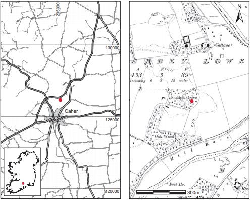
Fig. 4.61—Location map, Caherabbey Lower, Co. Tipperary
were noted in the spoil during the investigations described below. By 1906, the date of the second edition, a small quarry had been opened on the ridge and the area within the field boundary is noted as the ‘Sandpit Grove’. The area around the ridge and the lower parts of the ridge itself have been extensively quarried in recent years, and the present landscape is not easily correlated to that depicted on the maps.
Description of site
The site consisted of a cemetery of extended inhumations, buried in simple shallow pits (Fig. 4.62). Because of the extensive quarrying and removal of topsoil that took place between the first and second visits to the site, it is not possible to directly correlate the earlier finds to the burials that were planned and excavated, though they may have been further west. Nevertheless, it is clear that there were a substantial number of burials on the top of the hill, probably in the southern part, overlooking the river.
The graves were dug into the underlying stony gravel, from which their fill was distinguished by being a darker, looser brown gravel and soil mix. The burials were unprotected and the graves were not very much larger than the bodies they contained. The floors of the pits were not completely level and may have risen up with the slope of the hill to the west. There appears to have been a preference for the head of the corpse to have been raised. The burials were aligned on an east/west axis with their heads to the west. Where burials were found in association they were very close or alongside each other, which would argue for a contemporary knowledge of each burial. No evidence for markers was noted, however, nor was there any evidence for grave-goods, coffins or other coverings. Many of the bones appeared to have been damaged by the weight of heavy machinery. Several individual
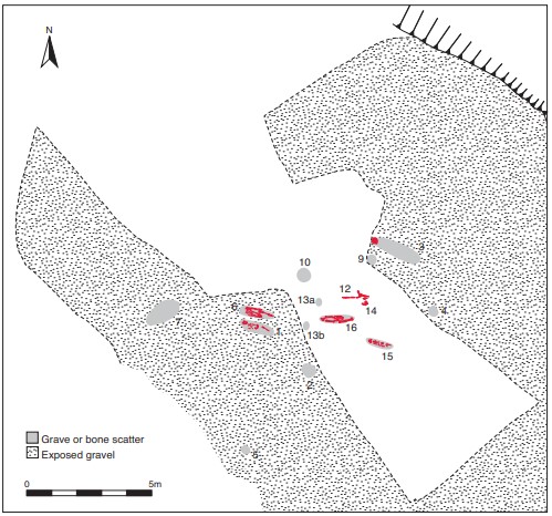
Fig. 4.62—Site plan, Caherabbey Lower, Co. Tipperary
burials were uncovered and a large quantity of disarticulated bone was also collected. Samples of bone from five different burials were submitted for radiocarbon dating and yielded dates corresponding roughly to the period 780–950.
Grave 1, 1988:231(1)
An extended inhumation of a male, aged 20–25 years and 173cm (5ft 8in.) in height, found partly exposed and removed by the quarry machinery. The skeleton lay in a supine position with the head to the west. The skull was raised; two naturally rounded limestone cobbles in the area of the nape of the neck may have supported the head. The arms lay beside the body, with the right hand upon the pelvis and the left beside the femur. This skeleton was in very good condition, although a lot of the bones were missing from the right side and very little remained of the skull. An isolated jaw lying to the south of the inhumation was identified as that of an individual aged 35–40. A sample of the human remains yielded a radiocarbon date of 1120±30 BP, which calibrates to 784–994.112
Grave 2, 1988:231(2)
This consisted of a heap of bones gathered by the quarry workmen and reported as having come from the approximate location noted on the plan. Buckley reports that this scatter represents the remains of at least three individuals.
Grave 3, 1988:231(3)
This consisted of a badly disturbed burial comprising a skull, fragmented but in situ, in the side of a bulldozer ‘cut’, with bones extending to the east in the cut surface. The skull was raised and lay upon a sloping gravel surface. When the remains were examined it was possible to separate out one individual, an older adult female of over 45 years with a stature of 160cm (5ft 3in.), but at least two other adults, three young adults (18–20 years old), three child skeletons (two aged 6–10 years and one aged 12–16 years) and one infant, representing at least ten individuals, were also present.
Grave 4, 1988:231(4)
This collection of scattered bones was gathered together by the quarry workmen and is reported to have been found in the location marked on the plan. At least two individuals are present.
Grave 5, 1988:231(5)
This scatter of bones was recovered from the surface of a heavily disturbed area at the southeasternmost part of the site. It consisted mainly of numerous fragments of skull bones, which appear to represent only one skull.
Grave 6, 1988:231(6)
This burial was an extended inhumation immediately to the north of, and on the same orientation as, grave 1. As was the case with burial 1, the skull and lower legs were damaged or missing as a result of the quarrying operations. The head may also have been supported on a stone. The arms lay beside the body and the left hand was upright beside the femur. Buckley has identified it as that of a male, aged over 45 years and 178cm (5ft 8in.) in height. Some bones collected with this burial appear to be from other individuals. A sample of the human remains was analysed and yielded a date of 1180±30 BP, which calibrates to 771–965.113
Grave 7, 1988:231(7)
This consisted of a scatter of disturbed remains, apparently mostly skull fragments, lying on the surface to the north-west of graves 1 and 6. The skull of at least one individual, an adult female aged 40–45, was identified.
Grave 8, 1988:231(8)
This consisted of a small scatter of disturbed bones to the south of grave 1. It is possible that these remains were removed, perhaps deposited over burials 1 and 6 by machinery, and consisted of a few fragments of long bone too small to be identified.
Grave 9, 1988:231(9)
Grave 9 was represented by an area of bone c. 1.5m south-west of grave 3. The few small skull fragments appear to be those of a male.
Grave 10, 1988:231(10)
An area of scattered and disturbed bones, including skull fragments, lying on the surface. These would appear to be those of an individual probably aged 20–25 years at death.
Grave 11, 1988:231(11)
This number was used as a general identification number for all bones collected from the surface of bulldozed spoil, excluding those burials or areas of exposed bone thought to be burials and which were numbered individually. There were at least two individuals represented, including one less than 25 years of age at death.
Grave 12, 1988:231(12)
This consisted of an extended inhumation, somewhat disturbed but almost complete. The remains were those of an adult male, 23–30 years of age and some 170cm (5ft 9in.) in height. A sample of the human remains was radiocarbon-dated and yielded a date of 1080±35 BP, which calibrates to 893–1019 (see note 113).
Grave 13, 1988:231(13)
This consisted of two separate scatters of bone (13a and 13b) representing two individuals. The remains were found at slightly different levels.
Grave 14, 1988:231(14)
Grave 14 was a crushed skull to the west of grave 9 and adjacent to grave 12. It has been identified as that of a female aged 35–45. Other bones were found in the area, including foot bones close to the head.
Grave 15, 1988:231(15)
This consisted of an extended inhumation aligned west/east, located approximately 1.5m south-east of grave 16. The remains have been identified as those of a single individual aged 20–25 at death. It was not possible to determine the sex of the individual. One infant bone was also found in association with the adult remains. A sample of the human remains yielded a date of 1200±35 BP, which calibrates to 693–941.114
Grave 16, 1988:231(16)
A male skeleton in good condition, aged 40–45 years and 159cm (5ft 2in.) in height. A sample of the human remains was submitted for radiocarbon dating and yielded a date of 1125±35 BP, which calibrates to 782–993.115
Grave 17, 1988:231(17)
Two bags of unlabelled scattered bones were also examined by Laureen Buckley, who reports that the minimum number of individuals was six, based on the minimum number of mandibles present. The mandibles were identified as follows: an adolescent of 18–20 years old, and adults of 20–25, 25–30, 30–35 and 45+.
Comment
This cemetery is dated to the early medieval period on the basis of the radiocarbon dates obtained from the burials. Like many of the sites in this chapter, it appears to be an early medieval ancestral burial ground, unassociated with a church, which was in use up to the eleventh century. This is unusual, as most examples of this type of cemetery fell out of use in the eighth or ninth century (O’Brien 2003, 67).
HUMAN REMAINS, 1988:231(1–14)
LAUREEN BUCKLEY
Introduction
This was a collection of skeletal material discovered during quarrying operations at Caherabbey Lower, Co. Tipperary. Only six inhumations were excavated in situ, most of the skeletons being scattered while bulldozing was in process. As a result the bones are in bad condition, being extremely fragmented. Nevertheless, they must have been in an extremely good state of preservation originally, as most joint ends are present in excellent condition and there is little decay in the small bones of the hands and feet. They did, however, appear to be light in weight. The various bone scatters were numbered by the museum staff, and each numbered bag is dealt with separately in this report. Where a scatter was located close to an inhumation, it was always checked to see whether any of it belonged to the burial, but invariably this was not the case.
Individuals were isolated from the bone scatters where possible; otherwise the minimum number of individuals in the scatter was determined.
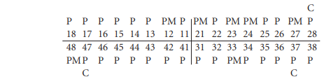
Grave 1: young adult male, 173cm
This skeleton was in very good condition, although a lot of the bones were missing from the right side. Very little remained of the skull except for a few fragments of cranium and most of the mandible. All the cervical, thoracic and lumbar vertebrae were present and virtually complete. The body of the sternum was complete and there were fragments of twenty ribs. Both scapulae were present and virtually complete and both clavicles were complete. The left humerus was complete but the medial part of the distal joint surface was broken off the right bone. The left radius was complete and only the head was missing from the left ulna. The hands consisted of the left lunate, left triquetral, left and right trapezium, left and right trapezoid and left capitate, three metacarpals and 26 phalanges.
Most of the pelvis was present. The left ilium and ischium were complete but only a small portion of the right ilium and ischium remained. Part of the right pubis showing the pubic symphysis was present. The body of the first sacral vertebra was present and the coccyx was complete. The left femur was complete but there was only a small fragment of the distal extremity of the right femur. The proximal end of the left tibia was present but broken and there was a fragment of the shaft of the left fibula. There were no foot bones.
Pathology
Schmorl’s nodes were present on the upper and lower surfaces of the sixth, seventh and eighth thoracic vertebrae.
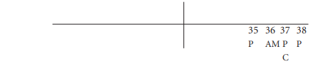
Dentition
Occlusal attrition was slight. Dentine was exposed on all cusps of the first molars, 36 and 46, and there was some slight dentine exposure on the second molars.
There was some slight calculus formation on the lingual surface of all teeth, especially the incisors. It was also present on the mesial and distal surfaces of all teeth and on the buccal surfaces of the left canine, premolars and first molar.
Enamel hypoplasia in the form of slight horizontal lines was evident on the lower half of the crowns of the incisors and left second molar.
Skeleton 1A
The left side of a mandible was also present in this bag. This has been labelled skeleton 1A. Teeth present were the left second premolar and all the left molars. Occlusal attrition was severe on all these teeth, consistent with an individual aged 35–40 years. There were heavy calculus deposits on the lingual surfaces of all teeth and a moderate amount on the buccal surfaces. There is some slight alveolar resorption around the sockets of the molars, indicating periodontal disease.
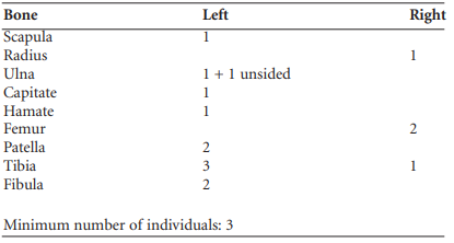
Grave 2: bone scatter
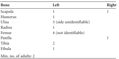
This scatter represented the remains of at least three individuals. The skull fragments were mostly of frontal and parietal bone, but there were three temporal bones present, two from the left and one from the right side. In addition, there was a fragment of mandible and a fragment of maxilla. The minimum number of postcranial bones present is shown in Table 4.14.
Table 4.14—Minimum number of postcranial bones, burial 2
Dental pathology
The mandible and maxilla were from two separate individuals, as the occlusal wear patterns on the molars did not match.
Individual A:
Occlusal attrition was quite severe on the second premolar, 35, with exposure of dentine over the entire surface, but was slight on the second molar, 37, with only patches of dentine exposed; attrition was very slight on the third molar.
There was a small caries cavity on the buccal surface of the crown of the second molar at the occlusal edge.
The first molar had been lost during life and the socket was almost closed. There was considerable alveolar resorption around the roots of the second molar.
Individual B.

Occlusal attrition was severe, with exposure of dentine on all teeth over the entire occlusal surfaces.
There was slight calculus formation of the palatal surfaces of the canine, 23, and first premolar, 24, and on the buccal and distal surfaces of the second premolar, 25.
The socket for the first molar was closed, so it was lost a considerable time before death.
Grave 3: inhumation and scatter
It was possible to separate out one individual from these bones but there were at least two other adults, three young adults (18–20), three juvenile skeletons and one infant, representing at least ten individuals.
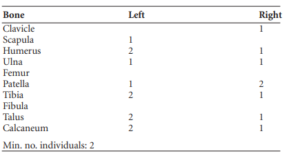
Skeleton 3A: older adult female, 160cm
The skull from this skeleton was virtually complete and intact, including the maxillae and mandible. The post-cranial skeleton included a complete left scapula and a fragment of the right, the complete left clavicle and the medial half of the right clavicle. The left humerus was complete (L 30.5cm) but the head was missing from the right. There were fragments of thirteen ribs and the manubrium of the sternum was present. Some foot bones were present. These included left and right calcanea, left and right cuboid and left and right medial cuneiform. All the metatarsals except the fifth right were present.
Pathology and anomalies: cribra orbitalia was present in the left orbit. There were severe arthritic changes on the sternal end of both clavicles and on the acromial end of the left (right not present). The corresponding articular areas of the manubrium and left scapula also showed severe arthritic changes. The first metatarsal had an articulation area for the second metatarsal, which, although not normal, is not an uncommon anomaly.
Dentition:

The alveolar bone around 28 was damaged, so it was not certain whether this tooth had been lost ante-mortem or not. The sockets for the lower left molars and the upper right first and second molars were completely closed over, so they must have been lost a considerable time before death. The sockets for the lower right molars were only partially healed, so they were probably lost shortly before death.
There was a moderate amount of alveolar resorption around the roots of all the remaining teeth and some slight resorption around the sockets of the lower incisors, 41, 31 and 32. Occlusal attrition was very severe on all remaining molars and premolars, except for the first mandibular premolars.
There were moderate calculus deposits on the lingual surfaces of the upper premolars and first molar and also on the buccal surfaces of the upper left premolars. The deposits were heavy on all the surfaces of the lower right second premolar and the lower left canine, and also on the buccal surface of the lower left lateral incisor, the lingual surface of the lower left first premolar and the buccal and distal surfaces of the upper left first molar. There were moderate calculus deposits on the lingual surfaces of the upper premolars and first molar and also on the buccal surfaces of the upper left premolars. The deposits were heavy on all the surfaces of the lower right second premolar and the lower left canine, and also on the buccal surface of the lower left lateral incisor, the lingual surface of the lower left first premolar and the buccal and distal surfaces of the upper left first molar.
There was a small caries cavity on the distal surface of the upper right third molar, above the cervical margin. This tooth also has an abscess cavity at the buccal root.
Enamel hypoplasia was present in the form of two grooves on the lower half of the crown of the mandibular canine. The effects of hypoplasia may not be visible on other teeth owing to heavy attrition and heavy calculus deposits.
Other bones
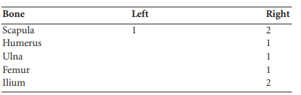
In addition to the main adult burial 3A, the following adult bones were present.
Table 4.15—Additional adult bones present.
There were a number of younger adult/adolescent and juvenile skeletons mixed in with this bag. These have been differentiated according to the state of epiphyseal fusion.
There were also numerous hand bones: left and right lunate; left and right capitate; left and two right scaphoid; left and right hamate; left and right trapezium; nine metacarpals and five phalanges. There were also four cervical, five thoracic, four lumbar and three sacral vertebrae.
Table 4.16—Individuals aged 18–20 years
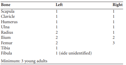
Table 4.17—Juvenile skeletons.
Juvenile skeletons
Owing to the different size of the bones, it is estimated that at least three juveniles are present. In the absence of skulls, juveniles may be aged by comparing bone lengths and size to other skeletons of known age. This should be done using other skeletons from the same site. As this is impossible in this case, a rough estimate of age was obtained by comparison with skeletons from another Irish site (Buckley 1991). On that basis it was estimated that two of the skeletons were between six and ten years and one was between twelve and sixteen years of age at the time of death.
Skulls
Apart from the skull belonging to skeleton 3A, four other skulls were present.
Skull B: young adult, probably female
Although very fragmented, much of the cranium was present here. Most of the occipital, parietal and temporal bones were present, along with the right zygomatic and a small amount of the frontal, including the left orbital part. The basi-sphenoid synchondrosis was not fused. The mandible was virtually complete and most of the maxilla was present.
Skeletal pathology: The left upper orbit exhibited cribra orbitalia.
Dentition:
Attrition was slight on the first molars, with virtually no wear evident on the second and third molars. This individual was probably a young adult. Slight calculus deposits were present on the buccal surfaces of the upper right canine, premolars and first and second molars and the lower canines and first premolars, and also on the lingual surfaces of the lower second premolars and first and second molars as well as the upper right second molar.
There was a small caries cavity on the centre of the occlusal surface of the lower right second molar and another on the middle of the buccal surface. The left second mandibular molar, 37, also had a small caries cavity on the buccal surface. The third left maxillary molar, 28, had three tiny cavities on the occlusal surface.
There were a few lines of enamel hypoplasia on all the incisors, the right mandibular canine and maxillary canine and right mandibular molars.
Skull C
This consisted of fragments of parietal, occipital and some temporal bones, but the mastoid processes were not present.
Skull D
This consisted of many fragments of frontal, parietal and occipital bones. There was some temporal bone, but the mastoid processes were not present. A fragment of right maxilla from sockets 11–16 was present but there were only two teeth, 16 and 15. These were severely worn; wear was uneven, with the palatal surface of the crowns almost gone.
Skull E
This consisted of part of the temporal bone and mandible of a juvenile. The following teeth were present.

The second permanent molar was not erupted and this state of eruption indicates a juvenile aged eight to ten years. There was moderate calculus on the lingual surfaces of the deciduous and first permanent molars and slight calculus formation on the buccal surfaces of the lower right second deciduous molar and first permanent molar and the lower left first deciduous molar. Deposits were heavy on the buccal and lingual surfaces of the incisors. Linear enamel hypoplasia was evident on the lower parts of the crowns of the permanent first molars and the upper incisor.
Loose teeth found in this scatter included one deciduous molar and some newly formed incisors and canines, representing the dentition of at least one child aged seven to ten years.
Grave 4: bone scatter
This was a collection of scattered bones.
The postcranial bones are listed in Table 4.18.
In addition, there were some hand bones, including a trapezium, a scaphoid, a capitate, four metacarpals and seven phalanges. The foot bones consisted of the left cuboid, medial cuneiform and four metatarsals.
Vertebrae present included the upper five cervical vertebrae and the upper two thoracic vertebrae. There was also a fragment of ischium from the pelvis.
Table 4.18—Postcranial bones, burial 4.
Pathology
The fragment of scapula showed marginal lipping around the glenoid fossa.
The distal end of the left ulna also exhibited signs of degenerative joint disease, with bony lipping on the articular area for the radius and build-up of bone on the styloid process.
Skull (no. 4)
There were two skulls mixed here. One had a fragmented but almost complete calvarium. The posterior parts of the temporal bones were also present.
The other skull consisted of small fragments of frontal parietal and the right temporal bone. Also present was a left zygomatic bone and some fragments of a sphenoid. There were two maxillae present.
Maxilla A:

Occlusal attrition was very slight even on the first molar, where only small islands of dentine were exposed. There were very slight calculus deposits on all surfaces of the right canine and third molar and on the buccal surface of the left canine and mesial surfaces of the first premolar and first molar.
Maxilla B:

Occlusal attrition was severe. Secondary dentine was exposed and all cusp detail was lost over all the teeth except the right first premolar, 14, on which, although secondary dentine was exposed, cusps could still be distinguished. The crown of the right first molar, 16, was almost worn down to the root. There was a small caries cavity on the distal surface of 27 just above the cervical margin. There were slight calculus deposits on the buccal surfaces of the premolars and right second molars, the mesial surfaces of the right second premolar and molars and on the distal surfaces of the first right premolar and both second molars. Hypercementosis was present on the roots of 16, 15 and 24, with some slight cementum build-up on the roots of 17 and 25. Also present was a mandible

Only the sockets for the incisors and left canine remained; the other sockets were completely closed and therefore the teeth must have been lost a considerable time before death.
Grave 5: bone scatter
This consisted mainly of numerous fragments of skull bones, which appear to represent only one skull. Both parietals and occipital bones were present but incomplete. The left temporal was incomplete and there was a fragment of the right. Both zygomatics were present and there were fragments of sphenoid bone. There was a fragment of maxilla from the right side. No teeth were present, only sockets for 16, 14 and 13. The socket for 15 was closed over, so this tooth was lost ante-mortem. In addition, there were fragments of three ribs, a fragment of ulna, a left trapezium and eleven phalanges from the feet. There were also four cervical vertebrae, including a first and a second.
Grave 6: older adult male, 178cm
Although very fragmented, this skeleton was in good condition down to knee level.
The skull was very fragmented and consisted mainly of fragments of occipital bone, although some fragments of frontal and temporal bone were present. The body of the mandible was present through fragmented. The vertebral column was in poor condition, but there were fragments of the first two cervical vertebrae, six thoracic, five lumbar and two sacral. Fragments from at least twelve ribs remained.
Most of the lateral half of the left scapula was present and there was also a fragment of the right scapula. The left humerus was virtually complete and the distal half of the right was present. The left radius was complete (L 25.2cm) but the right was fragmented in mid-shaft. Both ulnae were almost complete, but the distal end was missing from the left and the right was fragmented in mid-shaft. There were a number of hand bones, most of which were complete, and only the right pisiform and the right fifth metacarpal were missing. There were eleven hand phalanges.
The pelvis was virtually all present although it was fragmented. The left ilium was complete and most of the right was present. The right ischium and pubis were complete; the left ischium and pubis were fragmented. The left femur was complete (L 470mm); the head and neck only remained from the right, the shaft being in fragments. The proximal end of the left tibia and a fragment of shaft of left fibula were also present.
Pathology
There is an oblique fracture in the distal third of the right ulna. Both hip joints are severely arthritic. There is gross osteophytic growth around the head of the right femur, which is distorted, and an area of pitting on the posterior part of the head. The right acetabulum exhibits a large area of pitting and eburnation in the inferior and medial part. The superior edge exhibits marginal lipping and pitting of the surface. The left femur head shows similar arthritic changes, as does the left acetabulum.
There were early degenerative changes at the left knee (the right was not available).
The vertebrae were broken up, but some degree of lipping could be seen on the lumbar articular surfaces. There was slight osteophytic growth on at least one lumbar vertebral body.
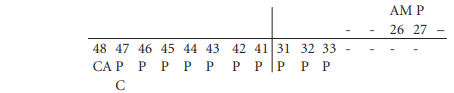
Dentition

The mandible was fragmented and not all sockets were available for inspection. Only the third right molar, 48, was in situ. The first right molar, 46, was present but the socket was damaged. The stump of a premolar, 44, was also present.
There was considerable wear on the occlusal surface of the molars. There was a large caries cavity on the distal root of 48 and another on the mesial surface of 46, extending over the root and what was left of the crown. This cavity penetrated to the pulp chamber. There was some moderate alveolar resorption around the root of 48. The third molar on the left side, 38, had been lost shortly before death, as the sockets were starting to close over. The socket for the second molar on the right was closed, so it had also been lost during life.
Other bones
Part of a right ulna, a first metatarsal and two foot phalanges from other burials were present. There were also some teeth (five incisors and three premolars) that did not belong to skull 6.
Grave 7: bone scatter, late middle adult female
This consisted mainly of small fragments of skull. There were numerous fragments of frontal, parietal and occipital bone, which could not be pieced together but appeared to represent only one skull. There was a small portion of maxilla and most of the mandible.
Dentition
There were also two unidentifiable root stumps, which exhibited hypercementosis. The incisoral edges were worn flat with exposure of dentine, and there was also moderate wear on most of the other teeth. There were slight calculus deposits on the buccal surfaces of the right incisors, both canines and right second premolar, and on the lingual surfaces of the central incisors, right lateral incisor, canine and first premolar. There was a small caries cavity on the buccal surface of 47 at the cervical margin. Grooves of hypoplasia were present on the canines and premolars and linear hypoplasia was present on the incisors and right second molar. This individual had obviously suffered from disease constantly through childhood and early adolescence.
The condyle of the left mandible exhibited marginal lipping. Osteoarthritis of the temporo-mandibular joint can occur when the diet is very abrasive, owing to heavy mastication, although there can be other causes.
Other bones
In addition to the skull, there were fragments of one cervical vertebra, some small fragments of femur and fibula shafts, and small fragments of ilium.
Grave 8: bone scatter
This merely consisted of a few slivers of long bone too small to be identified.
Grave 9: bone scatter This consisted of a few small skull fragments, mainly of occipital and parietal bones. There was a part of the left temporal around the area of the mastoid process, which appears to indicate that this is a male skull. One cervical vertebra and one thoracic vertebra were also present.
Grave 10 There were two bags, one marked ‘No. 10’ and one marked ‘No. 10?’
Bag no. 10 contained numerous tiny fragments of skull and a small part of a humerus shaft. Also present was the first cervical vertebra and the body of one other cervical vertebra. The following teeth were present:
Occlusal attrition was not severe, with slight wear on the first molars, some polishing of the second molar and no wear on the third molars. This individual was probably aged 20–25 years. There were slight calculus deposits on the labial and lingual surfaces of the incisors and the labial surface of the canine, and there were moderate calculus deposits on the buccal and lingual surfaces of the premolars and canines.
There was a slight caries cavity on the occlusal surface of the right third molar where the fissures meet, and there was root caries just below the cervical margin of the right first molar. Enamel hypoplasia in the form of lines was evident on the crowns of the incisors, canine and molars.
Bag no. 10? contained mainly hand bones. There were two complete carpals, the right trapezium and capitate, part of another unidentifiable carpal, the second and fourth metacarpal of the right hand plus two other incomplete metarcarpals which could not be identified and five hand phalanges. Three vertebrae were present, the first cervical vertebra and the bodies of one other cervical vertebra and one thoracic vertebra. There were also a few fragments of long bone shaft.
Grave 11: bone scatter
This was a scatter of bones. There were numerous fragments of one skull, which probably represented one cranium, but the fragments were too small to reconstruct. There were fragments of frontal, parietal and occipital bone, including the basilar part. Both temporal bones were also present. The state of the basi-sphenoid synchondrosis suggests that this individual was less than 25 years old at death. There is one tooth present, which appears to be a second mandibular molar from the left, which has an extra cusp. There was very little wear on this tooth. Other bone fragments from the site include a fragment of humerus shaft, a fragment of ulna and a few fragments of femur shaft. There was also the lower end of a left fibula and part of the first sacral vertebra. Another tooth, the right third molar, 48, from another skull was present. There was a moderate degree of attrition on this tooth and a heavy calculus deposit on the lingual surface.
Grave 12: early middle adult male, 170cm
This skeleton was almost complete but was very fragmented. The skull, in particular, was in many small fragments and could not be reconstructed, although probably most of the cranium was present. There were fragments of occipital, parietal, frontal and temporal bones. The mastoid processes were very prominent and the posterior root of the zygomatic process extended beyond the external auditory meatus. The right side of the maxilla was present. Most of the mandible was present although it was broken up. The vertebral column was in bad condition. There were fragments of five cervical, nine thoracic, five lumbar and two sacral vertebrae. Most of these were bodies only; all spines and articular processes were broken off. There were fragments of about twelve ribs.
Both clavicles were present; the left clavicle was almost complete. There were fragments of both scapulae from around the glenoid fossae and the lateral borders, but the spines were broken off. The distal halves of both humeri were present. The ulnae were virtually complete (L 29.9cm) and so was the left radius. The distal end of the right radius was present. There were a number of hand bones: left and right scaphoid and lunate, right triquetral, left pisiform, right trapezium and left and right trapezoid, as well as five incomplete metacarpals and fifteen phalanges, eight proximal, six middle and one distal.
The pelvis was very broken up, but there were parts of the left and right ilia showing the acetabulum and sciatic notch. Both ischial tuberosities were also present and parts of both pubic bones, although the pubes symphyses were broken.
The distal half of the left femur and most of the shaft of the right femur were present. Both tibiae were present and complete (L 390mm), although the right tibia was badly smashed at the upper end. The left patella was present. Both fibulae were present but broken. Most of the foot bones were present from each foot, although a few were fragmented. All the tarsals were present except for the right second cuneiform and left and right third cuneiforms. All the metatarsals except the third left were present. There were also nine proximal phalanges, three middle phalanges and six distal phalanges.
Pathology
Schmorl’s nodes were present on the bodies of all the lumbar vertebrae.
There was an extensive area of periostitis extending over most of the posterior surface of the distal half of the right femur shaft. There was possibly another area of periostitis on the medial surface of the proximal end of the shaft of the right tibia. This particular bone was badly decayed, however, so there is the possibility of pseudo-pathology here.
Dentition

There was a moderate amount of attrition on the molars, consistent with the early middle adult age group. There were slight calculus deposits on all teeth, and linear enamel hypoplasia was evident on the lower incisors, right canines, the right second premolar, all the left premolars and the remaining molars, apart from the first molars.
Grave 13: bone scatter
This consists mostly of numerous fragments of shaft and the distal joint surface of what appeared to be one femur. There were also some tibia fragments and one carpal, the right lunate.
No. 13A
There were two bags, each labelled No. 10?/No. 13A; these represented two separate individuals. (1) This consisted of very fragmented long bones, probably representing one inhumation. Most of the shaft of the right humerus and most of the right ulna were present. There were some fragments of radius shaft. The right ilium was present from the pelvis. The shafts of both femora were present but the distal ends were very fragmented. The left patella was present but incomplete and there was a fragment of the proximal end of a tibia (2) This represented the remains of another skeleton, also totally fragmented. Only one tooth, an upper left second molar with very light wear, was present from the skull, and only the body of one cervical vertebra remained from the vertebral column.
There was a fragment of the right scapula, as well as the middle section of a clavicle. Some of the shaft of the right humerus was present and there were shaft fragments of both radii and ulnae. Some carpal bones were present: left scaphoid, right lunate, left and right triquetral, left pisiform, right capitate and left hamate. There was also a left first metacarpal and six hand phalanges. Only a tiny piece of acetabulum remained from the pelvis. Both femur shafts were present but the joint ends were fragmented. The left patella was present but fragmented. The proximal ends of both tibiae were present, as well as the shaft and distal end of the left fibula. Some foot bones were present but were very fragmented. These included a talus, a calcaneus, a navicular, two cuneiforms and three phalanges.
Grave 14: skul
l This consisted of a skull, which, although fragmented, could be reconstructed. The calvarium was mostly complete. Also present were both temporal bones, both zygomatic bones, the sphenoid bone and fragments of palatine bone. Most of the mandible and a large portion of the maxillae were present but fragmented. The axis, the atlas and bodies of four lower cervical vertebrae were also present.
Non-metric traits
There were two lambdoid ossicles present. The frontal (metopic) suture was retained. There was a supraorbital foramen on the right.
Skeletal pathology
There was a circular lesion on the left frontal, 2cm superior to the supraorbital notch. This lesion measured 4.5mm in diameter and there was an area of swelling immediately above it. The cortex was exposed but the bone was not perforated.
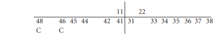
Dental pathology
There was light attrition of the incisors, canines, premolars and third molars but heavy wear on the second molars. Calculus deposits were heavy on all teeth, especially the molars. There was a large caries cavity on the mesial surface of 48 and the distal surface of 47. Only root stumps of the first molars, 36 and 46, remained, the rest of the crowns being presumably destroyed by caries. There was a small caries cavity on the distal surface of the lower right second premolar, 45. In the maxilla there was a large caries cavity on the buccal surface of the upper right third molar, 18, below the cervical margin, and there were also slight caries cavities on the mesial surfaces of the upper left premolars, 24 and 25, at the cervical margin. Three other root stumps, which could not be placed, were also present.
Additional bones Other bones present included a fragment of left scapula, the lateral half of a right clavicle, fragments of humerus shaft and one humerus head. There was also a left trapezoid and two foot phalanges.
Grave 14A: bone scatter
This consisted of five proximal, two middle and two distal foot phalanges.
Grave 15: young adult, unsexed
This represented one inhumation but it was in very bad condition, and there was insufficient evidence to enable the sex to be determined. The skull consisted of fragments of frontal, parietal and occipital bone, including the basilar part. Both temporals were present but the mastoid processes were broken off. The sphenoid was present but fragmented. There were also fragments of both maxillae and the right side of the mandible was complete. The first three cervical vertebrae were all that remained of the vertebral column. There was a fragment of the left scapula, some fragments of ulna and radius shaft and part of the sternum from the upper torso. The hand bones consisted of left and right scaphoid, pisiform and hamate as well as eight metacarpals. Only a small fragment of the left ilium remained from the pelvis. Most of the femurs, tibiae and fibulae were present but they were too fragmented to reconstruct. A lot of foot bones survived. These included left and right talus, calcaneus, cuboid, the cuneiforms, five metatarsals and seven phalanges.
Pathology and anomalies
Cribra orbitalia was present in both orbits. There was a third trochanter present on the right femur.
Dentition

Occlusal attrition was light on the first molars and there was virtually no wear on the second molars. There were very slight calculus deposits on the buccal surfaces of the upper first molars and right second premolar and on the lingual and interstitial surfaces of the incisors and canines. Enamel hypoplasia was evident on all teeth, so this individual had suffered extensively throughout childhood from various infections and dietary deficiency.
Extra bones
A calcaneum and talus and the distal end of a fibula were present from another burial. There were also two severely worn incisors. A shaft of an infant (<3 months) long bone was present.
Grave 16: older adult male, 159cm
This skeleton was in quite good condition and almost complete. The skull was very fragmented but the calvarium was mostly complete. Both temporals were present, as well as the sphenoid, both zygomatics and fragments of maxilla and mandible. The vertebral column was very fragmented but there were six cervical vertebrae, including the first and second, nine thoracic, five lumbar and two sacral vertebrae. There were fragments of both scapulae and both clavicles. Both humeri were present but the middle parts of the shafts were smashed. Both radii and ulnae were complete. All of the carpal and metacarpal bones except the pisiforms were present, as well as five proximal, six middle and six distal phalanges. The ilia from the pelvis were present but incomplete. There were fragments of both ischia and the pubic bones were also present. Both femurs were complete, as well as both patellae and the left tibia. The proximal end of the right tibia was smashed. All the tarsal bones and most of the metatarsals were present, as well as seven phalanges. Pathology Osteophytosis was present throughout the vertebral column. Of the lumbar vertebrae, L3 was the most severely affected, with osteophytes extending 5mm beyond the edge of the centrum on both the inferior and superior surfaces. The superior surface of L4 and the inferior surface of L2 were affected to a lesser extent. Of the thoracic vertebrae, T9, T10 and T11 were severely affected on both the inferior and superior surfaces, especially T10. There was a moderate amount of osteophytosis on T8 and on all the cervical vertebrae. Osteoarthritis of the posterior joints was also widespread throughout the vertebral column, although not all the articular surfaces were available for inspection. The cervical region was most affected, with eburnation of the inferior articular surfaces of C2 and C5, and on the facet for the odontoid process on C1. Some of the thoracic vertebrae showed severe arthritic changes, but as they were fragmented they could not be assigned to any particular vertebrae. There was eburnation of the superior and inferior articular surfaces of two vertebrae, and two others with eburnation of the upper surface only. The left inferior articular surface of L1 and the left superior articular surface of L2 showed arthritic lipping and slight eburnation. The right inferior articular surface of L4 and the right superior articular surface of L5 also had some slight eburnation.
Degenerative joint disease was present to a slight extent in nearly all other major joints that were available for inspection. Both shoulder joints were affected, with marginal lipping around the glenoid fossa of the scapulae. The right humerus displayed marginal lipping and some pitting of the head (the left humerus head was fragmented). Both elbow joints had DJD, with slight marginal lipping of the trochlea of the humeri and trochlear notch of the ulnae. There were also early degenerative changes at both wrists, with slight marginal lipping of the distal radii articular surfaces. There was marginal lipping and a small area of pitting on the superior surfaces of both acetabula (hip joint).
The knee joints also showed early degenerative changes, with slight marginal lipping of the anterior portion of the medial condyle of both femora and the medial surface of both patellae. The distal articular surfaces of both tibiae (ankles) also had slight marginal lipping, as did the superior articular surfaces of the tali. The soleal line on the left tibia was very pronounced. This suggests that the soleus muscle, which is important in walking, was well developed.
Dentition

The molars were very severely worn down. Secondary dentine was exposed on all surfaces of the first and second molars and wear was uneven, with no enamel left on the lingual surfaces. Attrition was moderate on the third molars.
There were very heavy calculus deposits on the buccal surface of the right first molar and both third molars, on the lingual surfaces of the first and second molars and left third molar, and on the distal surfaces of the left second and third molars.
There was a large caries cavity on the buccal surface of 48 below the cervical margin, and another on the occlusal surface of 46 near the meso-lingual corner.
Considerable alveolar resorption was evident around the roots of the left first molar. The mandible was considerably damaged, so it was not possible to estimate resorption around all the roots of the teeth. Hypercementosis was evident on the roots of the right molars and left second and third molars.
There were also ten teeth, presumably from this skeleton, which were worn down completely to the root. Two incisors had only a small piece of crown remaining on the labial surface, one of which had the pulp cavity exposed. The other teeth could not be identified, except that they were either incisors or premolars. Some of the roots exhibited hypercementosis.
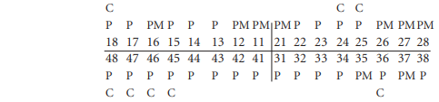
Unlabelled bone scatter
Apart from the numbered burials and scatters previously examined, there were two bags of scatter, which were not labelled. Most of these bones were in poor condition, being badly fragmented, so the best that could be done was to ascertain the minimum number of each bone, to enable the minimum number of individuals to be determined.
The numbers of each adult bone present were as follows.
Table 4.19—Adult bone present
Only one left femur, two left tibiae and one right radius were complete.
Also present were a few tarsals and metatarsals, five second cervical vertebrae, seven lower cervical, eleven thoracic and seven lumbar vertebrae. The minimum number of adult skulls was also six, based on the minimum number of mandibles present.
Pathology
One femur, a right femur, probably male, had an area of periostitis at the proximal end, around the area of the greater trochanter, partially extending on to the neck.
One of the left adult scapulae had severe osteoarthritis, with marginal lipping and eburnation of the lower third of the glenoid fossa.
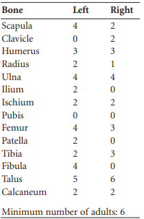
Dental pathology
Very few teeth remained from the six skulls; some of the mandible portions consisted of bone only.
Mandible 1, adolescent/young adult: the following teeth were present.
There was slight wear on the occlusal surfaces of the first molars only. There were moderate calculus deposits on the lingual surfaces of all teeth. Linear enamel hypoplasia was evident on the incisors and the left canine and premolars.
Mandible 2, middle adult: this consisted of the right side of the body and the ramus of the mandible. Only two teeth, the right first and second molars, 46 and 47, were present. There was a moderate degree of occlusal attrition and moderate calculus deposits on all teeth.
Mandible 3, middle adult: only the right three molars, 46, 47 and 48, were present. There was a moderate degree of attrition on all teeth but the cusps could still be distinguished. Calculus was moderate on the buccal surfaces but heavy on the lingual surfaces.
Mandible 4: there was only one tooth, the left permanent first molar, 36, ‘in situ’, and occlusal attrition was heavy.
Maxillae: there were three maxillae present, two from the right side and one from the left. Only two

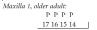
Occlusal attrition was very heavy and uneven, with the crown completely worn away on the lingual surfaces. A moderate degree of calculus was present on the buccal surfaces and the interstitial surfaces
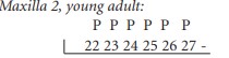
There was a moderate degree of attrition on the occlusal surfaces of 25, 26 and 27, with exposure of dentine on the palatal cusps. In addition, ten other teeth were present, which could not be assigned to any skeleton.
Summary
The minimum number of individuals present was 30. This is made up of 26 (86.6%) adults over the age of eighteen, three juveniles and one infant. The minimum number of adults was based on the number of skulls present.
Age and sex
There were only six discrete ‘in situ’ burials, the rest being found in the various scatters. Four of these burials were male, one was female and the other was indeterminate. Of the remaining scattered adults, only four were possible to sex by skull fragments; one was male and three were female.
Age range determination was possible for only nineteen of the adults. Nine (47.4%) were young adults, three (15.8%) were early middle adults, three were late middle adults and four (21%) were older adults.
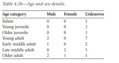
The four juveniles represent 13.3% of the total number of burials. Obviously this does not reflect the true infant mortality rate, which may have been as high as 40–50% in past societies. The paucity of infant bones may be due to the fact that preservation of infant bones is poor compared to adult bones after they have been disturbed; in addition, when bones are scattered to such a degree as in this site, infant bones are difficult to pick out and retrieve. Age and sex details are summarised in Table 4.20.
Dentition
Twenty-six adult skulls alone should have yielded 832 teeth. Only 221 teeth were available from all the individuals, however, which, combined with thirteen that were lost ante-mortem, represents less than one quarter of the potential total. Given this small sample size, it is probably unwise to draw conclusions about the state of dentition of these individuals.
Some general observations can nevertheless be made. Calculus was present on virtually all individuals with dental remains. In general this was slight on younger individuals, increasing with age, with a few individuals over 35 years exhibiting quite heavy calculus deposits. One juvenile aged 9–10 years had quite heavy calculus deposits on the incisors. Caries was present in seventeen teeth spread over nine adult individuals, i.e. 34.6% of adults had caries. This is a high caries rate compared to the 30.2% at Gallen priory (Howells 1941) and the 20.3% of the Early Christian population at Castleknock (McLoughlin 1950), but again it must be emphasised that it may not be accurate owing to the small sample size. Five of these individuals had caries on more than one tooth. Three of these were more than 35 years of age and two were young adults. There was a difference in the type of caries, however, with the majority of crown caries occurring in younger individuals and the majority of root caries occurring in older individuals.
Caries in the twentieth century is caused by a high level of sugar in refined foods in the diet. In earlier societies sugary foods such as honey were not unknown, but caries was also probably caused by retention of starchy food particles in the mouth long enough to be broken down into sugars. Food debris would tend to be trapped near the cervical margin and give rise to caries at the neck or root of the teeth.
It seems likely, therefore, that there was a carbohydrate content in the diet of the individuals from this site, but this must have included some sugars, as crown caries is present in young adults, with one individual in particular (3(b)), aged 18–25 years at death, having caries on three molars. The dentition retrieved from two juveniles had no caries present. Occlusal attrition was quite severe in this group of skeletons, some of the older individuals having crowns completely worn away. This could be caused by a highly abrasive diet such as one containing roughly ground cereals.
Dental abscess: only one abscess cavity was found, which was at the root of a third maxillary molar that had a caries cavity at the cervical margin. This older individual also had severe occlusal attrition and periodontal disease.
Periodontal disease: periodontal disease is an infection of the gums and alveolar bone that causes recession of the bone, with the result that there is less support for the tooth, which eventually falls out. Lack of dental hygiene, calculus deposits and attrition can contribute to this disease. In the older individual already mentioned, periodontal disease was severe, with the loss of eight teeth ante-mortem. The total number of teeth lost was thirteen, or 5.5%. This is quite low but is probably not a true reflection of the ante-mortem tooth loss on this site, as many teeth were found loose with no supporting bone and the number of maxillae found in particular was quite low. All the teeth lost were molars.
Enamel hypoplasia: hypoplasia is a defect in the enamel of the teeth as a result of an illness or dietary deficiency during the time when the enamel is being formed. These defects may be microscopic but only the larger macroscopic defects visible to the naked eye are considered here. Usually hypoplastic defects are in the form of horizontal lines or grooves on the surface of the enamel. There were nine individuals from this site with enamel hypoplasia evident. Generally this involved the incisors plus various other teeth. A few individuals had hypoplastic defects on nearly all the teeth present. It is not possible to make detailed analysis of hypoplasia from this population owing to the low proportion of teeth retrieved and the fact that hypoplastic defects may be obscured by calculus and sometimes removed by the heavy attrition. All that can be said is that acute illness and dietary deficiency occurred throughout infancy and early childhood in the population.
Pathologies
(1) Cribra orbitalia: this condition, characterised by pitting of the orbital roof, is of uncertain origin; the most recent theory is that it is caused by iron-deficiency anaemia, but only if the deficiency occurs in infancy. It was present in three skulls; in two cases it occurs in the left orbit and in the other case it was bilateral. A lot of skulls from this site were fragmented and the orbital roofs were not always available for inspection, so the true incidence of this condition may be higher.
(2) Osteoarthritis: this is a degenerative joint disease generally caused by the minor traumatas of everyday use, i.e. wear and tear on the joints. It would therefore increase in severity with age. At the onset of degenerative disease the joints exhibit bony lipping around the edges. This has the effect of increasing the surface area and so relieving stress. As the disease progresses, the joint surfaces become pitted, finally becoming eburnated as bone moves upon bone. Occasionally osteoarthritis can be brought on at an earlier age by trauma of the joint caused by a fracture.
Three skeletons from this site had arthritis in various joints. All these individuals were over 45 years old at the time of death. Two were male and one was female. Unfortunately the female was very incomplete, consisting mainly of the upper torso. This individual exhibited severe osteoarthritic changes at both sterno-clavicular joints and the left acromio-clavicular joint (the joint from the right side was not available for inspection). Arthritis of the shoulder is rare today, but its presence in earlier societies is probably due either to the carrying of heavy loads or to the constant use of the arms in some heavy or vigorous task. Both the male skeletons had osteoarthritis of the hip joint. In the case of skeleton no. 6 this was very severe, with gross changes of the femur head and eburnation of the acetabulum. This individual had early degenerative changes at the left knee. Possibly arthritis would have been more extensive in this skeleton but it was incomplete and many joints were missing. Skeleton no. 16, however, was very complete, with nearly all joints available for inspection, and degenerative joint disease was extensive. There were early changes at the shoulders, elbows, wrists, hip, knees and ankles. The shoulders and hips were the most affected, but this was still only slight compared to skeleton no. 6. The vertebral column of this individual also showed osteoarthritis of the posterior joints in all regions but was particular severe in the neck area. The soleal line of the left tibia was very pronounced in this individual. The soleus is a muscle used for flexing the feet and is important in walking, but the fact that one soleal line is more developed than the other suggests that some severe strain or damage to the muscle attachment occurred.
(3) Osteophytosis: skeleton no. 16 also had extensive osteophytosis throughout the vertebral column. This condition, though influenced by the same factors that affect osteoarthritis, is not true arthritis because the joints between vertebral bodies are not diarthodial. The vertebral bodies react to strain in a similar way as other joints by developing bony margins known as osteophytes. The severity of the condition increases with age. Not all vertebrae are affected to a similar extent, and in skeleton no. 16 the fourth lumbar and tenth thoracic vertebrae were the most severely affected.
(4) Periostitis: periostitis arises as a consequence of inflammation of the outer covering of bone, the periosteum. It can arise as a result of low-grade chronic bacterial infection of the overlying tissues or may be secondary to trauma. It is not always possible to determine the cause on dry archaeological bone specimens.
Skeleton no. 12 had areas of periostitis on the posterior surface of the right femur shaft and the medial surface of the right tibia. Periostitis of the tibia is generally believed to be caused by the repeated minor traumas of an everyday working life.
Another femur retrieved from some unlabelled scatter had an area of periostitis around the region of the great trochanter. It is difficult to see how this could be caused by trauma, but an X-ray would be required to see whether the inner layers of bone were infected. If this was the case, the cause would probably be bacteria carried in the bloodstream from the primary source of infection elsewhere in the body.
Conclusions
The 30 individuals from this site probably existed on a diet that contained a fair proportion of roughly ground cereals. They were subject to a number of acute illnesses and periods of dietary deficiency in childhood. There is a possibility that chronic infections still dogged their adult life. The evidence from a few older individuals suggests that they, at least, had a fairly strenuous working life.
It is difficult to draw conclusions from such a small sample of individuals in such a bad state of disarray. Probably a lot more information about the lifestyle of these individuals could have been retrieved if the bones had not been fragmented and scattered to such an extent.
109. Further bones were also collected by Patrick Holland on 25 October and handed over to the NMI.
110. Parish of Caher, barony of Iffa and Offa West. SMR TS075-041——. IGR 205398 126175.
111. The descriptions of the burials are by PH and MC, while the osteoarchaeological analyses are by Laureen Buckley. The latter are given in summary and a fuller description can be found in Buckley’s report.
112. GrN-17620.
113. Grave 6, GrN-17621. Grave 12, GrN-17622.
114. GrN-17623.
115. GrN-17624.