1987:078 - BALLYBUNNION, CO. KERRY, Kerry
County: Kerry
Site name: BALLYBUNNION, CO. KERRY
Sites and Monuments Record No.: SMR KE004-061
Licence number: E1075
Author: RAGHNALL Ó FLOINN
Author/Organisation Address: —
Site type: Iron Age and early medieval graves, c. 300 BCc. AD 1200
Period/Dating: —
ITM: E 486500m, N 640398m
Latitude, Longitude (decimal degrees): 52.503155, -9.671805
Introduction
In April 1987 a long cist containing an inhumation burial was discovered at Ballybunnion, Co. Kerry, during the digging of trenches for drainage pipes for new County Council houses off Sandhill Road. During the digging operations, at least one complete skeleton had been found lying approximately south-west/north-east, with the head to the south-west. It appeared to have been fully extended. The workmen at the site could not be certain whether this grave had been stone-lined but many slabs were visible scattered around the sides of the trenches. The site was reported to the NMI by Paddy O’Donovan, director of the North Kerry Archaeological Survey, and a rescue excavation was undertaken by Raghnall Ó Floinn. Examination of the northern trench, which ran east/west, uncovered human remains in situ at a depth of 0.6m, set tightly against a stone slab. This grave was excavated and revealed two further successive inhumation burials—a lintel grave overlying an unprotected inhumation.28
The human remains were analysed by Barra Ó Donnabháin.
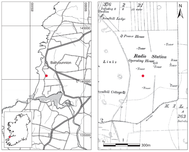
Location (Fig. 4.12)
The site was in Ballybunnion townland, approximately 1km south of Ballybunnion town, north Co. Kerry.29 It was less than 1km from the coast, at an altitude of 0–30m above sea level, and 1km north of Killehenny Church. Approximately 1km south-west of the churchyard, burials were exposed in the sand-dunes in 1934 and 1940, and a cist was examined at the same site at Ballyeagh in 1979 (this volume, pp 41–3). A large number of artefacts have been recovered from the sandhills in the area over the past century, including a Roman coin of Hadrian (AD 117–138), worked flint, bronze pins and bone pins.
Description of site
Grave 1
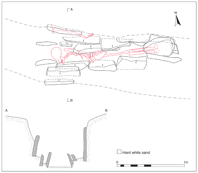
This grave was represented by two edge-set slabs, their bases at a depth of 0.6m, one at each side of the trench (Fig. 4.13). It is probable that these were part of a slab-lined cist, aligned north-west/south-east, the remainder of which had been destroyed. Some human remains were found set tightly against this slab, which measured 0.55m long by 0.5m high. No capstones were found, although the cist had clearly been badly disturbed. The floor of the grave was not paved.
The bones recovered at this level consisted of a left clavicle, humerus, radius, ulna and finger bones, all of which were articulated (1987:55). As relatively few bones were recovered, it was only possible to state that they were possibly the remains of a female. A sample of the human remains was submitted for radiocarbon dating and yielded a result of 1405±45 BP, which corresponds to the period 558–684.30
Grave 2
Between the two slabs represented by grave 1, at a depth of 0.5m below ground level, the tops of a series of side slabs were uncovered (Fig. 4.13). When fully excavated, they formed a long cist measuring 2.1m long by 0.6m wide by 0.25m high (approx.). The cist was aligned west/east and was narrower at the eastern end, being roughly trapezoidal in plan. Many of the side stones inclined inwards and in some cases occurred in pairs. These slabs were all thin flags, 0.2–0.5m in length and averaging 0.05m in thickness. Only one lintel or capstone, 0.5m long by 0.3m wide, survived at the eastern end. There was no trace of a closing slab at the western end, nor were there any floor slabs. The closing slab at the eastern end was a thin, slate-like slab measuring 0.15m long by 0.01m thick. No evidence of a pit was uncovered, although the sand inside the cist was grey while the surrounding sand was buff-coloured.
The cist contained a fully extended inhumation of an adult male (1987:54). The body lay on its right side with the skull at the west, facing south (Pl. 77). The left arm was flexed at the elbow, the hand placed in front of the pelvic region. The legs were fully extended, the left lying over the right. The right arm was straight, with the hand placed underneath the right pelvis. Some of the bones of the left hand, the left femur and pelvic bone and both patellae were missing, presumably dislodged in the course of digging the trench. A sample of the human remains was submitted for radiocarbon dating and yielded a result of 1400±35 BP, which calibrates to 583–674.31
Grave 3
This grave was identified as such by the presence of human remains.32 It was located 0.5m west of grave 2 and was aligned south-west/north-east. There was no evidence for stone or any other protection in the vicinity of this interment.
The burial consisted of a partially articulated skeleton, probably extended and lying on its back with the head to the south-west. This burial was aligned south-west/north-east (240º SW–60º E) and therefore at an acute angle to grave 2 (275º W–90º E).
Comment
Excavation of this burial demonstrated that the site was reused on a number of occasions for burial. It is possible that some of the lintels from grave 2 were reused as side slabs for grave 1, as was seen at Millockstown (Manning 1986, 145). The date range of the sixth to seventh centuries is comparable to dates from other lintel graves discussed in this chapter. It is possible that this cemetery was more extensive, but excavation was limited to the burials that were exposed.
HUMAN REMAINS
BARRA Ó DONNABHÁIN
Introduction
The contents of one long cist grave were examined. The grave contained the remains of two adults. The skeleton of one individual was represented by four bones, none of which was complete. The remains are reasonably well preserved.
Skeleton 1 (1987:54)
This burial from grave 2 consists of the mostly complete skeleton of an adult male. All of the skull was recovered, albeit in a fragmentary state. The vertebral column was complete except for the absence of the first and second thoracic elements. All of the long bones are present with the exception of the left femur. The left hipbone is missing. Most of the ribs, all of the bones of the feet and some of the bones of the hands were recovered.
The morphology of the right hipbone and the bones of the skull, along with the general rugosity of the remains, indicates that this was a male. Tooth wear is moderate to heavy and the presence of an ossified costal cartilage is suggestive of a middle-aged or older adult.
Stature
Using the regression equations formulated by Trotter and Gleser (1952; 1958) it is possible to estimate that the maximum living stature of this person was about 169cm.
Teeth
All of the teeth present at the time of death were recovered. These were:

The presence of a healed socket indicates the ante-mortem loss of the upper left central incisor.
A chronic dental abscess occurs at the upper right lateral incisor. The abscess drained through the buccal side of the alveolar bone of the maxilla. Only the root of the associated tooth remains and it seems likely that the tooth was broken during life, with the abscess developing owing to infection of the exposed pulp cavity. All of the teeth have a moderate amount of calculus formation, while the degree of resorption of alveolar bone at the roots of the molars is indicative of the occurrence of chronic periodontal disease. Linear enamel hypoplasia occurs on

This defect is caused by the occurrence of serious illness or malnutrition during the period of tooth development. In this case, such disturbances must have occurred when this individual was aged about 4–5 years. Dental caries did not occur in any of the teeth from this burial.
Pathology
A number of pathological changes are to be seen in the vertebral column and other joints of this individual. All are degenerative changes whose progress and severity are directly related to both age and lifestyle. Schmorl’s nodes occur on the central of the eighth, ninth and eleventh thoracic vertebrae. These lesions result from the herniation of the core of the intervertebral discs. This core solidifies in early adulthood. Herniation can only occur, therefore, during childhood or adolescence and is usually due to the disc being subjected to severe strain, usually by compression forces from carrying heavy loads. This straining and compression of the vertebral column, and especially of the intervertebral disc, is also responsible for the occurrence of vertebral osteophytosis on the eighth to twelfth thoracic and second to fifth lumbar vertebrae. This is mild on all but the fourth and fifth lumbar vertebrae, on which the condition is moderately severe. A mild degree of osteoarthritis also occurs on the posterior joints of the second and third lumbar vertebrae. Related to this vertebral pathology are the arthritic lesions observed in the ribs of the eleven costocapitular joints available for inspection; four are arthritic, while of the twelve observable costotransverse joints two have lesions.
The right hip was also arthritic: mild lipping occurs on the head of the right femur and on the acetabulum of the right innominate. The left hip was not available for inspection. Mild arthritis also occurs in both temporo-mandibular joints. The arthritic changes occur only on the temporals—the mandibles are not affected.
All of these degenerative changes combine to suggest a lifestyle characterised by strenuous physical labour. Neither their occurrence nor their severity is unusual relative to other early skeletal series.
Cribra orbitalia occurs in both orbits of this individual. The lesion in the right orbit appears to have been healing at the time of death, while that on the left side was mild/moderate in severity. Cribra orbitalia is a condition of unknown aetiology, though it is commonly accepted as evidence of anaemia owing to an inadequate diet or chronic illness.
There is a ‘button’ osteoma on the ectocranial surface of the frontal on the right frontal boss.Benign osteomata such as this are the most common type of tumour found in archaeologically retrieved skeletal material (Brothwell 1981). Their dense osseous form gives them a high archaeological visibility. These tumours are usually less than 20mm in diameter and are found most commonly on the frontal and parietals (Ortner and Putschar 1981). In this instance the osteoma has a diameter of 13mm and rises 3mm above the surface of the skull.
Skeleton 2 (1987:55)
The second individual from grave 1 is represented by only four bone fragments. These are portions of a left humerus, left radius, left clavicle and a mid-thoracic vertebra. These fragments are more gracile than the remains of skeleton 1 and may be from a female.
No pathological changes or anomalies are noted in these remains.
28. The uppermost grave (that represented by two slabs) has been called grave 1; that immediately underneath (the lintel grave) is called grave 2, and the burial to the west of the latter is called grave 3.
29. Parish of Killehenny, barony of Iraghticonnor. SMR KE004-061——. IGR 086525 140351.
30. GrA-24468.
31. GrA-29049.
32. The remains from this grave were not retained.