1985:069 - DURROW DEMESNE, CO. OFFALY, Offaly
County: Offaly
Site name: DURROW DEMESNE, CO. OFFALY
Sites and Monuments Record No.: . SMR OF009-006
Licence number: E1147
Author: RAGHNALL Ó FLOINN AND ELIZABETH OBRIEN
Author/Organisation Address: —
Site type: Iron Age and early medieval graves, c. 300 BCc. AD 1200
Period/Dating: —
ITM: E 632192m, N 730452m
Latitude, Longitude (decimal degrees): 53.323352, -7.516788
Introduction
In February 1985 human remains were discovered during bulldozing at Sheean Hill, Durrow, Co. Offaly. The land had been purchased by the landowner, Mr Michael Egan, nine months previously and levelling was taking place in advance of sowing wheat. The discovery was reported to the Gardaí at Tullamore, who informed the NMI. An excavation undertaken by Raghnall Ó Floinn, assisted by Elizabeth O’Brien, revealed evidence of successive burial and also possibly burial in rows. The excavated human remains were analysed by Laureen Buckley. Ploughing in the same field had revealed traces of a D-shaped vallum, roughly 500m in diameter, enclosing the standing remains of the monastery.
Location (Fig. 4.43)
The site was in the townland of Durrow Demesne, north Co. Offaly (Pl. 80).89 The standing remains of the monastery of Durrow were located in the same field. The site comprised three
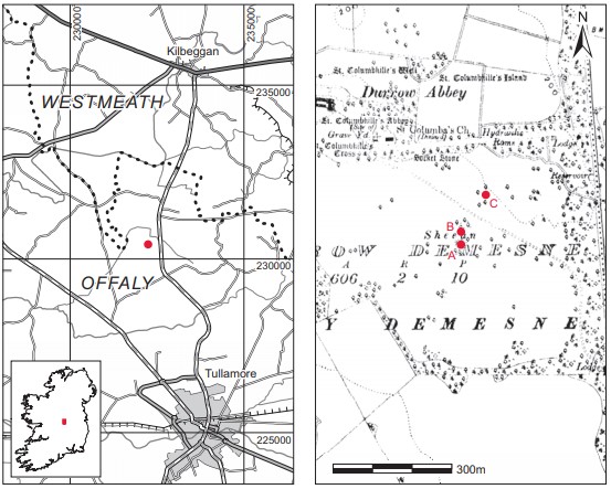
Fig. 4.43—Location map, Durrow Demesne, Co. Offaly
possible cemeteries and the monastic vallum.
The site excavated by Ó Floinn (‘A’ on map, Fig. 4.43) was on a low rise known as Sheean Hill. It consisted of a cemetery of extended burials surrounded by a bank and ditch, c. 30m in diameter. This was almost completely destroyed at the time of excavation. According to O. Davies, there was a small mound to the north of it (marked ‘B’ on the map). This (site B) was not visible at the time of this excavation and no archaeological features were noticed in the vicinity.
The site marked ‘C’ on the map (approximately 100m north-east of site A) consisted of a cemetery of extended skeletons on a low hillock approximately 30m in diameter. This had not been deep-ploughed by the landowner and was subsequently grassed over.
Despite planting and re-ploughing, the monastic enclosure was still visible as a double arc of yellow clay against the darker clay of the surrounding field. It measured c. 200m east/west and 120m north/south from the field fence that marks the edge of the Williams’ property at Durrow Abbey. There was a possible entrance to the south-east.90
Description of site
Two areas of burials were exposed in the cutting by the bulldozer in the region of Sheean Hill (60–90m OD)—one in the north quadrant and a second in the south quadrant (Fig. 4.44).
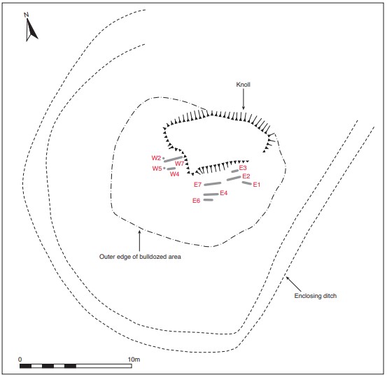
Fig. 4.44—Overall site plan, Durrow Demesne, Co. Offaly
Pegs were laid out on east/west and north/south axes. The burials appeared to be confined to north and east of these axes but bone scattered on the surface extended to the south of the east/west line. Two surface collections of bone were made, one on the eastern side and one on the west. Excavation revealed evidence for successive burial as well as possible burial in rows. Remains of adults and children were found. A curvilinear feature filled with dark soil was exposed, which appears to represent an enclosing ditch. No associated artefacts were discovered, although a bronze ringed pin (1985:89) was later found on the surface of the site.
The burials in the eastern quadrant are labelled with the prefix E and those on the western with the prefix W. The western burials will be described from the uppermost burials down, i.e. level I represents the first excavated (i.e. the latest deposited) burials, while level III contains the lowermost (and earliest) burials found. The eastern burials appear to have been on one level.
Western burials, 2010:92–98 (Fig. 4.45) These burials extended into a raised area that had been partly removed by the landowner. From this area a curvilinear feature filled with dark soil was visible on the northern, western and southern slopes, enclosing the cemetery.
At least seven burials (W1–W7) were noted from three different levels. The burials consisted of simple dug graves, aligned west–east. The grave-cuts do not appear to have been clearly defined, and their extent was not found in any of the graves in this area.
Level I
Grave W1: the burial consisted of an extended, supine inhumation (2010:92) aligned west/east (260°–80°). The skull had been crushed, the left arm was in a fragmented state and the left hand rested inside the left pelvic bone. The right hand was placed over the hip. The left leg was 0.07m above the right but these met at the same level at the toes.91 An arm bone and fingers were found lying diagonally across the left humerus but these were not part of W1. The total height from shoulder to chin was 1.34m. When this burial was lifted, it was found to immediately overlie another skeleton (W5) (see below, level II). The remains were those of an adult male aged 25–34 years at death. A sample of the human remains was submitted for radiocarbon dating and yielded a date of 940±40 BP, which corresponds to the period 1019–1185.92
Grave W3: this burial was found approximately 0.3m west of burial W1 at the same level. It consisted of the in situ remains of a skull and upper vertebrae of one individual (2010:94). It was not possible to determine the sex of the individual, but it would appear to be a young or middle adult. From the position of the skull, the burial would appear to have been in a supine position.
Level II
Grave W4: this consisted of a skull and upper vertebrae, apparently at the same level as W5 (i.e. directly underneath W1 and W3), some 0.5m west of the burials. The remains were those of a middle adult male (2010:95). The burial was overlain by large quantities of disarticulated bone.
Grave W5: this burial lay underneath burials W1 and W3 and overlay W2. It consisted of an extended supine inhumation (2010:96) aligned west/east (80°–260°). The arms rested on the pelvis and in settling the radius of the right arm had slipped under the right pelvis, while the ulna remained above it. The finger bones lay at an angle, dipping below the pelvis (the finger bones of the left hand rested slightly under the pelvis). The feet were crossed. The length
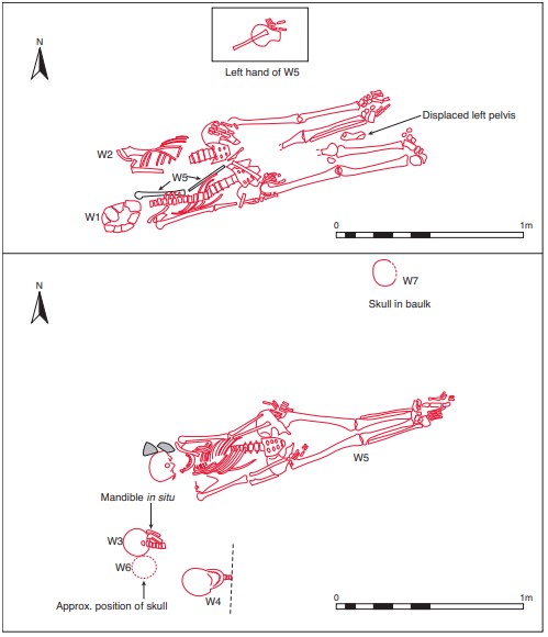
Fig. 4.45—Plans of western graves, Durrow Demesne, Co. Offaly
of the burial was 1.46m. The remains were those of an older adult female (see below). A sample of the human remains was submitted for radiocarbon dating and yielded a date of 895±40 BP, which corresponds to the period 1035–1217.93
Grave W6: this consisted of a single skull (2010:97) and was located immediately south of W3. No other remains were found with this.
Grave W7: this consisted of a single skull (2010:98) found in the baulk, approximately 0.5m north of the feet of burial W5. No other associated remains were found.
Level III
This represents the earliest phase of burial found during these excavations. Grave W2: this burial was also aligned west/east (260°–80°), and the right side of the body had been destroyed by the insertion of burial W5. It consisted of an extended supine inhumation (2010:93), with the skull missing. The lower vertebrae, left ribs and left scapula were preserved in situ. The left arm was missing but the left hand lay over the left pelvis; the right arm was intact and lay straight, with the right hand over the pelvis. The feet were close together and the leg bones appeared to be at a lower level than the foot bones and the lower vertebrae. The height from shoulder to shin was 1.46m. The remains were those of an adult female, aged 25–34 years at death. A sample of the human remains was submitted for radiocarbon dating and yielded a date of 1115±40 BP, which corresponds to the period 782–1017.94
Eastern burials, 2010:99–104 (Fig. 4.46)
The burials on the eastern side of the site all appear to have occurred at the same level and provide evidence for burial in rows in this cemetery.
Grave E1 This is the most easterly burial found on the site. The body was aligned west-north-west/eastsouth-east (300°–120°) and, from the remaining portion, appears to have been deposited in a supine and probably extended position. The skull was crushed but the upper and lower jaws were partly in situ. The arms lay parallel to the torso; the hands lay over the pelvic region. Only the upper portion of the left leg was intact. The length of the right arm was 0.29m. The remains were those of an adult female (2010:99), aged between 25 and 29 years at death. A fragment of a juvenile tibia was also found associated with these remains (see below).
Grave E2 (Pl. 81)
This burial was located north-west of burial E1 and was aligned west/east (270°–90°). The body lay extended in a supine position with the hands by the sides and the legs extended (2010:100). The skull had settled in such a way that the lower jaw touched the cervical vertebrae. The pelvis lay at the lowest level, the skull and legs both being slightly more elevated. The lower parts of the legs were missing, as were the feet. The back of another skull was found between the femora. The remains were those of a juvenile aged twelve to sixteen years at death. The skull of a juvenile was also found immediately south of the skull of this burial. The burial lay in grey clay bordered on the south and west sides by a band of darker clay 0.1m wide. This may have been the remains of a grave-cut.
Grave E3
This burial was found close to the north baulk, 0.35m north of E2. It consisted of the partially preserved upper torso of one individual (2010:101) lying in a supine position. The remains consisted of the lower vertebrae and right pelvis of an adult, again aligned west/east (270°–90° approximately), and were identified by Buckley as those of a female, probably aged less than 25 years at death. Some ribs and arm bones were also found.
Grave E4
This burial was located south of E1. It consisted of the skeleton of a young child (aged six to ten years at death; 2010:102), aligned west-north-west/east-south-east (300°–120°). The body lay extended in a supine position, with the right arm by the side and the left arm flexed at the elbow and placed over the pelvis. The legs were extended and the foot bones were missing. The skull was also missing.
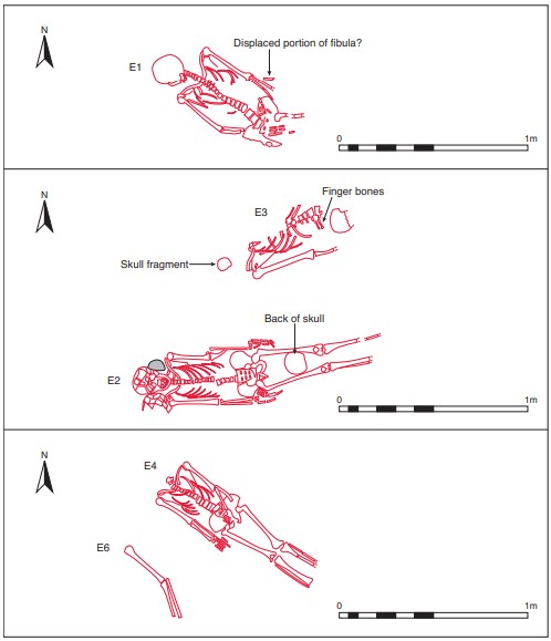
Fig. 4.46—Plans of eastern graves, Durrow Demesne, Co. Offaly
Grave E5
This consisted of a group of disarticulated bones to the south of E4 and included both adult and juvenile remains (2010:103).
Grave E6
This consisted of the left arm of an individual (2010:104), located just 0.3m south-west of E4. The arm lay on the same axis as E4, i.e. west-north-west/east-south-east.
Grave E7
This consisted of a large group of disarticulated long bones (2010:105) lying parallel to one another and forming two groups. The northern group lay on an axis parallel to E2 and the southern group were aligned like E4. At the eastern end of the latter, a ‘cache’ of hazelnuts and dark humus was exposed and sampled.
Comment
Three separate phases of burial were identified within the western burial group. The earliest interment was burial W2, which is dated to 780–1020. The next sequence was burials W4, W5, W6 and W7. A radiocarbon date from this phase places it between 1020 and 1230. The uppermost burials were W1 and W3, and a radiocarbon date places them between 1010 and 1190. The eastern group of burials all appear to have occurred around the same time and provide evidence for burial in rows in this cemetery.
HUMAN REMAINS
LAUREEN BUCKLEY
Skeleton W1 (grave W1): early middle adult male, 170cm
This skeleton was well preserved but in a very fragmentary condition. Only a fragment of the right temporal bone remained from the skull. The upper vertebrae were missing. Nine thoracic vertebrae survived but most consisted of the bodies only, with only the two lower vertebrae being complete. The lumbar vertebrae were all present but fragmented. There were three ribs from the left side and six from the right, and the manubrium and body of the sternum were present but in a fragmentary state.
Only the distal two-thirds of the left radius survived from the left arm. The left hand consisted of the scaphoid, lunate, trapezoid, capitate and hamate, all the metacarpals and three proximal, four middle and one distal hand phalanges.
The glenoid area and acromion of the right scapula were present and the right clavicle was complete, apart from the lateral end. The proximal shaft of the right humerus was fragmented but the bone was complete. The right ulna was also complete and the proximal half of the right radius was present. Only the right triquetral, lunate and hamate remained from the right carpal bones; the fifth metacarpal was present and there were three proximal, three middle phalanges and one distal hand phalanx.
The ilia, ischia and pubes were present from the pelvis but they were in a fragmentary condition. The sacrum was virtually complete. Only the distal third of the left femur remained but the patella and left tibia were complete. The right femur, patella, tibia and fibula were present and complete. All the tarsals apart from the left calcaneum and left second cuneiform were present, but only the left first and second and right fourth and fifth metatarsals remained and were incomplete. None of the foot phalanges survived.
Age and sex
The sciatic notch of the pelvis was narrow, indicating that the individual was male. In the right pubic bone, the ventral arc, sub-pubic concavity and sub-pubic angle were all of the male type. The metric indicators—diameter of head of femur, bicondylar width, humerus head diameter and radius head diameter—were also all definitely in the male range. All the epiphyses were fused but the epiphysis at the proximal tibia was still vaguely visible. The surface of the pubic symphysis was still ridged, and the auricular surface of the ilium indicated an age of 25–34 years. Therefore the individual was an early middle adult. The stature was estimated at 170cm, using the length of the femur and fibula.
Non-metric traits
Vastus notches were present in both patellae and squatting facets were present on both tibiae.
Skeletal pathology
There was a small facet for a lumbar rib on the right side of the first lumbar vertebra. The facet was just lateral to the superior articular surface. The lumbar rib was not present. On the inferior surface of the right clavicle near the lateral end there was an additional articular facet. This may have occurred as a result of a slight dislocation of the bone. Unfortunately the proximal right humerus was fragmented and it was not possible to see the full joint surface to ascertain where the false joint on the clavicle articulated.
Schmorl’s nodes were present on all the vertebral body surfaces from the inferior surface of the sixth thoracic vertebra through to the inferior surface of the eleventh thoracic vertebra. They were also present to a very mild degree on the upper four lumbar vertebrae. At the right ankle there were very prominent muscle attachments for the tibio-fibular ligament on both the tibia and fibula. This was probably caused by some minor trauma to the joint. Spina bifida occulta was present in the three lower sacral vertebrae.
Additional bones
Fragments of a left tibia shaft, possibly from W2, were present and there was also a right ischium.
Skeleton W2 (grave W2): early middle adult female, 158cm
This skeleton consisted mainly of the lower half only. Only the lateral border of the left scapula, the lateral third of the left clavicle, the left hand and eight left ribs remained from the upper half. The left hand consisted of the scaphoid, trapezium, trapezoid and capitate, all the metacarpals and four proximal, three middle and one distal hand phalanges. The four lower lumbar vertebrae were complete and there was a fragment of the body of the first lumbar. Only the complete left ilium and ischium, both incomplete pubic bones and a virtually complete sacrum remained from the pelvis. The left femur was complete but only the distal third remained from the right femur. Both patellae and both tibiae were complete and only the proximal ends were missing from both fibulae. All the tarsals and metatarsals except for the left second metatarsal were present from the foot bones, and there was one proximal phalanx from the left foot and three proximal phalanges and one middle phalanx from the right foot. There was also a sesamoid bone associated with each foot.
Age and sex
All the observable features of the pelvis, including the ventral arcs, sub-pubic concavities, subpubic angles and sciatic notch, were of the female type.
The first sacral vertebra was not fully fused to the second sacral vertebra at the centrum, indicating that the individual was less than 27 years of age (Scheuer and Black 2000). Examination of the auricular surface of the ilium and the pubic symphyses indicated an age of between 25 and 34 years. It is safe to assume that this individual was an early middle adult aged 25–34 but more likely to be on the lower side of this estimate, 25–29 years. Stature was estimated, using the lengths of the femur and tibia, as 158cm.
Skeletal pathology
Deep Schmorl’s nodes were present in the superior surface of the body of the second lumbar vertebra, and associated with this was a slight crack running to the posterior left side of the body. A crack on the lower surface near the anterior edge had occurred post-mortem. The fourth lumbar vertebra was slightly compressed on the left side of the body. Schmorl’s nodes were also present on both surfaces of the body of the third lumbar.
Non-metric traits
Squatting facets were present on both tibiae and there was an acetabular crease in the left acetabulum.
Skeleton W3 (grave W3)
This consisted of the skull and upper vertebrae only. The skull was very fragmentary and only part of the right side of the frontal bone, fragments of both parietal bones, most of the occipital bone, the mastoid area of the left temporal bone and the left zygomatic bone survived from the cranium. Part of the maxilla was present and most of the mandible was present but fragmented. The cervical vertebrae and one middle thoracic vertebra also survived. There was only one rib present from the left side.
A fragment of acromial spine of the left scapula and the distal end of the left humerus were all that remained of the rest of the skeleton.
Age and sex
It was not possible to determine the sex of this individual. The external occipital protuberance was of the male type but the mastoid process seemed to be of the female type. The mental eminence of the chin was also probably female, but the root of the zygomatic arch was male. Since there was an equal number of features that could be male and female, it was not possible to sex this skeleton reliably. The only method of ageing available was the wear on the teeth. Molar attrition was very light so this was probably a young adult or a middle adult.
Skeletal pathology
The frontal bone appeared to be enlarged and the diploe had the appearance of having extra layers of bone within it. Layers of new bone are usually added to the external surface of bone as a result of an infection or inflammation of the periosteum. It is possible that these layers represent healed periostitis from episodes that occurred in childhood.
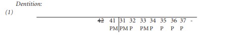
Anomalies: there was an enamel pearl on the lingual surface of the root of the lower right third molar, 48.
Attrition: there was very light wear on all teeth.
Calculus: deposits were mainly light on the buccal surfaces of the upper teeth and light to moderate on the lingual surfaces. The lower teeth had moderate calculus deposits on the buccal surfaces of the canine and first molar and light deposits on the buccal surfaces of the other teeth. The lingual surfaces had light deposits except for the first premolar and third molar, where deposits were moderate.
Caries: there were two small cavities on the occlusal surface of the upper left second molar and very small pinholes on the lower right third molar in the lingual distal quadrant.
Periodontal disease: there was slight alveolar recession around the roots of the maxillary premolars and second and third molars on the left side. There was also slight recession around the roots of the lower right third molar.
Hypoplasia: linear enamel hypoplasia was present on the upper left and lower right canines.
Skeleton W4 (grave W4): middle adult male
This skeleton consisted of skull and upper vertebrae only. The skull was fragmented but virtually complete, although part of the occipital bone and the orbital area of the frontal bones were missing. The sphenoid bone was also absent. The maxilla and mandible were virtually complete. All the cervical vertebrae were present, with the upper four being complete.
Age and sex
The mastoid processes, right supraorbital ridge and mental eminence of the mandible were all of the male type. The sutures of the skull were well fused but not obliterated, so the individual was an adult but probably not an older adult.
Skeletal pathology
Mild marginal lipping was noted on the posterior articular surfaces of the third and fourth cervical vertebrae. There was no osteophytosis of the cervical bodies, which is common in old age.
Abrasion: there was a chip out of the bucco-mesial quadrant of the upper right second premolar, and also out of the same quadrant of the adjacent first molar. This may indicate that the teeth were used as tools. The damage had occurred early in life as calculus was deposited over the chipped area.
Attrition: there was heavy wear on the upper incisors, canines, premolars and first and second molars. There was also heavy wear on the lower first and second molars and moderate wear on all other teeth.
Calculus: there were light deposits on the lingual surfaces of most of the teeth in the maxilla and on the buccal surfaces of the lower premolars. Deposits were moderate on the buccal surfaces of all the upper teeth except the second and third molars, where deposits were heavy, and on the lingual surfaces of most of the mandibular teeth. There were also heavy deposits on the buccal and lingual surfaces of the lower left second and third molars and on the lingual surface of the lower right third molar.
Caries: there was a small cavity on the distal surface of the crown of the upper right canine. The upper left second molar had a moderate-sized cavity on the distal surface at the cervical margin and there was a small cavity on the mesial surface of the crown of the adjacent third molar.
In the mandible there was a moderate-sized cavity on the distal surface of the crown of the left second premolar and a large cavity on the mesial surface of the adjacent first molar.
Periodontal disease: there was a slight degree of alveolar recession around the roots of the molars and premolars on the right side of the maxilla and mandible, with recession being moderate around the lower left premolars and molars and upper left molars. The alveolus on the left side of the maxilla was damaged around the anterior teeth.
Skeleton W5 (grave W5): older adult female, 155cm
This skeleton was virtually complete and was in a good state of preservation apart from the left lower arm and hand, which were very decayed. The skull was fragmented but most of the frontal, parietal, temporal and occipital bones were present. The sphenoid and left zygomatic bones were present but were incomplete. The maxilla and mandible were also present. All the vertebrae were present but only the first two cervical and first thoracic were complete, the remainder consisting of the neural arches only. Nine ribs from the left side and twelve from the right survived.
Both scapulae were virtually complete. The shaft of the left clavicle was present and the right clavicle was complete. All the arm bones were present and complete. Only the left and right lunate and capitate, right triquetral, trapezoid and hamate remained from the carpal bones but all the metacarpals were present and complete. There were also five proximal phalanges from the left hand and three proximal and two middle phalanges from the right hand.
Both ilia and ischia from the pelvis were present and complete and the sacrum was virtually complete. All the leg bones were complete apart from the left patella and the right patella, which were missing. All the tarsal bones were present apart from the left cuneiforms and right third cuneiform. The metatarsals were all complete and there were four proximal phalanges from each foot, one middle phalanx from the right foot and the first distal phalanges from both feet. Two sesamoid bones were also present.
Age and sex
All the observable features of the skull, the sciatic notch of the pelvis and the measurements of the heads of the femur and humerus were of the female type.
Only the auricular surface of the ilium could be used to estimate age and the degree of lipping and erosion on them indicated that this was an older adult.
Stature, based on the lengths of the humerus, femur and tibia, was estimated at 155cm.
Non-metric traits
There was a septal aperture in the right humerus and a squatting facet on the left tibia.
Skeletal pathology
Cribra orbitalia was present in both orbits; it was mild in the left orbit and moderate in the right. The frontal bone was very decayed but there appeared to be expansion of the diploe and thinning of the outer cortex. This would indicate porotic hyperostosis, which, together with the cribra orbitalia, is indicative of iron-deficiency anaemia.
Osteoarthritis was present in the vertebral column, with severe osteophytes and polishing of the right inferior articular surface of T5. There were also moderate degenerative changes with lipping and porosity of the right superior surface of T5 and right inferior surface of T4. The right superior surface of T6 was similarly affected. The posterior surfaces of the lumbar vertebrae were also mildly affected by marginal lipping.
Degenerative changes were also present in the right hip, with mild lipping and porosity of the inferior part of the right acetabulum and moderate lipping of the head of the right femur. There was mild lipping present on the head of the left femur. The heads of both femurs also had surface osteophytes. There was indication of degeneration of the left shoulder, with slight porosity of the lateral end of the left clavicle. There was a chondral defect in the proximal articular surface of the first proximal phalanx of the left foot.
Attrition: there was heavy wear on the first molars and moderate wear on all other teeth.
Calculus: there were light deposits on the buccal surfaces of most of the teeth in the maxilla and the lower incisors, canines and first right premolar. Deposits were moderate on the buccal surfaces of the lower premolars and molars, the lingual surfaces of the lower incisors, canines and left premolars, and on the lingual surfaces of most of the upper teeth. There were heavy deposits on the buccal surface of the upper left second molar and on the lingual surfaces of all the lower molars and lower right premolars.
Periodontal disease: there was a slight degree of alveolar recession around the roots of most teeth, with recession being moderate around the upper canines and the upper left premolars and molars.
Hypoplasia: linear enamel hypoplasia was present on all the incisors and the upper left canine.
Stray bones found with skeleton W5
Adult long bones: these included the proximal end of a humerus, the proximal third of a right ulna, the distal third of a left radius, the lesser trochanter of a right femur, a fragment of lateral condyle probably also from a right femur and small fragments of femur shaft.
Vertebrae: the arches of five lumbar vertebrae, including a fifth lumbar, were present but were probably not all from one individual. A fourth and fifth sacral vertebrae were present, along with the dorsal surface of the third sacral vertebra from the same sacrum. There was also a first coccygeal vertebra. Spina bifida occulta was present in the third sacral vertebra.
Pelvis: a left ischium from a young individual with the epiphysis at the tuberosity just fused was present, and there was also a fragmented right ilium and ischium from an older adult male.
Scapulae: a fragment of glenoid fossa, part of a lateral border and the end of the acromion from a right scapula were present. There was also a fragment of clavicle shaft.
Hand bones: the right hamate, lunate and triquetral, left triquetral and five proximal, two middle phalanges and one distal phalanx were present.
Ribs: a minimum of one left and one right rib were present and the left rib had osteoarthritis at the transverse articular process.
Skull: skull fragments included a right orbit from a frontal bone, a fragment of left sphenoid bone, a basilar occipital bone and a fragment of inferior margin from a mandible. One loose upper left central incisor was also present. There was moderate cribra orbitalia in the right orbit.
Juvenile bones: these consisted of a fragment of the medial end of a clavicle, two fragments of long bone, probably femur, and one small rib fragment.
Skeleton E1 (grave E1): early middle adult female, 159cm
This skeleton lay in a supine extended position, aligned north-west/south-east with the head turned to the right side. Only the upper half of the skeleton was present. The skull was almost complete but was very fragmented, especially the facial bones. The frontal, parietal, occipital and temporal bones were all present but incomplete. The left wing of sphenoid and the right zygomatic bone were present and the maxilla and mandible were incomplete. Part of the hyoid bone was also recovered. Most of the cervical vertebrae were complete but bodies and fragments of arches were all that remained of the thoracic and lumbar vertebrae. Twelve ribs from the left side and eleven from the right remained, and part of the manubrium and body of the sternum survived.
Both scapulae were almost complete and both clavicles were complete. The humeri were complete although the proximal end of the left bone was fragmented. The proximal two-thirds of the left radius and ulna survived and the proximal halves of the right radius and ulna remained, although the head was missing from the right radius. All the metacarpals of the left hand were present and there were five proximal, four middle and two distal phalanges. Only four proximal phalanges survived from the right hand. Both ilia and ischia survived from the pelvis but were very incomplete. Only the upper two sacral vertebrae were present. The proximal third of the left femur was all that remained from the leg bones.
Age and sex
All the features of the skull, the external occipital protuberance, the mastoid processes, the supraorbital ridges, the right orbital rim and the mental eminence, were of the female type. The sciatic notch of the pelvis was wide, which usually indicates a female. The diameter of the humeri heads, the radius head, the widths of the glenoid fossae and the clavicle lengths were all within the female range. Epiphyses of the sternal ends of the clavicles were not completely fused. This state of fusion is usually present in an individual aged 24–29 years. S1 was not fully fused to S2 at the anterior body and this usually indicates an age of less than 27 years. The auricular surface of the ilium indicated an age of 25–29 years. The indications are that this individual was aged 25–29 years but may have been less than 27 years at the time of death. Stature was estimated, using the length of the humerus, as 159cm.
Non-metric traits
The metopic suture was retained.
Skeletal pathology
The orbits were incomplete but moderate cribra orbitalia was noted in the left orbit. This may be caused by iron-deficiency anaemia. A small lesion of osteochondritis dessicans was present on the posterior surface of the trochlea of the right humerus.

Attrition: there was light wear on the incisors, canines, premolars and first molars, moderate wear on the lower left second molar and no wear on the third molars.
Calculus: there were light deposits on the lingual surfaces of most of the teeth in the maxilla except the molars, where deposits were moderate. Most of the maxillary teeth also had light deposits on the buccal surfaces apart from the right first and second molars, where deposits were moderate.
There were light deposits on most of the buccal surfaces of the lower teeth and moderate deposits on most of the lingual surfaces. The occlusal surface of the lower left second molar had moderate calculus deposits and there were considerable deposits on the distal surface of the lower left third molar.
Hypoplasia: linear enamel hypoplasia was present on all the incisors except the lower right. It was also present on all the canines, the upper right first premolar and upper left second molar.
Additional bones
A fragment from the mid-shaft area of a juvenile tibia was also present.
Skeleton E2 (grave E2): juvenile, 12–16 years
This was the skeleton of a juvenile. It was almost complete, with most skeletal elements present, but most of the bones were fragmentary. Fragments of the occipital, both parietal, both temporal and the frontal bones remained from the skull. The sphenoid, both zygomatic bones, part of the maxilla and most of the mandible were also present. The upper four cervical vertebrae were complete but the rest were fragmented. There were twelve thoracic vertebrae and five lumbar vertebrae present but they were totally fragmented. There were ten ribs from the left side and eleven from the right side present, and the manubrium and body of the sternum were present but fragmented.
Both scapulae were present but incomplete and the lateral ends were missing from both clavicles. The left humerus was complete but only the shaft remained from the right humerus. The proximal halves of the left radius and ulna were present and the proximal two-thirds of the right radius and ulna survived.
The left hand consisted of the scaphoid, trapezoid, capitate and hamate, all the metacarpals and four proximal, two middle and one distal hand phalanges. The right hand consisted of the lunate, trapezium, capitate and hamate, and the third, fourth and fifth metacarpals.
Both ilia were present but incomplete and both ischia were complete. The pubic bones were also present but they were in a fragmentary condition. The sacrum was virtually complete. The proximal two-thirds of both femurs remained and the patellae were complete; the proximal epiphysis and fragments of shaft of the left tibia were present as well as some fragments of shaft of the right tibia, and the shafts of both fibulae were virtually complete. There were no foot bones remaining.
Epiphyseal fusion
All the epiphyses at the ends of the long bones were unfused, the acetabulum was unfused and the lower sacral vertebrae were only partially fused.
An individual with this state of dental eruption was probably aged between twelve and sixteen years.
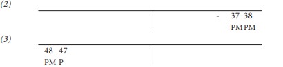
Anomalies: there was a gap between the lower left premolars 34 and 35 and the teeth were leaning in towards each other.
Attrition: there was light wear on the incisors, canines and first molars. There was no wear on the premolars and second molars.
Calculus: there were light deposits on the buccal surfaces of the upper lateral incisors, canines, left second premolar and first and second molars. In the mandible there were light deposits on the lingual surfaces of most teeth and moderate deposits on the buccal surfaces of the central incisors. The buccal surfaces of the premolars also had light deposits.
Hypoplasia: linear enamel hypoplasia was present on the upper incisors and canines and the lower canines. There were also pits of hypoplasia on the upper left first molar. Also present were three unformed crowns, probably from three partially formed molars, possibly two upper and one lower deciduous second molars.
Additional skull found between the legs of E2
This skull from a juvenile consisted of most of the occipital bone, the posterior parts of both parietal bones and a fragment from the right frontal bone.
Skeleton E3 (grave E3): young adult female
This consisted of the middle of an adult skeleton found near the north baulk. Some infant bones were also found and are described separately below as E3a.
Only the posterior part of the right parietal bone remained from the skull. The vertebral column consisted of the arches only of the lower three thoracic vertebrae and five lumbar vertebrae. There were five ribs from the left side and eight from the right side. The right scapula and clavicle were present and almost complete. The distal half of the shaft of the left humerus was the only bone to survive from the left arm. The right arm consisted of the almost complete shaft of the right humerus, a fragment of right radius shaft and the proximal two-thirds of the right ulna. There were no hand bones present. Both ilia were present from the pelvis but were incomplete. The left ilium consisted of the posterior part near the rim only. The right superior articular surface was all that remained from the sacrum. Only a fragment of mid-shaft and lower shaft of the left femur survived from the leg bones and the left talus was the only foot bone present.
Age and sex
The only features to help identify sex were the sciatic notch, which was very wide, and the width of the glenoid fossa of the scapula, which was within the female range.
The medial epiphysis of the clavicle was unfused and the iliac crest was partially fused. These indicate that the individual was probably less than 25 years of age at the time of death.
Stray bones
There were a few stray bones present, including a fragment from a left ilium, a lumbar arch, a right talus and a left fourth metatarsal.
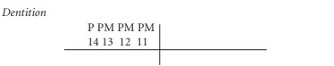
Three loose teeth, the upper right lateral incisor, 12, upper right first premolar, 14, and lower right lateral incisor, 42, were present. There was light wear on the upper teeth and moderate wear on the lower incisor. Calculus deposits were light.
Skeleton E3a: infant
The lower half of one infant was found with the female skeleton E3. Bones present included the complete right ilium, complete left and right femurs, and complete right tibia.
Age
Using the regression equations of Fazekas and Kosa (Kosa 1991), the body length of the infant was estimated using all the bones, and from this the age of the infant was determined. It was found that this was a foetus aged 30–34 weeks in gestation. While a foetus is viable at 28 weeks, it is unknown whether this infant had been born and died during or shortly after birth or was still in the womb when the mother died.
Skeleton E4 (grave E4): juvenile, 6–10 years
This was the almost complete skeleton of a juvenile, although the skull was missing. Only one small fragment of occipital bone and one upper central incisor, 21, was present from the skull. The lower four cervical vertebrae, twelve thoracic vertebrae and five lumbar vertebrae were present from the vertebral column. Nine ribs from the left side and ten from the right remained. The glenoid fossae, acromial spines and lateral borders of both scapulae were present, as well as the lateral two-thirds of the left clavicle and the complete right clavicle. The left humerus was complete, the left ulna was almost complete apart from the distal end but only the mid-shaft area remained from the left radius. All the bones of the right arm were complete. There were five metacarpals and six phalanges from the left hand and two carpals, five metacarpals and eight phalanges from the right hand.
The pelvis consisted of the incomplete ilia, complete ischia and left pubic bone and four incomplete sacral vertebrae. The leg bones consisted of both complete femurs, the proximal two-thirds of the left tibia, complete right tibia and proximal two-thirds of both fibulae. There were no foot bones present.
Age
The vertebral half-arches were fused together and fused to the centrum, indicating that the juvenile was over six years of age. Since no other epiphysis was fused, the individual was less than thirteen years. There were no teeth present to enable an accurate assessment of age, so the only other way of ageing the skeleton was to compare measurements of long bone lengths with those from juveniles whose dental age was known. Using this method the age was estimated as between eight and ten years.
One central permanent incisor was found; assuming that it belongs to this skeleton, it can be said that, since the tip of the root was not 100% formed, the juvenile was probably less than eight years.
In view of the fact that there is conflicting evidence, it is probably safer to age this juvenile as between six and ten years of age, although it is likely that he/she is around eight years.
Skeleton E5 (grave E5)
Only a few fragments of bone were present with this label. These included two small fragments of unsided adult fibula, one fragment of adult tibia shaft, the lower right part of a neural arch from an adult lumbar vertebra and a fragment of transverse process from a thoracic vertebra. There were also a very decayed fragment of juvenile skull, a few fragments of juvenile rib, a fragment from near the proximal end of a juvenile ulna, one juvenile vertebral body and a few other fragments of juvenile bones.
Loose bone from surface, eastern area of site
Skull fragments
There were a minimum of two frontal bones, consisting of two left and one right orbit and two fragments from the left squamous frontal. A small fragment of right posterior parietal bone was present and also a fragment of the anterior part; there was also some anterior of a left parietal bone as well as the posterior part of a left parietal. A large fragment of left and right parietal bone from another individual was also present.
At least three almost complete squamous occipital bones were present and there were two right and one left temporal bones from male individuals. There were fragments from at least two left sides and two right sides of mandible.
Attrition was moderate and there were moderate calculus deposits on the lingual surfaces of most teeth. There was also a moderate degree of recession in the alveolus. Linear enamel hypoplasia was present on the lateral incisor and the molars
There was moderate attrition and moderate calculus on the remaining molar but there was considerable recession around the observable molar sockets.
The other fragment of right mandible had no sockets or teeth observable.
Long bones
Table 4.11—List of long bones present
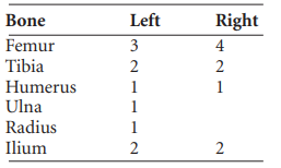
Also present were some fragments of fibula shaft, the proximal articular surface of a femur, two cervical and two mid-thoracic vertebrae with moderate osteophytosis, a few tarsal bones, a metatarsal and a metacarpal, as well as an incomplete left ilium and a right ischium from a juvenile.
Notes
One of the left tibiae and one of the right tibiae were a match and seemed to belong to the same individual as the complete left femur. The proximal end of a right tibia had severe eburnation of the posterior part of the lateral condyle. One of the left ilia and one of the right ilia were from a female and the female was a late middle adult aged 35–44 years.
Loose bone from surface, south/west of site
These included a right fibula shaft, three fragments from another fibula shaft, and a small fragment from the mid-shaft of a left humerus. Also present were a few small fragments of tibiae and femur, a fragment of pelvis rim, small fragments of rib, a few metatarsals and one fragment of cervical vertebra. Skull fragments included a few small fragments of frontal bone, the basal occipital bone, one petrous temporal bone and a fragment of maxilla.
There was only slight polishing of the occlusal surface of the premolar and slight calculus deposits on the buccal and lingual surfaces.
Disturbed bone from surface
This consisted of the proximal third of a left radius, the distal third of a right fibula, a left talus and part of a right calcaneum.
Skull fragments of W1 or W2 (disturbed)
These included one fragment of left parietal bone with a small part of the sagittal suture and a tiny fragment of a right parietal, a fragment of left temporal bone and a left zygomatic bone.
W1 (disturbed)
Skull fragments included a right orbit from a female with mild cribra orbitalia, other fragments of frontal bone and four fragments of parietal bone, a fragment of occipital bone from near lambda and the mandibular fossa from a left temporal bone.
Other bones from this context included the left medial end of a clavicle, a fragment from the mid-shaft of a left humerus, the proximal two-thirds of a right radius, the distal half of the shaft of a right ulna, the distal third of a right femur, and a few fragments of vertebral arches and fragments of ribs.
An almost complete juvenile fibula and a fragment of shaft from a juvenile femur were also present. Skull fragments on pelvic area of W3 These consisted of fragments of squamous frontal bone, one left orbit, small fragments of parietal bone and most of a left temporal bone. There was also a fragment from the right side.
Skull fragments on pelvic area of W3
These consisted of fragments of squamous frontal bone, one left orbit, small fragments of parietal bone and most of a left temporal bone. There was also a fragment from the right side of the mandible and a second cervical vertebra as well as a right calcaneum, left navicular and a hand phalanx.
Some juvenile bones were also present. These consisted of a very decayed right ilium and a partial thoracic vertebra arch.
Notes
There was mild cribra orbitalia in the left orbit.
The temporal bone was probably from a female.
There was very heavy wear on both teeth, with the teeth almost worn away to the roots, and moderate calculus deposits. Periodontal disease was present to a moderate degree and there was a mandibular torus.
Loose bone (unlabelled)
This included the proximal end of a femur, fragments of fibula shaft, the distal part of an ulna shaft, two metacarpals and two hand phalanges, a few metatarsals and phalanges, a few ribs, a complete first cervical vertebra and upper thoracic vertebra, fragments of two vertebral arches and a fragment of juvenile femur.
Skull bones included a left and a right orbit, posterior parts of two left and one right parietal bones, a right temporal bone, a left and a right zygomatic bone and the anterior part of a mandible.
Attrition was light to moderate; there were moderate calculus deposits on the buccal surfaces and considerable deposits on the lingual surfaces of the teeth. There was a slight degree of recession of the alveolus, and linear enamel hypoplasia was noted on the canines and right premolar.
Summary and conclusions
The total number of burials recovered from this site was nine, although one burial also contained the partial skeleton of a pre-term infant. A further four adult skulls were found among the disarticulated remains, but since one of them is thought to belong to skeleton W1 or W2 there are probably only three additional burials from the scattered remains. There were also some juvenile bones, from at least one juvenile, found in the disarticulated remains on the eastern side. The nine burials consisted of seven adults and two juveniles. Two of the adults were male and four were female; in one case the sex could not be determined. An additional two males were found in the disarticulated remains.
The juveniles consisted of an adolescent aged 12–16 years, an older juvenile aged 6–10 years and a foetus associated with burial E3 that was aged 30–34 weeks in utero. Although the foetus had reached a viable age, it is not known whether it had been born at the time of death of the mother as its exact position is not clear. It was associated with a young adult female, however. Most of the adults were in the early middle adult range, 26–34, with the male being a middle adult, 26–45, and one of the females being an older adult, more than 46 years of age.
Obviously the sample size is too small for a reliable statistical analysis and only an overview of the result can be given. Although this is a cemetery site, it is only a very small part of the overall cemetery and this sample may not be representative. There was not a high level of degenerative disease since the population was relatively young. The older female had degenerative joint disease in the hips and shoulders and the middle adult male had mild degenerative disease in the vertebral column. Two individuals had evidence for Schmorl’s nodes in the vertebral column. One individual had evidence for a healed infection from childhood. Cribra orbitalia, which may represent childhood anaemia, was present in two individuals and in three individuals from the disarticulated skulls. The only evidence for trauma was in skeleton W1, an early adult male, who had disarticulated his clavicle and damaged his ankle, both of which could have been caused by a fall. Despite the small size of this sample, some interesting pathologies and results were noted, indicating the important nature of this cemetery site.
Parish of Durrow, barony of Ballycowan. SMR OF009-006——. IGR 232249 230425.
A scatter of human remains was also later discovered on a ridge running north–south that delimited the western edge of the monastic enclosure.
89. Parish of Durrow, barony of Ballycowan. SMR OF009-006——. IGR 232249 230425.
90. A scatter of human remains was also later discovered on a ridge running north–south that delimited the western edge of the monastic enclosure.
91. The left leg of burial W1 lay 12cm above the right leg of burial W2.
92. GrA-24318.
93. GrA-24320.
94. GrA-24319.