1983:005 - SHERLOCKSTOWN, CO. KILDARE, Kildare
County: Kildare
Site name: SHERLOCKSTOWN, CO. KILDARE
Sites and Monuments Record No.: N/A
Licence number: E1090
Author: BREANDÁN Ó RÍORDÁIN
Author/Organisation Address: —
Site type: Graves of indeterminate date
Period/Dating: —
ITM: E 690037m, N 723322m
Latitude, Longitude (decimal degrees): 53.252620, -6.650745
In August 1983 human remains were discovered during bulldozing operations at Sherlockstown, near Sallins, Co. Kildare (Fig. 6.19).31 The remains were found at a depth of approximately 0.3m below ground level in sandy soil. The discovery was reported to the NMI by Gardaí at Naas and an investigation was carried out by Breandán Ó Ríordáin. The site was a raised area, possibly a mound, with a substratum of esker-like sand and gravel on which two houses were being built.32 The human remains that were discovered were piled on the freshly bulldozed sandy stratum and included skull and limb fragments of an adult (1983:44.2). Some bone noticed protruding from the face of the cutting was examined and found to be the remains of a young individual (1983:44.3). A small quantity of what appeared to be finely comminuted cremated remains (1983:44.1) were retrieved beside the unburnt remains.
Comment
In the absence of grave-goods or other evidence it is not possible to suggest a date for this site, but the presence of burnt and unburnt remains suggests that it may date from the prehistoric rather than the historic period.
HUMAN REMAINS
LAUREEN BUCKLEY
1983:44.1
This sample consisted of 299 fragments of cremated bone weighing a total of 12g. The bone was a white/grey colour and seemed well calcined. The largest fragment was 45mm in length but most of the fragments were very small, less than 10mm in length. The largest fragment
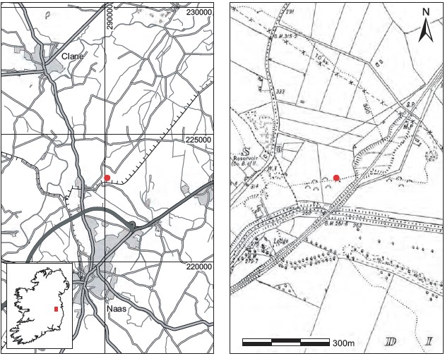
Fig. 6.19—Locationmap, Sherlockstown,Co. Kildare.
consisted of most of the shaft of an infant left femur, and most of the rest of the fragments consisted of very small and thin fragments of skull bone. Two petrous temporal bones from an infant were also present. Other bones present included several fragments of infant ribs, a tiny fragment of vertebral arch and the top of a crown of a deciduous canine. The tooth development suggests that these were the remains of a neonate. The estimated length of the femur, 55mm, and the length of the petrous temporal bone, 22.4mm, suggest that the infant was not full-term but between seven and eight months’ gestation. There may have been some shrinkage of the bone, however, and it could in fact have been a full-term infant.
1983:44.2
These bones were mainly from one juvenile, although some additional bone was present. The skull was fragmented but consisted of the frontal bone, both parietal bones, the occipital bone, both temporal bones, the sphenoid, left zygomatic and the mandible and maxilla. The only vertebra present was the first cervical vertebra. Part of the body of the sternum was present and there were eleven left ribs. Only the distal end of the left humerus, the proximal half of the left ulna and the distal two-thirds of the left radius survived from the left arm. The right arm consisted of the shaft of the right humerus and the distal two-thirds of the right radius. The scaphoid, lunate, trapezium and capitate, as well as five metacarpals, five proximal, three middle and one distal phalanges survived from the left hand, but nothing remained from the right hand. The left ilium and ischium from the pelvis were complete but the right ilium was incomplete. Only the complete left femur and distal epiphyses of the left tibia and fibula remained from the left leg, while the right leg consisted of the proximal two-thirds of the femur and distal halves of the tibia and fibula. All the tarsals and metatarsals were present from each foot and there were four proximal phalanges from each foot, as well as one right distal phalanx.
Epiphyseal fusion
All the epiphyses at the ends of the long bones were unfused. The anterior iliac crest epiphysis was formed but was unfused. The ilium had fused to the pubis and the ischium was partially fused to the ilium. The degree of epiphyseal fusion suggests an age of 13–16 years for this Juvenile.
Non-metric traits
There was an ossicle at lambda and at least two ossicles in the right lambdoid suture.
Skeletal pathology
Severe cribra orbitalia was present in the left orbit.
Dentition
Dental development: the crowns of the second molars were only half-formed, and the crowns of the lower incisors and canines were just complete. The crowns of the premolars were three-quarter-formed. A juvenile with this stage of dental development is consistent with an age of 5–7 years. Obviously the skull does not belong to the same individual as the rest of the skeleton.
Additional bones
Other juvenile bones present included a very decayed left femur shaft, the mid-shaft area of a left tibia and a very decayed fragment of another left tibia, the distal two-thirds of a left humerus, a very decayed left radius shaft, a fragment of fibula shaft and other decayed fragments of long bone.
Summary
At least two juveniles were represented in this bone collection. One was aged 5–7 years at the time of death and had probably suffered from iron-deficiency anaemia. The other was probably aged 13–16 years at the time of death.
1983:44.3
This consisted mainly of skull and long bones of one individual only. The skull was virtually complete, with the occipital, both temporal, both parietal, the sphenoid and most of the frontal bone present. The right zygomatic bone and the right side of the maxilla and mandible were also present. Only the left side of the first cervical and the body of the fifth lumbar vertebra survived from the vertebral column and there were only two fragments of ribs present. The distal two-thirds of the left humerus and left ulna remained from the left arm, while the right arm consisted of the distal half of the humerus, the proximal third of the ulna and the proximal two-thirds of the radius. Only the right third, fourth and fifth metacarpals and two proximal phalanges survived from the hand bones.
The left ilium and ischium were complete but there were fragments only of the right ilium and ischium. Part of the first sacral vertebra remained from the sacrum.
The left femur was almost complete apart from the distal end, and the shaft only remained from the right femur. The shaft of the left tibia was present but only the mid-shaft area remained from the right tibia. Fragments of both fibulae were present. Only the left talus survived from the foot bones.
Age and sex
The external occipital protuberance, the mastoid process, orbital rims and mental eminence were all of the male type. The sciatic notch was also narrow and the diameter of the femoral head was definitely in the male region. The auricular surface of the ilium indicated an age of 35–44 years, although the degree of wear on the teeth suggests an older adult.
Skeletal pathology
The inferior surface of the body of the fifth lumbar vertebra had mild porosity, suggesting degenerative joint disease.
The superior surface of the body of the first sacral vertebra had severe porosity and severe marginal osteophytes.
Dentition
Attrition: there was moderate attrition on the anterior teeth and heavy wear on the molars, especially the upper first molars. There were vertical grooves in the crowns of the upper right lateral incisor and upper right second premolar.
Calculus: deposits were light on the lingual surfaces of the lower teeth and were moderate on the buccal surfaces of the upper third molars but light on the distal and lingual surfaces of these teeth.
Caries: there were moderate cavities on the distal side of the root of the upper right first molar at the cervical margin and also on the mesial surface of the root of the adjacent second molar. The upper left first molar had a moderate cavity on the buccal surface of the root at the cervical margin.
Peridontal disease: there was a slight degree of alveolar recession around the roots of the upper incisors, and a moderate degree of recession around the remaining teeth on the right side of the maxilla and the molars on the right side of the mandible. Considerable recession was noted around the roots of the lower right canine and premolars.
Summary and conclusions
The remains from the site consist of the cremated remains of a neonate and the unburnt remains of a juvenile aged 5–7 years, a teenager aged 13–16 years and a late middle/older adult male. The juvenile had probably suffered from iron-deficiency anaemia. There was degenerative joint disease in the lower spine of the adult and this individual also had cavities in his teeth and periodontal disease in his jaws.
31. Townland and parish of Sherlockstown, barony of Naas North. OS 6in. sheet 19. IGR 290106 223294.
32. According to the finders, the area where the burials were found had originally been covered by 1.2m of soil.