1982:011 - DRUMLARGAN, CO. MEATH, Meath
County: Meath
Site name: DRUMLARGAN, CO. MEATH
Sites and Monuments Record No.: SMR ME043-052SMR ME043-037
Licence number: E1135
Author: FIONNBARR MOORE
Author/Organisation Address: —
Site type: Graves of indeterminate date
Period/Dating: —
ITM: E 685536m, N 746406m
Latitude, Longitude (decimal degrees): 53.460766, -6.711934
Introduction
In April 1982 human remains were discovered during deep ploughing near Summerhill, Co. Meath. The remains were found in a roughly circular raised area measuring 41.5m north/south by 31m east/west. The surface had apparently been ploughed for the last two years, prior to which it had not been ploughed within living memory. The remains occurred at as little as 0.02–0.03m below ground level. Possibly owing to previous ploughing, these exposed remains had been disturbed by visitors to the site. The discovery was reported to the Museum and the site was investigated by Fionnbarr Moore. The remains recovered by Moore were examined by Laureen Buckley and were found to represent the remains of at least five individuals.
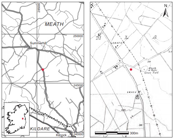
Location (Fig. 6.40)
The site was in the townland of Drumlargan, south-east Co. Meath.70 It is situated on high ground at an altitude of 122m above sea level, overlooking the valleys of the Boyne and Blackwater and two miles south-east of Summerhill. The ruins of a church enclosed by a bank and wall are located 15m from the site. Very little information is recorded as to the date of this church. It is marked on the first edition OS 6in. sheet, and the gravestones visible in the graveyard appear to be eighteenth-century in date.71
Description of site
The raised area in which the burials were found was approximately 2m above the surrounding
ground level at the highest point. The presence of shaly soil 0.4m below the present surface suggested that the rise was a natural feature, and a deep hollow to the west of the site seemed to suggest that the mound had been added to. The remains of at least five individuals were recovered. The burials appeared to be concentrated at the highest part of the mound. There was no evidence to suggest the protection of burials with stone or other material.
Most of the remains had been disarticulated and broken by disturbance to the site, but the original orientation of some of the burials was still discernible. There did not appear to be any fixed orientation, as burials were aligned both east/west and north/south. A scattering of burnt bones and flecks of charcoal were also visible on the surface, but not so much as to indicate intensive burning. Some horse teeth were also found. An iron hook-like object, two iron nails and some sherds of post-medieval pottery were found on the site, but no artefacts were found in direct association with the remains.72
Comment
Although the burials are likely to have been associated with the church site, it is not possible to associate them directly or to suggest a date, given the level of disturbance.
HUMAN REMAINS
LAUREEN BUCKLEY
Introduction
In addition to the ploughing it seems that the mound had been disturbed in antiquity, and in fact some of the photographs appear to show a collection of bones such as would be found in a charnel pit rather than disturbed burials.
Description of the sample (1935:35)
Right femur
(1) The head of a right femur (FeHd 43.5mm).
(2) The mid-shaft area of a right femur.
Left tibia
The proximal two-thirds of a left tibia (TiD1 35.3mm; TiD2 23.3mm; TiE1 75.7mm).
Fibula
(1) The proximal half of a left fibula.
(2) The proximal third of a left fibula.
(3) A section from mid-shaft, side unknown.
Left humerus
(1) The proximal half of a left humerus. The diameter of the head indicates that it could be from a female individual (HuHd 38.4mm).
(2) The proximal half of the shaft of a left humerus that was larger than the one described above.
Right humerus
(1) The proximal half of a right humerus, probably from a female (HuHd 44.0mm).
(2) The proximal two-thirds of a right humerus, also from a female individual. It may have been from the same individual as the left humerus (1) described above (HuHd 40.2mm).
(3) An almost complete right humerus shaft.
Left radius
(1) A complete left radius, which from the size of the head was probably from a female (RaL1 226mm; RaHd 19.5mm).
(2) The proximal third of a left radius, possibly also from a female (RaHd 18.1mm).
Left ulna
A left ulna with the tip of the olecranon missing and the distal third also missing.
Right ulna
An almost complete right ulna with only the top of the olecranon missing.
Ilium
An almost complete left ilium. The sciatic notch was narrow so it was probably from a male. There was some degenerative joint disease in the hip, with a moderate degree of marginal lipping in the superior part of the acetabulum.
Ribs
One left upper rib.
Phalanges
One proximal hand phalanx.
Skulls
(1) One complete face, with only a small part of the right frontal bone near the coronal suture missing and all the facial bones complete and fused together. The supraorbital ridges indicate that it was a male individual. Slight cribra orbitalia was present in both orbits.
(2) Mostly the left side of the frontal bone with a small part of the right just beyond the midline area.
(3) Most of the left squamous area of the left frontal bone.
(4) Part of the anterior of a left parietal bone with coronal and part of the squamous suture visible.
(5) A fragment of the left side of an occipital bone. Five mandibles and two maxillae were also present.
Mandibles
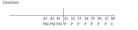
(1) This mandible was complete.
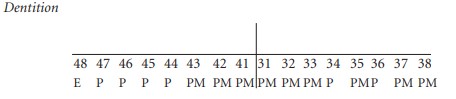
Dental development: The third molar on the right was in the process of erupting, so this was probably from a young individual aged 18–21 years. The third molar on the left had been lost post-mortem but was probably not fully erupted.
Attrition: there was a light degree of wear on the premolars and first molars but no wear on the second and third molars.
Calculus: there were slight deposits on the buccal surfaces of the first molar and moderate deposits on the lingual surfaces of the first left premolar and right second molar.
Periodontal disease: there was a mild degree of alveolar recession around the roots of the first molars and a moderate degree around the roots of the right second molar.
Hypoplasia: grooves of hypoplasia, which had occurred during the first year of life, were present on the first molars.
(2) This mandible was almost complete but the left ramus was missing.
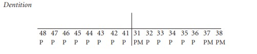
Attrition: there was light wear on the incisors, canines and premolars, moderate wear on the right molars and heavy wear on the left first molar.
Calculus: there were moderate deposits on the lingual surfaces of the right molars and left premolars and light deposits on the buccal surface of the left first molar.
Periodontal disease: there was light alveolar recession around the roots of the teeth on the left side, moderate recession around the right incisors, canine and premolars, and a considerable degree of recession around the roots of the right molars.
(3) This consisted of most of the left side of the bone and the anterior part.
Anomalies: the canine is rotated slightly clockwise and overlaps the adjacent premolar, 34, slightly.
Attrition: there was very light wear on most of the teeth, with a moderate degree of wear on the first molar.
Calculus: deposits were slight on the buccal surfaces of all teeth except the canine, where deposits were moderate. There were also moderate deposits on the lingual surfaces of all teeth except the second and third molars.
(4) This consisted of the right side and anterior of a mandible.

Calculus: there were light deposits on the lingual surfaces of the molars and second premolar and on the buccal surface of the first premolar. Deposits were moderate on the lingual surfaces of the incisor, canine and first premolar.
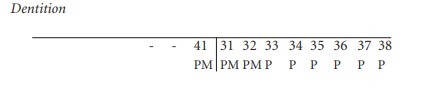
(5) This consisted of most of the left side and part of the anterior of the mandible.
The socket for 38 was visible although the tooth was missing. Nevertheless, from the shape of the socket it can be determined that the crown was just formed. An individual at this stage of dental development would be aged 15–16 years.
Attrition: there was very little wear on any of the teeth.
Calculus: slight deposits were present on the lingual surface of the first molar, there were moderate deposits on the lingual surface of the lateral incisor and considerable deposits on the lingual surface of the central incisor.
Hypoplasia: linear enamel hypoplasia was noted on the lateral incisor, canine and premolars.
Maxillae
(1) This was the maxilla with the face described as skull 1 above.
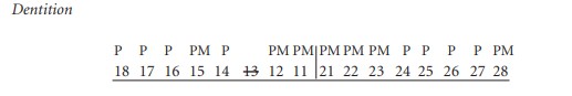
Attrition: there was moderate wear on the premolars and heavy wear on the first molars. Attrition was relatively light on the other molars.
Periodontal disease: there was a slight degree of alveolar recession around the roots of the right premolar and first molar.
Hypoplasia: linear enamel hypoplasia was noted on the left premolars, the first left molar and the third right molar.
(2) This consisted of the left side of a maxilla only

The third molar was not completely erupted. This maxilla could have come from the same individual as mandible (1), described above.
Attrition: there was no wear on the second and third molars and only slight polishing on the first molar and canine.
Calculus: deposits were moderate on the lingual surface of the second molar and considerable on the buccal surface of the same tooth.
Hypoplasia: grooves of hypoplasia were present on the canine and first molar.
Loose teeth
There were a number of loose teeth present that did not fit any of the above mandibles and maxillae. These included an upper right incisor and canine, and an upper left first and second premolar, all with very little wear. There were also an upper left and right second molar and an upper left third molar. The mandibular teeth consisted of a right central incisor, first, second and third molars from the left side and also from the right side, and another left third molar. There was very slight or no wear on most of these teeth. One upper left second molar had a large cavity on the distal side of the crown and root. Also present was one lower right central deciduous incisor.
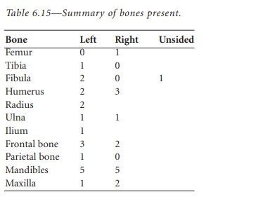
Based on the number of mandibles, the minimum number of individuals present was five.
Summary and conclusions
This sample from a burial mound consisted of at least five individuals. At least two of the individuals were females and one was male. There was at least one adolescent aged around 15–16 years and one young adult aged 18–21 years.
The male had some degenerative joint disease in the left hip. The complete face from the male individual had cribra orbitalia in both orbits, which could indicate iron-deficiency anaemia. Apart from the male, who seemed to be older than the other individuals, there was very little attrition of the teeth. Most individuals had evidence for nutritional deficiency or acute disease during childhood. Although only adult or adolescent bones were collected, there was a deciduous molar present among the loose teeth collected so there must have been juveniles buried in the mound.
70. Parish of Drumlargan, barony of Deece Upper. SMR ME043-052——. IGR 285604 246383.
71. SMR ME043-037——.
72. A sample of the human and animal remains from the site was recovered, 1982:17; two post-medieval potsherds were also found, 1982:18.1–2, as well as an oval iron object with a hook at one end, 1982:18.3, and two iron nails found in the surface fill of the mound, 1982:18.4–5.