1982:010 - TURREEN, CO. LONGFORD, Longford
County: Longford
Site name: TURREEN, CO. LONGFORD
Sites and Monuments Record No.: SMR LF017-030
Licence number: E1114
Author: FIONNBARR MOORE
Author/Organisation Address: —
Site type: Graves of indeterminate date
Period/Dating: —
ITM: E 602872m, N 765393m
Latitude, Longitude (decimal degrees): 53.638324, -7.956567
Introduction
In July 1982 human remains were discovered on the grounds of a newly constructed house near Lanesborough, Co. Longford, in the townland of Turreen (Fig. 6.30).49 A site visit was carried out by Fionnbarr Moore. The site, apparently a mound, had been levelled off by the time of Moore’s visit, but the remains of four or five individuals were visible scattered throughout the disturbed soil. They had apparently been lying in an east/west direction, and
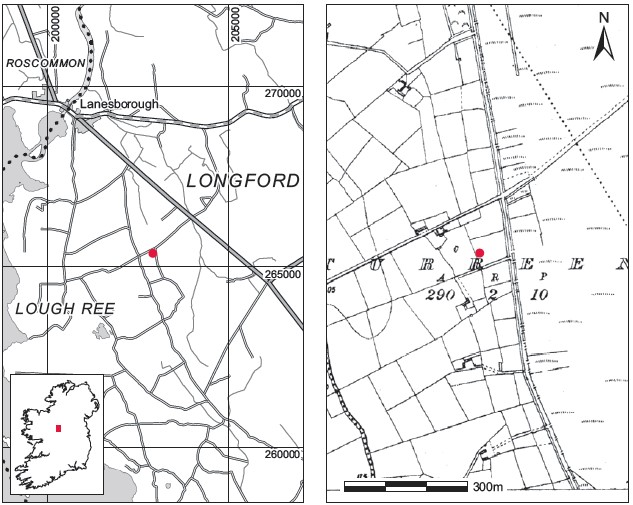
Fig. 6.30—Locationmap, Turreen, Co.Longford.
no grave-goods of any description had been found with them. According to reports, the mound had measured approximately 3m from north to south and 0.5m high. It was situated in the corner of a large field. There was no tradition of a church or graveyard in the area. Some of the skeletal remains were reburied by the Gardaí. The bones (2010:84) retained by the NMI are reported on below.
Comment
In the absence of associated finds or other dating evidence these burials must be regarded as Undated.
HUMAN REMAINS
LAUREEN BUCKLEY
Description of bones
The skull bones included a complete frontal bone, most of the left side of another frontal bone, a fragment of occipital bone and the left side of a mandible. The long bones consisted of the distal half of the shaft of a left humerus, the distal two-thirds of a right humerus and the mid-shaft area of a left femur. The two humeri were very slender and looked like a matching pair. The femur shaft was very large.
Sex and age
The supraorbital ridges of the complete frontal bone indicated that it was from a male individual and the incomplete frontal bone appeared to be of the female type. The female had evidence of hyperostosis frontalis interna (see below), which suggests that the individual may have been over 45 years of age. There was no way of ageing the male bone but the wear on the teeth on the mandible suggested that it was from a young adult or an early middle adult.
Skeletal pathology
The complete frontal bone had a small osteoma on the left side. This consisted of a small, oval, raised area of smooth bone approximately 1cm in length. This type of bone tumour is benign and is very common. The other incomplete frontal bone had a small fleck of bone on the internal surface, which may represent the initial stages of hyperostosis frontalis interna. In this condition, which affects post-menopausal women, extra bone is formed on the internal surface of the frontal bone.
Attrition: there was slight wear on the molar teeth.
Calculus: deposits were moderate on the buccal and lingual surfaces of the first two molars and the distal surface of the third molar. There were considerable deposits on the lingual surfaces of the third molar.
Periodontal disease: there was some slight recession around the roots of the molars.
Hypoplasia: pits of enamel hypoplasia were present on the second and third molars.
Summary
The bones collected represent the remains of at least two adult individuals, one female and the other male. The female was probably an older adult, at least 45 years old at the time of death. It was not possible to age the male. There was one mandible present and molar attrition was light. Since the period of these burials is unknown, however, it is not possible to use this feature as a reliable indicator of age. The male had a small benign bone tumour on the frontal bone and the female had the early stages of a condition known as hyperostosis frontalis interna. This is thought to be caused by hormone imbalance and usually occurs in postmenopausal women.
49. Parish and barony of Rathcline. SMR LF017-030——. IGR 202922 265373.