1980:195 - BOHERAGADDY, CO. KILKENNY,, Kilkenny
County: Kilkenny
Site name: BOHERAGADDY, CO. KILKENNY,
Sites and Monuments Record No.: SMR KK024-113SMR KK024-06001
Licence number: E1097
Author: MARY CAHILL
Author/Organisation Address: —
Site type: Graves of indeterminate date
Period/Dating: —
ITM: E 657342m, N 649848m
Latitude, Longitude (decimal degrees): 52.596880, -7.153585
Introduction
On 15 August 1980 human remains were discovered during the digging of house foundations at Boheragaddy, near Bennetsbridge, Co. Kilkenny. The bones dislodged by the bulldozer were collected and later given to the local pathologist. The discovery was reported to the NMI and to the Garda Síochána a number of days later, and work was suspended pending a Museum investigation. The site was visited by Mary Cahill on 27 August 1980. The foundations for the outer walls of a house had been dug and the remains of at least three burials were visible as they had been cut through by the digger. The human remains have been examined by Laureen
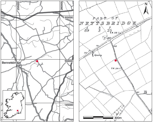
Fig. 6.22—Locationmap, Boheragaddy,Co. Kilkenny.
Buckley and were found to represent three individuals, a young adult female, an early middle adult male and an early middle adult female.
Location (Fig. 6.22)
The site was in the townland of Boheragaddy, east Co. Kilkenny.35 It was close to the side of a road, at an altitude of c. 30–60m above sea level, just 200m north-east of an enclosure marked on the SMR.36 In the same year an early medieval spiral-headed ringed pin37 was discovered in the adjacent townland of Bishopslough-Newtown (Cahill 1981, 253).
Description of site
Most of the bone (2010:83) was removed from its context before the investigation, and no structural remains were noticed on the site. The graves appear to have been laid out as follows. Grave 1, containing a young adult female, had been exposed on the north-west side of the trench. Grave 3, containing an early middle adult female, was about halfway along the trench on the west side. Grave 2, which contained an early middle adult male, was located on the south-eastern face. Most of the exposed bones had been removed by the finder around the time of discovery.
Comment
According to the original report of the discovery by Ellen Prendergast, the field in which the burials were found was known locally as ‘Killeen’. There was nothing in the skeletal remains, however, to indicate that the site had been used for the burial of babies or very young children.
HUMAN REMAINS
LAUREEN BUCKLEY
Introduction
Bone assemblage 2010:83 was disarticulated and stored in two boxes. It was possible to divide the contents of box 1 into two burials, skeleton 1 and skeleton 2, based on size, sex and epiphyseal fusion, although some of the smaller bones could not be allocated. The second box contained burial 3 only.
Skeleton 1 (from grave 1): young adult, female, 151cm
This consisted mainly of pelvis and legs, although some skull fragments may belong to this individual. The skull fragments consisted of part of the left frontal bone, the anterior part of the right parietal bone and the basilar occipital bone. Only the fourth and fifth lumbar vertebrae remained from the vertebral column. The left ilium from the pelvis was complete and the right ilium was almost complete. Most of both ischia were also present and the sacrum was virtually complete. The left femur was absent but the left tibia and fibula were complete. The right femur and tibia were complete and the right fibula was almost complete, apart from the distal joint end. Only the right talus remained from the foot bones.
Age and sex
The sciatic notch was wide, and the diameter of the head of the femur and the bicondylar width of the femur were in the female range. Therefore this was a female individual. The epiphyses at the ends of the long bones were just fused but the iliac crest was unfused. The basio-sphenoid symphysis of the skull was also unfused. The first sacral vertebra was not fused to the second sacral vertebra. The auricular surface of the ilium indicated an age of 20–24 years. This is therefore the skeleton of a young adult female. Living stature was estimated, using the lengths of the femur and tibia, as 151cm.
Non-metric traits
Squatting facets were visible on both tibia and there was a third trochanter on the right femur.
Skeletal pathology
None.
Skeleton 2 (from grave 2): early middle adult, male, 168cm
This consisted of the lower half of a skeleton only. The pelvis consisted of the complete ilia and ischia, part of the left pubis and the upper three sacral vertebrae. The femora were complete; the proximal third and distal half of the left tibia were present, as well as the distal two-thirds of the right tibia. The left fibula was complete and the most of the right fibula was present. Both tali and both calcanea remained from the foot bones.
Age and sex
The sciatic notch was wide and the sub-pubic concavity, which is a female feature, was present on the pubic bone. There was no ventral arc on the pubic bone, however, which normally indicates a male. The bicondylar width of the femora was also above the male range. Overall, this was considered to be a male skeleton. All the epiphyses were fused and the auricular surface of the ilium indicated an age of 25–29 years. The living stature was estimated from the lengths of the femur and fibula as 168cm.
Non-metric traits
There was a third trochanter present on both femora and a squatting facet was present on the right tibia.
Skeletal pathology
Only the first three sacral vertebrae were present but spina bifida occulta was present in all three. Healed osteochondritis was present in the superior surface of the left talus. The lesion was 14mm long and had healed, but the healed part was rough and was raised above the smooth articular surface.
Skeleton 3 (from grave 3): early middle adult, female, 160cm
These bones were in one box and seem to be mainly from one skeleton. There was some decay on the outer surface of the arm bones and some mineral deposits on the internal surface of the skull but otherwise it was in good condition. The skull was fragmented but was mostly complete, with the frontal, both parietal, occipital and right temporal bones present and virtually complete. The sphenoid and right zygomatic bones were also present, most of the maxilla was present and the mandible was complete. Only the first two cervical vertebrae and part of the seventh cervical vertebra remained, but all the thoracic and lumbar vertebrae were present. There were four ribs from the left side and nine from the right side and most of the manubrium of the sternum was present. The glenoid area of the left scapula was present but most of the right scapula remained. The lateral halves of both clavicles were present. Both humeri were complete, the left radius and ulna were missing their distal ends, and the right radius and ulna were complete. Only the right scaphoid and third, fourth and fifth metacarpals from the right hand remained from the hand bones.
Both ilia were present from the pelvis and the right ilium was complete but decayed. Both ischia and the left pubic bone were incomplete. The first three sacral vertebrae also survived. Only the proximal half of the left femur and the proximal third of the right femur remained from the leg bones. Two fragments of fibula shaft and one tibia shaft were present but it was not possible to side them.
Age and sex
The external occipital protuberance, supraorbital rims and supraorbital ridges were of the female type but the temporal bones and the mandible were very male-like. All the features of the pelvis, the sciatic notch, sub-pubic concavity and sub-pubic angle, were of the female type. The measurements of the head of the femora, humeri and radii and glenoid cavity width were all within the female range. The auricular surface of the ilium indicated an age of 25–29 years and the epiphyseal line was still vaguely visible on the proximal femora, so the individual was probably aged around 25 years. The living stature was estimated from the length of the radius as 160cm.
Non-metric traits
The metopic suture was retained.
Skeletal pathology
Slight Schmorl’s nodes were present on the superior surface of T10, the inferior and superior surfaces of T11 and T12 and the upper three lumbar vertebrae.
Dentition
Attrition: there was a light degree of attrition on the incisors, first molars and lower right canine and premolars, but there was no wear on any of the other teeth.
Calculus: in the maxilla there were light deposits on the lingual surfaces of almost all teeth and on the buccal surfaces of the right second premolar and right molars and on the buccal and distal surface of the left second molar. In the mandible deposits were moderate on the lingual surfaces of the incisors, canines, left premolars and all the molars except the lingual surface of the left third molar, where there were considerable deposits.
Hypoplasia: linear enamel hypoplasia was noted on the upper left canine and the lower left canine as well as the lower right first premolar. The upper right first premolar and lower right second premolar had pits of hypoplasia on their surface.
Additional bone
There were a number of bones that could not be assigned to particular skeletons. These included the mid-shaft area of a right humerus, the distal thirds of a right radius and ulna and the distal half of a left femur. In addition, there was a left third metacarpal, a right capitate and right third and fourth metacarpals. Foot bones included a left first and a left fourth metatarsal, two proximal phalanges (unsided), all the right metatarsals from one foot and an extra second and third right metatarsal from a smaller foot. The smaller bones could belong to skeleton 1. The other bones could have belonged to any of the three skeletons already described.
Summary and conclusions
Three skeletons were recovered from this site: a young adult female, an early middle adult male and an early middle adult female. None of the individuals were very tall: the two females were 151cm and 160cm tall and the male was 168cm. The average stature of modern males is 174cm and of females is 161cm. In skeletal analysis of past Irish populations male stature is usually found to be around 170cm and female stature around 155cm. Spina bifida occulta was noted in the male. In this condition the dorsal surface of the sacrum does not fuse over completely but it does not usually cause any symptoms, as membranes and ligaments in this area adequately protect the spinal cord. It is thought to be present in 4% of the Irish population. This individual also had osteochondritis dessicans in the left talus. This lesion affects the cartilage and underlying bone of the joint surface, causing an area of bone death. The area affected is quite small and is roughly circular. Occasionally the lesion heals, as it has in this case, with the bone growing over the area of necrosis. The joint surface is not as smooth as it was originally, however, and this can cause pain and eventually osteoarthritis. The condition usually occurs in males and is thought to be caused by trauma to the joint. The early middle adult female had slight Schmorl’s nodes in the lower half of the vertebral column. These occur as a result of lifting heavy loads during adolescence. This skeleton was the only one with teeth remaining. They were in good condition with light wear, light to moderate calculus deposits and no caries. The individual had suffered from nutritional deficiency or acute illness during early childhood.
35. Parish of Boheragaddy, barony of Tullaherrin. SMR KK024-113——. IGR 257405 149804.
36. SMR KK024-06001.
37. 1980:85.