1978:033 - RATHMORE, CO. ROSCOMMON, Roscommon
County: Roscommon
Site name: RATHMORE, CO. ROSCOMMON
Sites and Monuments Record No.: SMR RO035-038
Licence number: E1152
Author: BARRY RAFTERY
Author/Organisation Address: —
Site type: Early Bronze Age graves
Period/Dating: —
ITM: E 587203m, N 771638m
Latitude, Longitude (decimal degrees): 53.694302, -8.193756
Introduction
In April 1978 a short cist containing a cremation and a bowl was discovered during ploughing on a farm near Roscommon town, Co. Roscommon. The capstone, apparently 0.15m below ground level, had been struck by the plough and was lifted to reveal the chamber. The landowner contacted the Garda Síochána at Roscommon, who reported the find to the NMI. A one-day rescue excavation was undertaken on behalf of the Museum by Dr Barry Raftery of University College Dublin. This report is based on Raftery’s account of the excavation. The human remains were examined by Laureen Buckley.
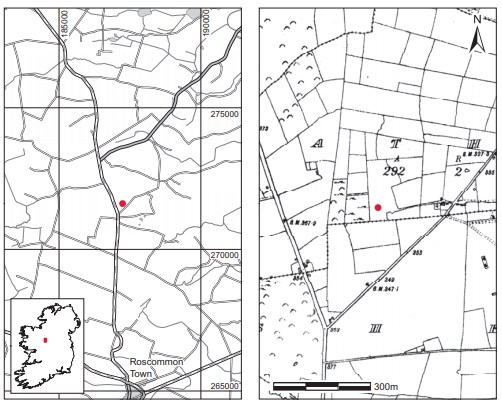
Location (Fig. 3.153)
The site was in the townland of Rathmore, approximately 7km north of Roscommon town, mid-Co. Roscommon.281 It was situated on slightly elevated ground between 100m and 200m above sea level. No sites of similar date are known from this townland.’
Description of site
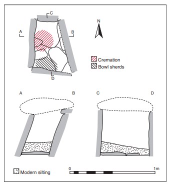
The cist was trapezoidal in plan, with its long axis running north/south. Internally it measured 0.58m long by 0.43m wide by 0.5m high (Fig. 3.154). It was composed of four main slabs, with one forming each wall. The northern end stone was vertical, and that at the south sloped slightly inwards. The western side stone sloped markedly inwards, while that at the east sloped outwards to roughly the same degree.
Two small stones had been placed on the upper edges of the side stones in the east and south-east. These slightly overhung the interior of the cist. No packing stones were found around the outside of the cist. The capstone was suboval in shape, measuring 0.63m long by 0.63m wide by 0.15m thick. The floor of the cist was paved with four flat slabs, but these did not cover the entire floor area.
The grave contained the cremated remains of an adult male (1978:346) and sherds of a ribbed bowl. The human remains lay in a pile on the floor in the north-western corner of the cist and covered an area of 0.25m by 0.2m. The bowl sherds were located in the south-western corner of the cist. These were in a brittle and partially disintegrated state, and represented less than half of the bowl. There were no traces of charcoal or any other burnt material. A layer of fine, silted material some 0.005–0.15m deep had formed over the burial deposit in modern
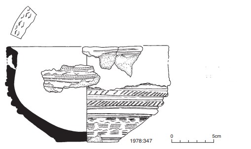
Ribbed bowl, 1978:347 (Fig. 3.155)
The surviving sherds form the base and approximately one third of the sides of a ribbed bowl, as classified by Ó Ríordáin and Waddell (1993, 129). It has a dark grey core with reduced greybrown internal surface and is brown externally except around the rim, which is grey-brown. The vessel has an upright neck and internal bevel on the rim. Its upper half, which is vertical, is encircled by four ribs. Below these the vessel narrows to a flat base. The vessel is decorated all over with false-relief ornament. This takes the form, on the neck and lower body, of a series of opposed impressed triangular shapes, leaving a raised, sinuous zigzag pattern. This is flanked by horizontal rows of comb impressions. The rim, ribs and base angle are outlined by pairs of continuous plain, incised horizontal lines. The two central ribs are decorated with rows of closely set diagonal impressions.
Dimensions: est. H 12cm; est. D rim 18cm; D base 7.5cm; T of body 0.8cm.
Comment
The human remains from this site have not been dated. The vessel is a fragmentary ribbed bowl which is similar to stage 1 bowls as defined by Brindley (2007, 172–3). This stage of bowl development is dated to 2160–2080 BC.
HUMAN REMAINS
LAUREEN BUCKLEY
Introduction
Sample 1978:346 consisted of 1,627 fragments of cremated bone, weighing a total of 1,479g. The bone was stained with charcoal but was basically a white colour and was efficiently cremated, with horizontal fissuring of the bone surface. Some fragments had a chalky texture. There were several fragments that were stained orange and some of the skull fragments were speckled with a sky blue colour.
Table 3.89—Fragmentation of bone, 1978:346.
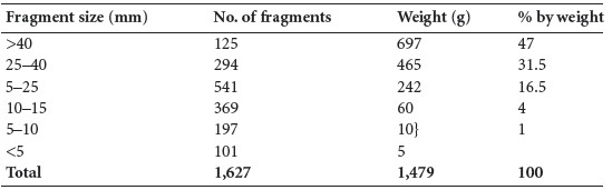
The fragmentation of the sample is shown in Table 3.89, with the largest fragment being 98mm in length. It can be seen that there is a very high proportion of very large fragments and that almost all the sample is made up of large fragments more than 15mm in length. The bone does not appear to have been deliberately crushed as part of the cremation ritual
Identifiable bone
As there was a high proportion of large fragments, a relatively high percentage of bone could be identified. A total of 1,038g (70% of the total bone) was identified. The amount and proportion of the identified bone are shown in Table 3.90.
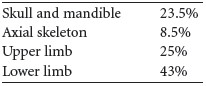
Table 3.91 summarises the main parts of the skeleton identified from this sample. It can be seen that the proportion of axial skeleton is very low, but that the other parts of the skeleton are slightly higher than expected.
Identifiable fragments
Skull
The right side of the squamous occipital bone was present and the external occipital protuberance was prominent. There were several fragments of parietal bone and squamous frontal bone, some of which had blue-green stains on them. A fragment of left orbit and the right orbit from a male were present. The left mastoid area of the temporal bone with part of the squamous temporal bone was present, and the posterior part of the zygomatic arch and the mastoid process were of the male type. The anterior part of the left temporal bone with the mandibular fossa was present. There were two petrous mastoid bones. Fragments of the left and right zygomatic bones were present.
Mandible, maxilla, teeth
Part of a left ramus and mandibular condyle were present, as well as a right condyle. There were also two fragments of the body with sockets present. The right side of a maxilla was present.
Dentition
Sockets for the following teeth were present:
Vertebrae
The posterior part of the arch from a first cervical vertebra and the body of one lower cervical vertebra remained. There were also two partial arches from the thoracic vertebrae and at least four incomplete lumbar bodies and two lumbar arches. Mild osteophytosis was present on two fragments of lumbar vertebral bodies.
Ribs
There were several fragments of shaft.
Table 3.90—Proportion of identified bone, 1978:346.
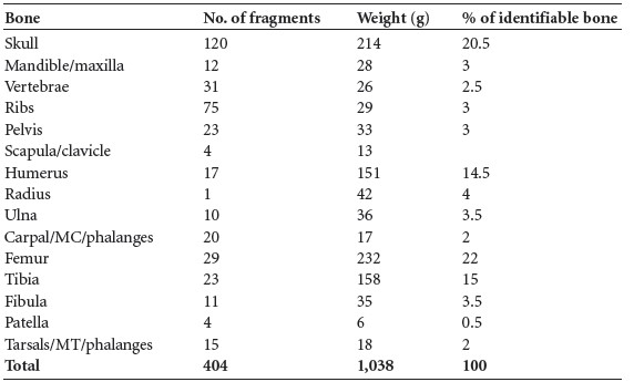
Table 3.91—Summary of identified bone, 1978:346.
Pelvis
There were several fragments of ilium, including the posterior part of the left and right ilium as well as the auricular surface of the left ilium. There were also some fragments of actetabulum.
Clavicle
An almost complete left clavicle with a green stain near the lateral end was present and there was a shaft of one other clavicle. The medial end of the shaft of a right clavicle was also present.
Scapula
One fragment of glenoid fossa was present.
Humerus
Proximal ends of shafts from a left and a right bone were present; there was also a fragment of the distal end of a right bone as well as a fragment from the distal end of another bone. Several large fragments of shaft were also present and there was one proximal articular surface.
Radius
The proximal end with the proximal articular surface of a right bone and some fragments from the middle of the shaft were present. The distal half of a right bone and the distal half of a left
bone were present and there was one fragment of distal joint end.
Ulna
This consisted of shaft fragments, including two proximal and two distal thirds of shaft. A right proximal surface and one other proximal surface were present.
Carpals, metacarpals, phalanges
The right lunate, scaphoid and hamate were present, as well as the left triquetral, seven metacarpals and also four proximal phalanges and one middle phalanx.
Femur
There were several large fragments of shaft, including the distal shaft and some of the proximal shaft. The neck, lesser trochanter and some of the shaft from the right bone were present, as well as the proximal shaft and lesser trochanter from the left bone. There were also two fragments of proximal joint surfaces and one fragment from the distal joint surface.
Tibia
Several large fragments of shaft were present, including the posterior shaft of the left bone near the proximal end with the nutrient foramen visible. There were fragments from the anterior surface of the bone, including the tubercle from the right bone. Most of the distal half of the shaft of the right bone was present.
Fibula
This consisted of fragments of shaft only.
Patella
The medial half of a left and the medial half of a right patella were present
Tarsals, metatarsals, phalanges
Fragments of two tali were present, as well as a fourth and fifth metatarsal shaft and shafts of two others and one distal foot phalanx.
Minimum number of individuals
There was no repetition of skeletal elements, so only one individual was present. This was an adult male.
Summary and conclusions
This was a relatively large cremation, consisting of 1,479 fragments of bone. The bone was efficiently cremated, consisting of white bone with a chalky texture, although some fragments were stained by mineral deposits. There was a high proportion of very large fragments, indicating that the bone had not been crushed as part of the cremation ritual. As a result of the large fragments it was possible to identify 70% of the bone with all skeletal elements represented, although the axial skeleton (vertebrae, ribs and pelvis) had not been totally collected. The remains represented one adult male. He had evidence of osteophytosis on some of the lumbar vertebrae.
281. Parish of Kilbride, barony of Ballintober South. SMR RO035-038——. IGR 187250 271620.