1977:073 - BLACK ABBEY, GARDENS, CO. KILKENNY, Kilkenny
County: Kilkenny
Site name: BLACK ABBEY, GARDENS, CO. KILKENNY
Sites and Monuments Record No.: SMR KK019-026021
Licence number: E1096
Author: RAGHNALL Ó FLOINN
Author/Organisation Address: —
Site type: Late medieval graves, c. AD 12001600, and post-medieval graves, AD 16001800
Period/Dating: —
ITM: E 650226m, N 656153m
Latitude, Longitude (decimal degrees): 52.654246, -7.257659
Introduction
In May 1977, during restoration work outside the east window of Black Abbey, Kilkenny, two stone coffin burials were discovered. A trench, 11.5m by 2m by 1.75m deep, had been cut across the eastern end of the church in order to clear soil away from the walls of the church, which had been periodically flooded in the past. This excavation uncovered the walls of the original chancel, which had been destroyed in the late eighteenth century. The burials were located inside the original chancel of the church. The site was reported to the NMI by Fr John Brennan of Jenkinstown, Co. Kilkenny. A rescue excavation was undertaken by Raghnall Ó Floinn. The human remains were analysed by Laureen Buckley.
Location (Fig. 5.9)
The site was located in Gardens townland in Kilkenny City.17 The burials lay outside the eastern end of the church, inside the original chancel. The Black Abbey was a Dominican abbey founded by William Marshall in 1225. The building consists of a nave and south aisle, which is probably thirteenth-century in date (Farrelly et al. 1993, 16–17).
Site stratigraphy
Before excavation, the general stratigraphy of the site was apparent (Fig. 5.10). In addition to
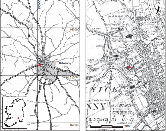 Fig. 5.9—Location map, Black Abbey, Gardens, Co. Kilkenny.
the chancel walls, the trench had revealed the footing of the tower (built c. 1500). At the level of the tower footing a floor level was visible which consisted of a layer of mortar overlying a layer of hard black clay with charcoal. Over much of the site this floor level formed a convenient sealing layer. Above it was a 1.2m-thick clay fill containing some large stone blocks. This reached to the top of the chancel walls on either side and was sealed by a 0.3m-thick upper layer of loose black garden soil containing animal bones and pottery of eighteenth-century date and later. The coffins were stratified under a mortar floor that had been set at the same level as the footing of the tower. Two of these occurred in coffin-shaped stone graves, and the third was in an unprotected pit. The fill in which the burials had been set, however, contained post-medieval pottery and tiles, suggesting a date after 1500 for the burials.
Wall
Running parallel to the south wall of the chancel and 0.5m inside (i.e. north of) it was a rough masonry wall containing robbed fragments of medieval carved stones. This wall rested on top of the mortar floor and was not bonded into the wall of the tower. It was 0.095m thick and 2.3m high. No corresponding wall was found on the northern side of the chancel and its function is thus unknown.
Finds
The finds consisted largely of pottery of post-medieval date. These were uncovered before the site was examined and are therefore unstratified but most probably came from the upper 0.3m of garden soil. A number of clay pipe fragments and a few pieces of pottery came from the mortar floor, indicating a possible late seventeenth-century date for its use. Around the
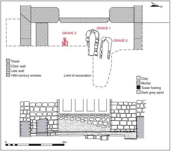
Fig. 5.10—Site plan and section, Black Abbey, Gardens, Co. Kilkenny.
burials below the mortar floor were small fragments of green-glazed red brick tiles. Other finds, including perforated roof slate fragments and fragments of window leading, came from the disturbed east end of grave 2 and may have filtered down from upper levels. The fragments of floor tile from the soil around the coffin burials indicate a sixteenth- to seventeenth-century date for the coffins and unprotected burials. Five fragments of stamped tiles of early fourteenth-century date were found in an unstratified position by workmen and indicate the possibility of a tiled pavement at a lower level. Owing to flooding of the area, however, this could not be investigated.18
Description of site
Grave 1
This had already been cleared out and then backfilled. According to the workmen who carried out this task, one of the skulls from this burial lay at the west end of the coffin, protected by a lintel.
This presumably represented the primary burial. The excavated coffin was wedge-shaped in plan, aligned roughly east/west (Fig. 5.11). It lay 0.36m below the mortar floor. It was semicircular at its wider, western end (max. Width 1.15m), narrowing towards the east, giving an overall wedge shape. Internally it measured 1.95m long by 0.55m wide by 0.24m high. The walls of the coffin were made of small, roughly squared pieces of stone measuring up to 0.3m in length. These were laid down in rough courses and mortared together. The walls included three pieces of carved stone: two
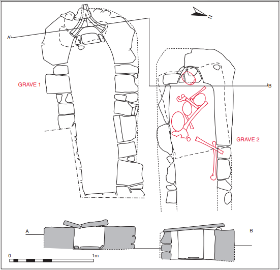
Fig. 5.11—Plans and sections of graves, Black Abbey, Gardens, Co. Kilkenny.
fragments of window moulding and a complete hood moulding, the latter providing the curve at the west end of the tomb. Part of a single capstone remained in situ at the western end. Others had apparently been removed by workmen. One displaced capstone lay to the north, measuring 0.55m by 0.55m. Some crude paving, perhaps pillow stones, occurred on the coffin floor at its western end.
As mentioned above, the contents of this burial had been removed and then replaced by the workmen. A skull (1977:2340) found at the western end of the grave is thought to have been from the original burial, but the rest of the remains had been disturbed.
Grave 2
This lay 0.25m north of grave 1 and was oriented along the same axis (Fig. 5.11). It lay 0.2m below grave 1 and 0.54m below the mortar floor. The capstone had been exposed and raised by the workmen, but otherwise it remained intact. It was constructed in a similar manner to grave 1 but had a more pronounced recess for the head at its western end, also formed of carved stone. The recess for the head was semicircular in shape, measuring 0.3m across. Pillow stones were also visible in this area. The eastern end of the coffin did not survive. The mortar floor that sealed all the burials was not apparent at the eastern end of this burial and this may be the reason why the burial was damaged at this point. Internally it measured at least 1.6m by 0.5m by 0.36m high, narrowing slightly towards the east end. The remaining capstone, which lay over the western end of the burial, was 1m long by 0.7m wide.
Four human skulls (1977:2339) lay underneath the capstone: a middle adult, an older adult, a middle adult female and a juvenile. One was set into the recess at the western end on the possible pillow stones and may have been the primary burial here. The other three skulls were inserted defleshed, as they occurred among a jumble of long bones and jaw bones. Other long bones were stacked against the sides of the coffin.
Unfortunately, owing to flooding of the coffin floor, it was very difficult to remove the bones in a systematic manner, but it was clear that only one of the skeletons was intact, lying extended with the arms crossed over the chest.
Grave 3
This was an unprotected burial, lying 1.75m south of burial 1 and 0.54m below the mortar floor (Fig. 5.10). The grave was a simple rectilinear pit, aligned east/west.
The grave contained an extended inhumation (1977:2341). The human remains, those of an adult female, were placed in a supine position with the arms crossed over the chest. The lower part of the burial was concealed under a bank of earth and it was decided to have this removed before further examination took place. To the east of this were three skulls and a number of long bones, not arranged in any order (1977:2341). These were found by Buckley to represent the remains of at least one male and one juvenile.
Site sequence
The site sequence appears to be as follows:
1 Construction of the chancel of the original abbey church, possibly with a tile pavement added in the early fourteenth century.
2 Construction of the tower c. 1500.
3 Deposition of burials—late sixteenth/early seventeenth century.
4 Mortar floor sealing layer laid down—seventeenth century.
5 Construction of wall on southern side of chancel using robbed carved stonework.
6 Filling of chancel, eighteenth century.
7 Destruction of chancel walls, c. 1780.
Comment
These burials have been interpreted by the excavator as dating from the late sixteenth or early seventeenth century. A carved stone reused to form one of the stone coffins was a window hood moulding dating from the fourteenth century. Most of the remains were disturbed, and only one burial was articulated. Earmuffs or pillow stones were noticed in graves 1 and 2. This feature is known from a number of medieval burial sites in Ireland, such as Ardfert, Co. Kerry, and Waterford City (Hurley et al. 1997, 193–5).
HUMAN REMAINS
LAUREEN BUCKLEY
Introduction
Apart from the third burial, which lay partially undisturbed, the burials had been disturbed either by the workmen on the restoration project or at some time in the past. The first burial contained more than one individual anyway, but was dug out and then filled back in again by the workmen. The second burial was undisturbed but contained a jumble of long bones and skulls. The third burial consisted of part of a skull and thorax in situ, with the lower half going under the baulk. There were a number of disarticulated bones associated with this skeleton also, however. In addition, a large quantity of unstratified bone seems to have been collected from the site.
In effect, apart from the main skeleton in burial 3, the bones from this site represent a large amount of disarticulated material. Although individual reports on each bone were carried out, it was felt that for the purposes of clarity in this report all the bone found should be categorised and summarised to enable the minimum number of individuals present to be determined.
Grave 1, 1977:2340
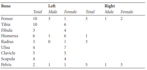
All the bone from this ‘burial’ was disarticulated. Nevertheless, it was well preserved, although some bones were fragmented. Six boxes and one bag of long bones were labelled as being from burial 1, and the results are summarised in Table 5.1.
Table 5.1—Summary of grave 1.
In addition, some vertebrae and a few bones of the hands and feet were also present.
Adult skull bones
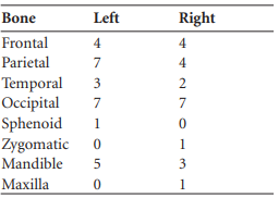
Table 5.2 presents the minimum numbers of skull bones present.
Table 5.2—Summary of all skull bones from grave 1.
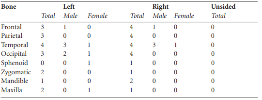
Table 5.2 summarises all the skull bones found from burial 1 and the upcast from burial 1, but since some of the skulls were labelled separately they are described below.
Skull 1: older adult, male
Virtually complete cranium although the facial bones are missing. The external occipital protuberance, mastoid processes and supraorbital ridges all indicate that it is a male individual. The sutures are fused so it is probably from an older individual. There was a slight ridge in the superior part of the metopic suture, probably caused by slight premature fusion of the suture.
Skull 2
This consisted of a right parietal bone only.
Other skull fragments
Other skull fragments were labelled as skull 3 but do not necessarily come from the same skull. They include the left and right sides of the squamous frontal bone with a small amount of both parietals near the coronal suture attached, fragments from the posterior part of a left parietal bone, fragment of occipital bone and part of a left and a right ramus of a mandible. Also present were two fragments of another squamous frontal bone and one fragment of frontal bone from a juvenile skull. In addition, there were other skull fragments found in the upcast from burial 1 and these have been included in the summary of skull fragments given above.
Juvenile bones
A few small fragments of juvenile bones were also present. These included a right frontal bone from the skull, two fragments of left humerus shaft, fragments of tibia shaft from one older and one younger juvenile, two left ilia and a right ilium and ischium, the distal two-thirds of a left femur, the medial end of a right clavicle, a left clavicle, two right ribs, one fragment of sternum and one cervical vertebra.
Skeletal pathology, ‘grave 1’
Spondylolysis had occurred in one lumbar vertebra, possibly L4. There was eburnation of the superior part of the arch. A fifth lumbar vertebra, possibly from the same individual, had spina bifida occulta, with the two unfused halves of the neural arch meeting at a pseudoarthrosis. Schmorl’s nodes were present in some thoracic and some lumbar vertebrae from at least one young adult and one older adult. There was also severe osteophytosis on one lumbar vertebra and moderate osteophytosis on another. A right acetabulum from a male had slight marginal lipping. A left tibia had a healed crack in the superior surface of the lateral condyle. The same bone had a large, smooth exostosis on the posterior surface of the bone in the midshaft. A complete sacrum was present and spina bifida occulta was present in S1. The right side of another sacrum was fused to the right ilium by ossification of the superior ligament with retention of the joint space. There was only one maxilla present but there was evidence of severe chronic infection, with sclerotic new bone formed on the internal surfaces, and on the left side there was some bone destruction, with the bone being almost destroyed to the floor of the orbit. This was probably a chronic infection of the maxillary sinuses owing to either sinusitis or chronic dental abscess.
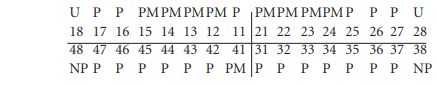
Dentition
1.
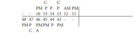
There was heavy wear on all remaining teeth, with a curved notch worn on the distal half of the lower premolar, 44, that might be due to clay pipe smoking. Calculus deposits were light on the buccal surfaces of all teeth. There was a small cavity on the mesial surface of the canine at the cervical margin and a moderate cavity on the mesial surface of the upper second premolar. The lower second molar had a moderate cavity on the distal surface at the occlusal edge of the crown. There was a large external abscess at the root of the lower first molar. There was a slight degree of alveolar recession around most teeth, with severe recession around the lower second premolar and first molar.
2.
The right canine was rotated 45˚ mesially, slightly covering the lateral incisor, 42. There was light to moderate wear on most teeth and moderate calculus deposits on the lingual surfaces of the incisor, right canine and right premolars.
3.
![]()
Dental attrition was moderate and there were light calculus deposits on the buccal surfaces of the teeth, with moderate deposits on the lingual surfaces of the roots. There was a moderate degree of alveolar recession around the first molar and slight recession around the premolars.
4.
![]()
There were also some loose teeth present, including an upper left canine and first premolar and an upper right canine. There was only slight wear and light calculus deposits on these teeth.
Grave 2, 1977:2339.1–5 A-E
1977:2339.1: middle adult, female
This consisted of the complete cranium of a middle adult female. The metopic suture was retained and there was a small benign osteoma, 15mm in diameter, in the posterior third of
the sagittal suture.
Dentition
Attrition: there was moderate wear on most teeth, with slightly heavier wear on the lower right molars.
Calculus: deposits were light on the buccal surfaces of most teeth apart from the lower premolars and first molars, where deposits were moderate; there were also moderate deposits on the lingual surfaces of the mandibular teeth.
Periodontal disease: there was a moderate degree of alveolar recession around the roots of the lower incisors, canine and first and second molars and also the upper right first and second molars, with a slight degree of recession around most of the other teeth.
1977:2339.2: older adult, probably male
This cranium was virtually complete but most of the facial bones were missing. Only the right zygomatic and part of the body of the right maxilla remained from the right side of the face.
The supraorbital ridges were not very prominent, but the external occipital protuberance and the mastoid processes were of the male type so it is probably the skull of a male. The degree of suture closure indicates that it is an older adult. Unfortunately female skulls acquire more male characteristics with old age, so it may not be safe to assume that this is the skull of a male.
1977:2339.3: adolescent, 14–16 years
This was the complete skull of a juvenile aged 14 1⁄2–15 1⁄2 years.
Non-metric traits
There were four ossicles in the left side of the lambdoid suture.
Dentition
There was no wear on the teeth, apart from moderate wear on the lower incisors. Calculus deposits were slight to moderate on the buccal surface of the upper teeth and the lingual surfaces of the lower teeth.
1977:2339.4: late middle adult, female
This consisted of the complete calvarium of a skull and the right side and a small part of the left side of a mandible. All the features of the skull were of the female type and the degree of suture closure indicated that it was probably a late middle adult.
Non-metric traits
There was an ossicle at lambda.
Dentition

There was moderate wear on the remaining molars and moderate calculus deposits on the lingual surfaces of the remaining teeth. Periodontal disease was present to a slight degree and there was linear enamel hypoplasia on the left canine.
1977:2339.5
This consisted of fragments of skull only, including a fragment of parietal bone from unknown side, a left temporal bone from a male, a right zygomatic bone, part of the left and right side of a maxilla and part of the right side of a mandible.
Skeletal pathology
There was a healed fracture of the right zygomatic bone. A direct blow usually causes this sort of fracture of the cheekbone in interpersonal violence.
Dentition
1
![]()
There was only slight wear on the teeth and light calculus deposits on the central incisor and first molar. Linear enamel hypoplasia was present on the incisors and canine. There was a slight area of abrasion on the distal corner of the central incisor on the buccal surface.
2
![]()
There was considerable alveolar recession around all the sockets.
3
![]()
The root of 47 was very shallow with considerable alveolar recession, and the sockets for 48 were completely closed. The other sockets were damaged post-mortem.
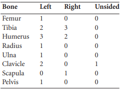
Long bones from burial 2
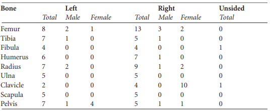
In addition to the four adult and one juvenile skulls present in burial 2, there were some long bones. These are summarised in Table 5.3. The pelvis and left femur belonged to a male Individual.
Also present were some vertebrae, a few metacarpals and hand phalanges and a few Metatarsals.
Juvenile bones
These included the proximal epiphyses of two left humeri of different sizes, the proximal third of a humerus shaft, the proximal half of an ulna shaft and the distal third probably from the same bone, the proximal half of a radius and one vertebra, a few ribs and a first metacarpal.
Table 5.3—Summary of long bones, grave 2
Skeletal pathology
The pelvis belonged to a male and there was a moderate degree of osteophytic lipping around the acetabulum. Degenerative joint disease was also present on the proximal and distal joints of a left tibia. Two of the thoracic vertebral arches had evidence for osteoarthritis, and there was severe osteophytosis and degeneration of the inferior surface of the body of a fifth lumbar vertebra. This vertebra also had some sclerotic new bone on the anterior surface of the body.
Grave 3, 1977:2341: late middle adult, female
This consisted of the upper half of a skeleton only. The cervical vertebrae were present and there were fragments of the first two thoracic vertebrae. Six ribs from the left side and four from the right side remained and there was part of the body of the sternum. Both scapulae and clavicles were present and almost complete. The left humerus was almost complete but the proximal end was fragmented. The proximal end was missing from the right humerus. Only the distal end was missing from the left ulna and the shaft of the left radius was present. The distal end of the right radius was all that survived. The left capitate, right hamate, third, fourth and fifth left metacarpals, first right metacarpal and two proximal phalanges were all that remained from the hand bones. The pelvis consisted of most of the right ilium, the right and left ischia and the upper three sacral vertebrae.
Age and sex
The sciatic notch was wide so it is probably the burial of a female. The auricular surface of the ilium indicates an age of 35–39, in the late middle adult range.
Additional bones
In addition to the burial described, there was another left scapula and a left and a right clavicle. There was also a left patella, fragments of a distal right femur and three metatarsals. There was osteoarthritis in this additional left shoulder, with eburnation of the acromial facet of the scapula and porosity of the lateral end of the left clavicle and also the right clavicle. Three skulls were found near burial 3.
1977:2341, skull A
This was an almost complete cranium but the maxillae were missing. All the morphological features indicated that it was a female skull.
Pathology
The frontal bone was very thick (1cm), and the internal surface appeared to be slightly porotic and thickened. This could possibly be the beginning of hyperostosis frontalis interna, a condition that affects post-menopausal women.
1977:2341, skull B
This was the almost complete skull of a juvenile. The frontal bone, most of both parietal bones and both temporal bones were present. The left zygomatic and the maxilla were also present. From the state of dental eruption the juvenile was aged 9–11 years. An additional left side of frontal bone from an older juvenile with retained metopic suture was present.
Dentition
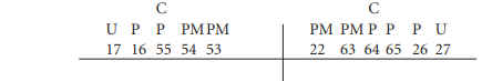
There was moderate wear on the deciduous left first molar, 64, but no wear on the other teeth. There were also moderate calculus deposits on the buccal surface of this tooth. There was a moderate caries cavity on the distal side of the crown on 64 and also a large cavity on the mesial side of the crown of the right second molar, 55.
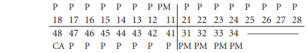
1977:2341, skull C
This consisted of fragments of both parietal bones, fragments of occipital bone, the left temporal bone, the left greater wing of sphenoid and most of the mandible. The mandible is possibly from a male individual. Part of an adult maxilla was also present and there were fragments of juvenile skull, including the parietal, occipital, left zygomatic and the right side of a mandible. Burial 3 consists of one late middle adult female, with additional skulls from one male and one juvenile also present.
Dentition
There was light wear on most teeth, with moderate wear on the first and second molars. Calculus deposits were light on the lingual surfaces of most teeth and the buccal surfaces of the molars. Linear enamel hypoplasia was present on the left incisors, both canines and right first premolar.
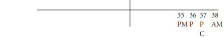
Maxilla
![]()
The sockets for the remaining teeth were very shallow.
Juvenile mandible

The juvenile was probably an adolescent, 12–18 years.
Unstratified remains, 1977:2343
The long bones found among the unstratified remains are listed in Table 5.4.
Table 5.4—Summary of long bones from unstratified remains.
At least three adults were male and three were female. A number of vertebrae, ribs, tarsals, metatarsals, metacarpals and phalanges were also present.
Adult skull bones
The minimum numbers of skull bones are presented in Table 5.5.
Table 5.5—Summary of skull bones found in unstratified remains.
All the skulls found in the unstratified material were included in Table 5.5, but it was possible to separate a few of the skulls as some of the bones were fused at the sutures.
Skull A
This consisted of a complete frontal bone, complete left and right parietal bone, most of the occipital bone, a complete left temporal bone and left greater wing of sphenoid, right zygomatic bone and part of the right side of the maxilla. All the observable features were of the female type and the sutures indicate a middle adult.
Dentition

There was light wear on the remaining teeth and light calculus deposits. There was a small cavity on the buccal side of the crown of the second molar.
Skull B
This consisted of most of a squamous occipital bone and the posterior halves of the left and right parietal bone. The sutures were obliterated so it was probably from an older adult.
Skull C
This consisted of the squamous occipital and most of the right parietal bone.
Skull D
This consisted of most of the occipital bone and part of the left and right parietal bones from around lambda and the posterior half of the sagittal suture. The occipital bone seemed to be of the male type and the sutures were all obliterated, so it may have been from an older adult.
Mandible 1

The mental eminence of this mandible appeared to be of the female type. There was severe osteoarthritis of the right mandibular condyle, with erosion of the surface and groove formation.
There was heavy wear on the molars and moderate calculus deposits. There was a moderate degree of alveolar recession around the roots of all the teeth. The second premolar and first molar appear to have been broken during life as there is a build-up of calculus over the broken enamel.
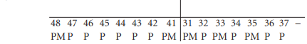
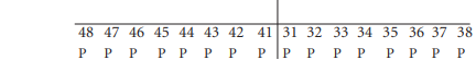
Mandible 2
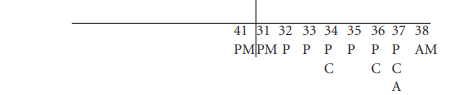
This mandible seemed to be of the male type. There was moderate wear on most teeth, with very heavy wear on the first molar. Calculus deposits were moderate on the buccal and lingual surfaces of most teeth except the premolars, where deposits were slight. There were moderatesized cavities on the mesial surfaces of the first premolar, 34, and the first molar, 36. The second molar, 37, had a large cavity, with the distal half of the crown destroyed. A large external abscess had developed at the root of this tooth. There was severe alveolar recession around the roots of the second molar and moderate recession around the other teeth. There was a pit of hypoplasia in the canine.
Mandible 3

The left third molar was impacted and was lying at an angle in the alveolus. There was light wear on most teeth, with moderate wear on the first molar. Calculus deposits were light. There was a moderate cavity on the buccal surface of the third molar.
Mandible 4

There was light wear on the third molar and light calculus deposits on the buccal, lingual and distal surfaces.
Mandible 5
There was very heavy wear on the first and second molars, and the socket for the third molars was not fully closed at the time of death. Calculus was heavy on the lingual surface of the first molar and there was a large cavity on the distal surface of the root of the second molar. There was considerable alveolar recession around the roots of the molars.
Mandible 6

There was slight wear on the remaining teeth and light calculus deposits. Linear enamel hypoplasia was present on the canine and first premolar.
Maxilla 1

There was a moderate degree of wear on the remaining teeth and light deposits on the buccal surfaces. The right canine had pits of hypoplasia.
Juvenile bones
There were a number of juvenile bones in the unstratified material. They were separated into adolescents, older juveniles, younger juveniles and infants, based on size and epiphyseal fusion.
Adolescent bones
These included the proximal third of a femur, the distal half of a femur shaft and a distal epiphysis, probably all from the one bone. There was also the distal half of a tibia shaft, a fragment of ilium with the ischium and pubis not present but had probably been just fused to the ilium, and a right clavicle.
Older juvenile
Bones present included one left and one right humerus shaft, the proximal epiphysis of a humerus, the posterior half of a right ilium, one left and one right scapula and the proximal third of a right ulna. There were also seven left and nine right ribs, four cervical, ten thoracic and one lumbar vertebrae, and a right clavicle.
Younger juvenile
Bones include the distal two-thirds of a right humerus, the proximal third and distal half of a right humerus shaft, the proximal half of a left femur shaft, the proximal third of a right femur, a proximal tibia epiphysis, distal halves of shafts of a left and a right tibia, and an almost complete matching left and right ilium. There were also four cervical vertebrae including a C2, one thoracic and two lumbar vertebrae.
Also present were matching left and right tibiae from a smaller juvenile as well as most of the shaft of another right tibia and a right ilium.
Infant
A complete right humerus from an infant was present (HuL1 63.9mm).
Juvenile skulls
There were a number of skull bones present, including fragments of squamous frontal bone and fragments of parietal bone, fragments of squamous occipital, a basal occipital and left and right lateral occipital bones, unfused, a complete left and a right temporal bone, left zygomatic bone, the right side of the maxilla, and the right side and part of the left side of the mandible. The bones appeared to be from one person.
Dentition

The crown of the upper central permanent incisors was formed, as were the crowns of the first permanent molars. The age of the juvenile is therefore 4–6 years. There was mild calculus on the lingual surfaces of the upper deciduous molars.
1977:2342 (see note 18)
This was a complete skull apart from the right side of the mandible, most of which was missing. It had been broken recently. The skull was completely black, though it is not known whether this was the result of 200 years of dust or whether it had been painted with a black substance when it was found. Bones can turn black if immersed in water, but this colour was only on the outside and the middle of the broken bone was the normal colour. All the morphological features of the skull were of the male type and the sutures were well fused, indicating that it was probably an older male. The only outstanding feature of this skull is that the bridge of the nose was very low on the face and the area at the glabella was very flat. There was a wide area between the orbits and the orbits appeared very square.
Dentition

The upper teeth were covered in the black substance and were impossible to examine. There was only slight attrition and light calculus deposits except for the buccal surfaces of the lower second premolar and first molar, where deposits were moderate. There was a slight degree of alveolar recession around the lower premolar and molars.
Summary of all bones found
The minimum number of long bones found in all the graves is summarised in Table 5.6 in order to allow an estimation of the minimum number of individuals present.
Table 5.6—Summary of long bones from all graves.
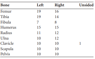
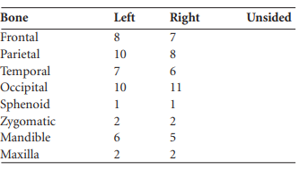
The minimum numbers of skull bones recovered are presented in Table 5.7. In addition, there were four adult skulls and one juvenile skull in burial 2. Burial 3 is assumed to be virtually complete and the female skull found in the grave probably belongs to it, but there was another male skull and two juvenile skulls in burial 3 and another juvenile skull in the unstratified material. This, together with the skull of ‘William Marshall the Younger’, gives a total of eighteen adult skulls and four juveniles. The minimum number of adults present in this site can therefore be taken from the number of left femurs and left tibia, i.e. nineteen adults. In addition, at least four juvenile skulls were present and there was also one infant present, so the minimum number of individuals present was 24. There was also an infant bone from the unstratified material.
Table 5.7—Summary of skull bones from all graves.
Summary and conclusions
The material from this site was mostly disarticulated and represented the remains of nineteen adults, four juveniles and one infant. Owing to the nature of the material it was not possible to identify individual burials, except for burial 3. The amount of information obtainable is therefore limited. It was estimated that there were at least six males and five females present among the adults, with the remainder indeterminate. Four of the males were older adults. Four of the females were middle adults, with two of these being late middle adults. It was not possible to age any of the other adults. The juvenile skulls consisted of an adolescent aged fourteen to sixteen years, one juvenile aged between twelve and eighteen years, one aged nine to eleven years and one aged four to six years. There was also one bone from a neonate.
There were 24 partial dentitions recovered, but the number of teeth present was quite low at only 140. As this is less than 20% of the teeth that would have been expected from this number of individuals, it is not possible to get a reliable indication of the dental pathology of this population. Some of the partial dentitions had no teeth remaining at all, and only four sets of dentition were close to being complete.
Nine individuals had moderate to heavy wear patterns on their teeth but nine had little or no wear. One individual had the pattern of wear associated with clay pipe-smoking, with heavy wear on his other remaining teeth. Five individuals had carious teeth, with the number per individual ranging from one to four. The total number of carious teeth was eleven (8%). The clay pipe notch indicates that this was a post-medieval population, however, and the rate of caries is low for that period. Again, it must be remembered that the number of teeth recovered was low and that this statistic is probably not reliable. Two of the individuals with caries had an associated dental abscess. Six individuals had lost teeth during life; this was only associated with caries in two individuals, but in five individuals it was associated with periodontal disease. Periodontal disease was noted in ten individuals and was severe in five cases.
Despite the large number of bones recovered, there was very little pathology noted. There was indirect evidence of early childhood illness or nutritional deficiencies in the form of enamel hypoplasia in only six individuals, although it must be remembered that the dentition was far from complete. There were a few bones with evidence for healed fractures. The right zygomatic bone of a skull in burial 2 had a healed fracture. This type of fracture is usually caused by a direct blow. One left tibia from burial 1 had a healed crack in the lateral condyle and some possible muscle damage in the calf area. A particular type of fracture known as spondylolysis was seen in the fourth lumbar vertebra of burial 1. This type of fracture is caused by strain on the vertebra, acting on a congenital weakness, causing the neural arch to become detached from the body of the vertebra. This type of fracture rarely heals and is generally quite stable. The individual must have lived with the fracture for many years, as there was eburnation of the pseudo-arthrosis between the broken arch and the rest of the vertebra. There was other evidence for instability in this vertebral column, with an adjacent vertebra, the fifth lumbar, probably from the same individual, having evidence of spina bifida occulta. A sacrum from the same ‘burial’ but not necessarily from the same individual also had spina bifida occulta at the first sacral vertebra. The only evidence for infection in this population was a chronic infection of the maxillary sinuses probably caused by either sinusitis or chronic dental abscess.
A few of the vertebrae had moderate to severe osteophytosis and osteoarthritis of the posterior joints. There were also a few with Schmorl’s nodes. Only two non-vertebral joints, the right hips from two males, had degenerative joint disease, and one older female had osteoarthritis of the right temporo-mandibular joint.
A female skull had some thickening of the internal surface of the frontal bone and this was probably the early stages of hyperostosis frontalis interna, a condition found in postmenopausal women.
In conclusion, this was a mixture of young and older adults and juveniles. The amount of information obtainable was limited by the disarticulated nature of the material but nevertheless some interesting pathologies were noted.
17. Parish of St Canice, barony of Municipal Borough of Kilkenny. SMR KK019-026021-. IGR 250287 156110.
18. A skull found in the nineteenth century, reputed to be that of William Marshall the Younger, was acquired and is registered as 1977:2342. However, there is no historical evidence to support this identification. Other unstratified bone is registered as 1977:2343.