1971:057 - TAWNAGHMORE, CO. MAYO, Mayo
County: Mayo
Site name: TAWNAGHMORE, CO. MAYO
Sites and Monuments Record No.: MA027-003
Licence number: E1125
Author: A.T. LUCAS AND ÉTIENNE RYNNE
Author/Organisation Address: —
Site type: Early Bronze Age graves
Period/Dating: —
ITM: E 494943m, N 821728m
Latitude, Longitude (decimal degrees): 54.133835, -9.607573
Introduction
In early July 1971 a short cist containing a cremation was discovered during the construction of a new road between Bangor Erris and Bellacorick in north-west Mayo. The grave was discovered at a depth of 1.3m below ground level when the capstone was hit by machinery. The site was reported to the NMI by Mayo County Council, who filled earth back into the cist and replaced the capstone pending a visit from Museum personnel. The site was visited on 12 July by Dr A.T. Lucas, accompanied by Professor Étienne Rynne, Department of Archaeology, University College Galway. This report is based on that filed by Lucas and Rynne. The human remains were analysed by Laureen Buckley.
Location (Fig. 3.124)
The site was in the townland of Tawnaghmore, near Bellacorick, north-west Co. Mayo.227 It was situated in flat bog land at an altitude of 60–90m above sea level. This is the only site listed in the SMR for this townland.

Description of site
The cist was rectangular in plan, with its long axis aligned north/south. It measured 0.495m long by 0.35m wide by 0.36m high internally (Fig. 3.125). The walls were composed of four flat slabs, with one forming each wall. The eastern slab measured 0.69m long by 0.12m thick by 0.38m high at its centre and 0.31m high at the sides (it stood in a shallow socket about 5cm

deep). The western side slab measured 0.62m long by 0.11m thick by 0.41m high with an irregular edge so that it projected about 0.1m deep into the bottom of the pit at one point. The northern slab measured 0.44m long by 0.1m thick by 0.23m high (this rested, for the most part, on top of a boulder occurring naturally in the bottom of the northern part of the pit). It had a possibly natural rebate at its eastern end, into which the northern end of the eastern slab had been set. The eastern end slab was slightly longer than the rest and extended out beyond the southern end slab. Apart from the eastern side, the walls of the cist were added to in height by a number of small slabs which were laid flat on them, one layer on the northern and southern slabs and two layers on the western slab. These stones,measuring between 0.002m and 0.003m thick, had been placed in position to ensure that the capstone rested level on the cist. The cist was sealed by a single large capstone measuring 0.86m long by 0.49m wide by 0.15m thick. There were several large boulders around and covering this capstone, most of which had been removed and thrown back later before the cist was excavated. The pit dug to receive the cist was roughly circular and flat-based, measuring 1m east/west by 0.95m north/south by 1m deep. It contained a number of large packing stones, approximately 0.2m in diameter, which supported the cist walls on the southern, eastern and western sides. All of the stones of the cist appeared to be of local sandstone.
The cist contained a large quantity of cremated bone representing an adult male and possibly one juvenile (1971:1042). No accompanying artefacts were found. The cremation had been placed on the floor of the cist. The rest of the cist fill had been disturbed by the finders, who apparently emptied a significant portion of the fill.
Comment
The human remains from this site have not been dated. In the absence of any associated finds, the site is assumed to be early Bronze Age in date based on its form and the cremated remains.
HUMAN REMAINS
LAUREEN BUCKLEY
Sample 1971:1042 consisted of 4,182 fragments of cremated bone, weighing a total of 2,095g. The bone was stained with charcoal but was basically a white colour and was efficiently cremated, with horizontal fissuring of the bone surface. There were one or two blue fragments where the bone was not so efficiently cremated.
Table 3.65—Fragmentation of bone, 1971:1042.
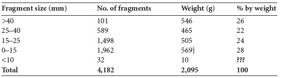
The fragmentation of the sample is shown in Table 3.65; the largest fragment measured 113mm. It can be seen that half the sample is made up of large fragments more than 25mm in length, although that means that almost half the sample consisted of medium-sized and smaller bones. The sample may have had a higher proportion of larger bones initially but was fragmented by the method of discovery and the backfilling of the cist.
Identifiable bone
A total of 1,039g (50% of the total bone) was identified (Table 3.66). This is relatively low
compared to other Bronze Age cremations but is due to the relatively small quantity of very
large fragments in the sample.
Table 3.66—Proportion of identified bone, 1971:1042.
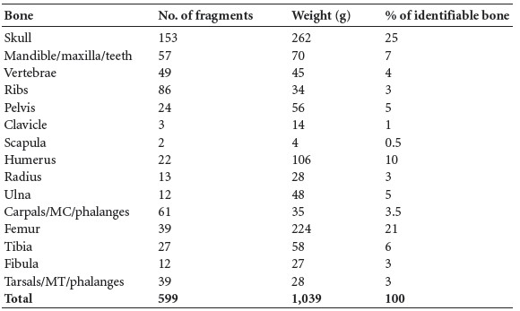
Table 3.67—Summary of identified bone, 1971:1042.
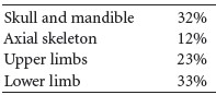
Table 3.67 summarises the main parts of the skeleton identified. It can be seen that the skull is overrepresented in this sample, as the proportion of skull is almost twice what it should be. The axial skeleton is underrepresented: only half of its normal proportion remained. The amount of upper limb was only slightly greater than it should have been and the proportion of lower limb bones was slightly smaller than it should have been.
Description of identifiable features of the bones
Skull
Part of the right frontal bone with the right orbit and supraorbital area was present. Both the supraorbital ridges and the orbital rim were of the male type. A left orbit was also present. There were several large fragments of squamous frontal and some parietal bone. Part of the left temporal bone with the anterior suture and the zygomatic arch was present and there was one petrous portion of a temporal bone. A fragment of the right zygomatic bone also survived, as well as the right greater wing of sphenoid.
Mandible and maxilla
The mandible was very complete, with the left and right sides of the body present as well as the left and right ramus. There were also two mandibular condyles. Fragments from most of the mandibular teeth were present. Most of the maxilla was also present; some teeth roots were in situ and some could be fitted into the sockets.
Dentition
The following sockets were present; where the roots of the teeth were also present, they are marked P.
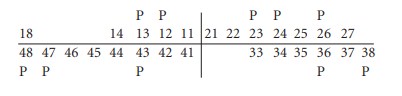
In addition, the roots of at least two molars and ten other teeth were present. Some of the tooth roots were blue internally where they had been protected by the alveolus and had not been fully cremated.
Vertebrae
The dens articulation area of the first cervical vertebra was present, as well as the dens and both superior articular surfaces of the second cervical vertebra, and five bodies and a few articular surfaces from the lower cervical vertebrae. Seven bodies of thoracic vertebrae and fragments of arches were present, and there were five lumbar bodies. One had mild and one had moderate osteophytosis. The dens area from another smaller, second cervical vertebra was also present.
Ribs
Several rib fragments from the shaft were present, and there were eight left and four right transverse processes.
Pelvis
Two fragments from two auricular surfaces were present, as well as other fragments of ilium, fragments of iliac crest and fragments of acetabula. A left ischium and the ramus of the left pubic bone were also present. The body of the first sacral vertebra, the right ala and part of the left ala were present.
Clavicle
The lateral half of a left clavicle was present, and the shaft of a right clavicle was almost complete. There was also one sternal end.
Scapula
Part of the acromion and a fragment of the right glenoid fossa were present.
Humerus
There were several shaft fragments, including fragments from the proximal and distal shafts of a left and a right bone as well as two proximal surfaces. Part of the capitulum and trochlea from a right humerus were also present.
Radius
The proximal third of one bone was present and the joint end was fused. The distal end of a right radius with fused joint end was present. There were also other fragments of proximal and distal shaft.
Ulna
There were several fragments from both the proximal and distal halves of shaft. There was also a fragment from the proximal end of a juvenile bone with unfused olecranon.
Carpals, metacarpals and phalanges
A right scaphoid, right lunate, right trapezoid and left hamate were present and there were ten metacarpal shafts, including two first metacarpals, two second metacarpals, two third metacarpals and a fifth metacarpal. There were ten proximal, eight middle and nine distal hand phalanges, most of which were complete.
Femur
Several fragments of shaft were present and some were very thick. The posterior part of the proximal shaft of a right femur was almost complete. Some fragments were from the middle shaft and the linea aspera was visible. There were fragments of two distal and two proximal articular surfaces.
Tibia
All the fragments were shaft fragments, including several from the proximal end of the shaft. One nutrient foramen was visible.
Fibula
Fragments of shaft and the left and right distal articular surfaces were present.
Tarsals, metatarsals and phalanges
A left and a right calcaneum, two fragments of talus bone, one cuboid and three cuneiform bones remained from the tarsals. Four metatarsal shafts and five heads, including the heads of two first metatarsals, were present, and there were four proximal and one middle phalanx and one sesamoid bone.
Minimum number of individuals
The majority of the remains appear to be from one individual, although there were two fragments that could have belonged to a juvenile.
Summary and conclusions
This sample consisted of the remains of one adult male individual and possibly one juvenile. Most of the sample appeared to be from the adult male and the bones were very thick and heavy. In fact, only two bone fragments may have been from a juvenile. Most of the bone was white, indicating very efficient cremation. It was not possible to estimate whether the bone had been fragmented as part of the cremation ritual, since it had been disturbed before it was excavated and this may have increased the fragmentation of the sample. Almost half the sample consisted of large fragments, however, so it is unlikely that it had been deliberately crushed. Most skeletal elements were recovered, indicating careful collection of bone from the funeral pyre. The skull seems to have been collected at the expense of the axial skeleton, since the proportion of axial skeleton is very low and the proportion of skull is high. The disturbance of the site may have affected the amount of juvenile bone that was collected.
The adult individual appears to be male and there was evidence of degenerative joint disease of the spine. It is not possible to age accurately by this alone but it could indicate that he was middle-aged or older.
227. Parish of Kilcommon, barony of Erris. MA027-003——. IGR 094970 321720.