1967:009 - TOWNPARKS SOUTH, EMMET STREET, TRIM, CO. MEATH, Meath
County: Meath
Site name: TOWNPARKS SOUTH, EMMET STREET, TRIM, CO. MEATH
Sites and Monuments Record No.: N/A
Licence number: E1143
Author: ELLEN PRENDERGAST
Author/Organisation Address: —
Site type: Graves of indeterminate date
Period/Dating: —
ITM: E 679934m, N 756541m
Latitude, Longitude (decimal degrees): 53.552704, -6.793685
Introduction
In August 1967 human remains were uncovered during digging operations at Emmet Street in Trim, Co. Meath. The burials were uncovered in foundation trenches at a depth of 0.45m below ground level. Loose bones that had been taken out of the filling of the trench were collected. The site was reported to An Garda Síochána at Trim, who informed the NMI. A rescue excavation was undertaken by Ellen Prendergast on 6 September 1967. This report is based on Prendergast’s plan and account of the site. The human remains were analysed by Laureen Buckley.
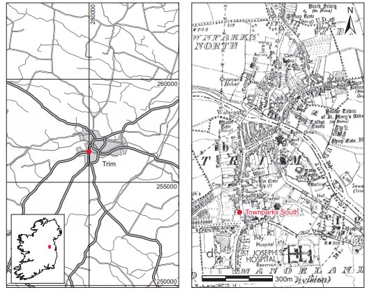
Location (Fig. 6.46)
The site was in the townland of Townparks South on the south side of Trim town,79 on the grounds of the new Vocational School adjoining the west side of Emmet Street, near the junction with Patrick Street. The level of this area had been greatly lowered in recent years, and up to about sixteen years prior to Prendergast’s visit a row of thatched cottages stood there. Such a row of buildings is shown on the 1837 edition of the OS 6in. map. Human remains were also uncovered on Castle Street in 1951 and investigated by P.J. Hartnett (this volume, pp 403–5).
Description of site
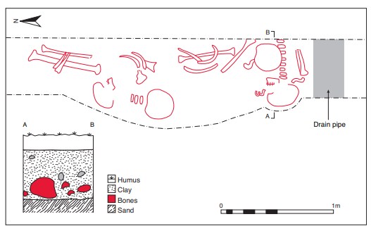
The trench in which the remains were exposed ran north/south and was bounded to the east by the cemented footpath.80 Several complete skeletons and portions of others were uncovered in situ. These lay on a sandy layer underneath mixed clay and stone at a depth of 0.5m below ground level, over which further burials had been inserted (Fig. 6.47). The orientation of the graves was not always evident but did not seem to be uniform, lying on both an east/west and a north/south axis. There was no evidence for any type of stone lining around the burials, and the individual grave-cuts do not seem to have been identifiable.
All of the in situ burials were extended inhumations, as mentioned above, aligned east/west and north/south. The minimum number of individuals present was five; three were identified as male and one as female. All of the remains were those of adults, two of whom appeared to be younger adults. Scattered stones embedded in the lower portion of the fill had crushed some of the other bones. Further portions of some burials extended to the east under the street and again to the west into the garden surrounding the new school. Four skulls and portions of several more skeletons (2010:87) were recovered.
Comment
There is no evidence to indicate the date of the burials, as no grave-goods were found with the bodies and no diagnostic grave structures were found. The skeletons were obviously deposited before the thatched cottages shown on the first edition OS 6in. sheet were built. These cottages were demolished around 1951. The ‘New Gaol’ building is shown on the first edition 6in. sheet, just east of the burials, and on the second edition the Fever Hospital, Graveyard and Union Workhouse are marked to the east of the New Gaol.
HUMAN REMAINS
LAUREEN BUCKLEY
Introduction
2010:87—the burials are described below under the labels given by Ellen Prendergast.
Skeleton 1
This consisted of a skull and a group of infracranial bones. As there was more than one individual present and the photograph does not appear to show an individual, they are treated here as disarticulated bones. The skull was fragmented but almost complete, with most of the frontal, complete left and right parietal, both temporal bones and the almost complete occipital bone present. The sphenoid, both zygomatic bones and most of the maxilla were also present. The mandible was virtually complete.
Age and sex
The external occipital protuberance was of the female type, but the right mastoid process, the supraorbital ridges, orbital rim and mental eminence of the mandible were all of the male type. As there were more male characteristics than female, it is very likely that this was a male.
Non-metric traits
The metopic suture was retained.
Skeletal pathology
The frontal and parietal bones of the skull were very porotic but there was no enlargement of the diploe.
Dentition
Ante-mortem loss: four teeth had been lost during life, the upper left first and second molars and the lower right first and second molars. The sockets for the upper molars were not completely healed.
Attrition: there was moderate wear on the incisors and first molars and light wear on the other teeth.
Calculus: there was a moderate degree of calculus on the buccal surfaces of the central incisors and on the upper premolars. Deposits were heavy on the buccal surfaces of the lower lateral incisors and on the lingual surfaces of all the lower incisors. The buccal and lingual surface of the remaining teeth had light calculus deposits.
Caries: there was a large cavity on the mesial surface of the upper right first molar, 16, with most of the mesial-buccal quadrant destroyed. The upper left second premolar had a moderate-sized cavity on the buccal surface of the crown at the cervical margin. The mesial surface of the upper left third molar had a large cavity that had destroyed most of the crown.
Periodontal disease:
there was a slight degree of alveolar recession around the roots of most teeth but recession was moderate around the roots of the upper left premolars and lower left molars. There was considerable recession around the roots of the upper left first and second molars and the lower right first and second molars.
Hypoplasia: linear enamel hypoplasia was present on the incisors, canines, upper right premolars and lower right second premolar.
Hypercementosis: in an attempt to hold the tooth in the socket while the alveolus was receding there was hypercementosis of the root of the upper right first molar.
Periostitis: the destruction of the alveolus around the roots of the upper left first and second molars appeared to be due to infection. The bone was very porotic and periostitis was evident. Some sclerotic new bone was visible on the internal surface of the maxilla and in the lingual root socket of the second molar.
Infracranial bone
The bones present included seven cervical vertebrae and five upper thoracic vertebrae. Eight ribs from the left side and four from the right side were present, and the manubrium of the sternum was present but incomplete. There were two almost complete right scapulae and the acromion and lateral border of another right scapula. A complete right clavicle and parts of two other right clavicles were also present. The proximal end of a right humerus, the proximal two-thirds of a left radius and the proximal third of another left radius remained from the arm bones. There was a small fragment of left ilium.
Age and sex
A lot of the bone came from an adolescent or younger adult. The complete clavicle had unfused epiphyses, the acromial epiphysis of a right scapula was unfused, and the iliac crest of the ilium was only partially fused. The proximal epiphysis of the humerus was also unfused. All the vertebrae and the ribs were from a young person.
Skull 2: young adult, male
This was the very fragmented but almost complete skull of a young individual. The frontal bone, both parietal bones, both temporal bones and the occipital bone were present and almost (but not quite) complete. The sphenoid bone was present and the left zygomatic bone was complete but only a fragment remained from the right zygomatic bone. The maxilla was almost complete and the mandible was complete. The first two cervical vertebrae were also present.
Age and sex
The external occipital protuberance, mastoid processes, supraorbital ridges, orbital rims and mental eminence all indicated that this was a male. The sutures were open and there was very little wear on the teeth, so it was probably the skull of a young individual.
Dentition
Anomalies: the lower left deciduous second molar, 75, had been retained and the permanent second premolar had not developed. The lower right deciduous molar had also been retained.
Attrition: there was light wear on the incisors and first molars and virtually no wear on the other teeth except the deciduous right molar, where there was considerable attrition.
Calculus: there was a moderate degree of calculus on the buccal surfaces of the central incisors, upper right canine and first premolar. There were also moderate deposits on the buccal surfaces of the lower right lateral incisor and canine and the lingual surfaces of the lower right premolars and first and second molars. Deposits were light on the lingual surfaces of the upper incisors and right canine and were moderate on the lingual surfaces of the upper left lateral incisor and canine.
There were considerable deposits of calculus on the buccal surfaces of the upper left canine, premolars and molars. The upper left premolars and molars also had considerable deposits of calculus on their occlusal and lingual surfaces. The lower central incisors, left premolars and molars had considerable deposits on their buccal surfaces and the premolars and molars also had considerable deposits on their occlusal and lingual surfaces.
The pattern of calculus deposits on the biting surfaces of the teeth in the left side of the jaw suggests that this side of the jaw was painful and was not used for chewing food. The pain may have been caused by caries.
Caries: there was a large cavity on the occlusal surface of the upper left first molar, 26, with most of the crown destroyed. The upper left second premolar had a small cavity on the distal surface of the crown at the cervical margin. The buccal surface of the upper right third molar had a small cavity at the cervical margin.
Periodontal disease: there was a slight degree of alveolar recession around the roots of most teeth.
Hypoplasia: linear enamel hypoplasia was present on the upper left canine only.
Skull 3: middle adult, male
This skull was fragmented and incomplete. The frontal bone was present but the supraorbital area was missing. Both parietal bones and the left temporal bone were complete and the occipital bone was also complete. The left zygomatic bone and the right ramus of the mandible were present. The first and second cervical vertebrae and an arch from a thoracic vertebra were also present.
Age and sex
The external occipital protuberance was more like the female type but the left mastoid process and posterior part of the zygomatic arch were of the male type. It is probable that this was a male skeleton and from the suture fusion it was probably a middle adult, 26–44 years.
Dentition
Only one tooth, the right mandibular third molar, 48, was present along with its socket. There was no wear on the tooth and there was a small cavity on the buccal surface at the cervical margin.
Skull 4: adult, female
This consisted of a skull and long bones. The skull was fragmented but virtually complete, with the frontal, parietal and occipital bone complete, both temporal bones almost complete and the left zygomatic bone, maxilla and mandible complete.
Age and sex
The supraorbital ridges and the external occipital protuberance were all of the female type and the mastoids were probably but not definitely female. The skull sutures were mainly open.
Dentition
Attrition: there was slight wear on the upper first molars and virtually no wear on any other teeth.
Calculus: there was a moderate degree of calculus on the buccal surfaces of the incisors, canines and upper first premolars. Deposits were also moderate on the lingual surfaces of the lower incisors, all the teeth in the left side of the mandible and the lower right first and second molars. The buccal and lingual surface of the remaining teeth had light calculus deposits.
Hypoplasia: linear enamel hypoplasia was present on the upper incisors, canines, premolars and first right molar, as well as the lower canines, first premolars and right second premolar. There were also pits of hypoplasia on the lower left second and third molars.
Disarticulated long bones
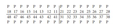
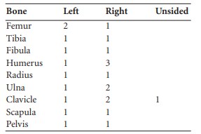
The minimum numbers of long bones present are listed in Table 6.16. Also present were three cervical vertebrae, C1, C2 and C3, five thoracic vertebrae and one lumbar vertebra. These all seemed to be from one younger adult individual as the epiphyses of the vertebral bodies were just fused. The first sacral vertebra was almost complete and there were five ribs from the left side and six from the right, also from a younger adult. There were also a few carpals, metacarpals and phalanges present, as well as a few tarsal bones. The left ilium was almost complete and was from a female individual but there was also a pubic bone from a young male. An additional bag of bone from this skeleton contained nine lower cervical vertebrae (including two C7s), fourteen thoracic vertebrae from a young adult, four lumbar vertebrae and one sacrum. There were also fourteen left and eleven right ribs and some tarsals, metatarsals, carpals, metacarpals and phalanges. From the quantity of bone recovered, there were at least three individuals present. Two of them were probably younger adults and at least one was male and one was female.
Table 6.16—Minimum numbers of disarticulated long bones present.
Skeletal pathology
A complete right radius had a healed Colles fracture in the distal third of the bone. The bone was overlapped and had healed well, with a rough callus around the site of the fracture. The distal third of the right ulna was present and also bore evidence of a fracture with considerable overlap of the bone. Both of these fractures could have occurred at the same time and would typically be caused by a fall on an outstretched hand.
Another right ulna was distorted in the distal third of the bone, with the distal end slightly flattened and bent out of shape. There is no callus visible but this may be a possible greenstick fracture that occurred during childhood, with the bone being bent out of shape rather than being snapped in two.
Bone group A
This consisted of the proximal end of a right tibia, a complete right radius, three left ribs and one right rib.
Bone group B
This group included a complete left humerus, complete left and right radii, a complete left ulna and almost complete right ulna, the distal two-thirds of a left femur, the proximal twothirds of a right femur, almost complete left and right tibiae and one fibula shaft.
Also present were fragments of two scapulae, two thoracic and five lumbar vertebrae and a complete sacrum. A complete pelvis from a young male was also present, as well as some carpals, metacarpals and phalanges.
Skeletal pathology
The left tibia had healed periostitis in the distal half of the shaft on the medial surface. A fragmented distal end of a right femur had osteochondritis on the medial condyle. The lesion was 11mm in length. There was also a defect of the area for the medial head of gastrocnemius.
Scattered bone
This bag contained at least one skull, with part of a right frontal, the orbital area of a left frontal, the posterior part of a left parietal bone, fragments of sphenoid bone, the basilar occipital bone, the left temporal bone and an almost complete maxilla and mandible. The brow ridges seemed to be male and the mastoid processes seemed to be female, so there may have been two skulls present. Other bones present included the proximal end of a right femur, the distal end of a left tibia, the distal two-thirds of a left fibula and the distal end of a right fibula, the distal two-thirds of a left humerus and an unsided proximal end of humerus. There were also two left and one right clavicles, two left and one right scapulae, two cervical, ten thoracic and one lumbar vertebrae from one young individual, a few ribs and some metatarsals and foot phalanges.
Dentition
Attrition: there was slight wear on the upper first molars and virtually no wear on any other teeth.
Calculus: deposits were slight on the buccal surfaces of most of the upper teeth and on the lingual surfaces of the lower right premolars and first and second molars. There were moderate deposits on the buccal surfaces of the upper left premolars and first molar and on the lingual surfaces of the lower right incisors and canine and lower left second premolar and first and second molars.
Caries: there was a very small cavity on the buccal surface of the lower left third molar at the cervical margin.
Periodontal disease: there was a slight degree of alveolar recession around the roots of the upper premolars and first and second molars and the lower second premolars and right first and second molars. There was moderate recession around the roots of the lower left molars.
Hypoplasia: linear enamel hypoplasia was present on the upper incisors, canines, premolars and first right molar, as well as the lower canines, first premolars and right second premolar. There were also pits of hypoplasia on the lower left second and third molars.
Summary of bone found on entire site
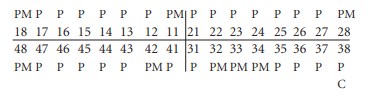
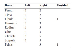
The minimum numbers of bones found are listed in Table 6.17.
In addition, there were five skulls present. The minimum number of individuals present was therefore five. They all appeared to be adult and at least three males and one female were present. There were a lot of bones from younger individuals and it is estimated that at least two of the adults were younger adults. The only pathology noted was trauma. One individual had suffered a fractured right radius and ulna as a result of a fall, while another individual had probably suffered the same injury during childhood. One adult had evidence of trauma to the right knee.
Three of the adults had caries in their teeth and it was severe in two cases, with one individual having suffered tooth loss probably as a result of caries. Another individual had suffered so much pain from caries that he had stopped chewing on one side of his jaw.
Table 6.17—Minimum numbers of bones present from site.
79. Parish of Trim, barony of Moyfenrath Lower. OS 6in. sheet 36. IGR 280001 256520.
80. The graves are described as a unit here according to Prendergast’s report.