1958:008 - BALLEALLY WEST, CO. DUBLIN, Dublin
County: Dublin
Site name: BALLEALLY WEST, CO. DUBLIN
Sites and Monuments Record No.: SMR DU008-019
Licence number: E1066
Author: BREANDÁN Ó RÍORDÁIN AND ÉTIENNE RYNNE
Author/Organisation Address: —
Site type: Iron Age and early medieval graves, c. 300 BCc. AD 1200
Period/Dating: —
ITM: E 721344m, N 753020m
Latitude, Longitude (decimal degrees): 53.513141, -6.170407
Introduction
In 1958 land reclamation work near Lusk, Co. Dublin, resulted in the discovery and damage of a burial mound containing a number of partially stone-lined graves. The mound, which had measured 25m in diameter and approximately 2m in height, had been removed to within 0.5m of ground level. Human bones and stone slabs were only noticed when the area was being ploughed subsequent to the bulldozing activity and, according to the landowner, appeared mainly in the area which would have been under the southern portion of the mound. Some of the slabs had been removed and placed at the edge of the field. On 18 February the landowner, Mr Thomas Leonard, reported the discovery to the local Garda Síochána, who informed the NMI. The site was investigated by Breandán Ó Ríordáin and Étienne Rynne. A small area formerly covered by the southern portion of the mound was selected for investigation.16 Excavation revealed three graves, two of which were partially stone-protected. This report is based on Ó Ríordáin and Rynne’s excavation report.
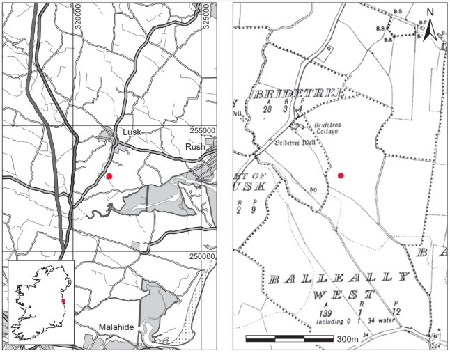
Location (Fig. 4.7)
The site was in the townland of Balleally West, just over 1km south of Lusk village, north-east Co. Dublin.17 The mound had been constructed on a natural hill between 30m and 60m above sea level and just north of Rogerstown Estuary.
Description of site
Grave 118
The cist was rectangular in outline, with its long axis running east/west. Internally it measured 1.4m long by 0.4m wide by 0.2m high (Fig. 4.8). When excavated, the grave was found to be formed of three main slabs, two long side slabs (at the northern and southern sides) and one short end slab (at the eastern end). The western end of the grave was not closed and therefore the grave may originally have been longer than 1.4m, although as far as could be established the long side stones (north and south) of the cist did not extend beyond this point. The slabs were of shale and appeared to have been regular in form, measuring approximately 0.15m thick. The eastern end stone was 0.33m long and the side stones were a maximum of 1.3m long. The grave had been dug into a layer below what appeared to be the old ground level, and the tops of the side and end stones lay c. 0.1m below old ground level. There was no evidence for packing stones surrounding the cist slabs, nor were any covering stones noted. The gravepit does not seem to have been identified. The floor of the grave was not paved.
The grave contained the disarticulated remains of two adult males (1958:37.1) and no accompanying artefacts were noted. The bones lay in a heap at the approximate centre of the cist and their disposition suggested that the individuals had been ‘unceremoniously deposited in the grave, possibly after disarticulation of the limbs’. Samples from each of the individuals identified here were submitted for radiocarbon dating.

Sample 1958:37.1A yielded a date of 1495±40 BP, which calibrates to 436–646.19 Sample 1958:37.1B yielded a date of 1545±45 BP, which calibrates to 418–605,20 indicating that the two burials were approximately coeval.
Grave 2
This was situated immediately east of grave 1 (Fig. 4.8) at the same level as the tops of the cist slabs of grave 1, approximately 0.1m below original ground level. It was a simple pit aligned east/west. No slabs were noted in association with this grave.
The grave contained the extended inhumation of an adult male (1958:37.2) aged over 25 years without any accompanying artefacts. The skeleton lay in a supine position with the head to the west, the arms extended by the sides and the legs parallel and slightly apart, perhaps indicating that the body had not been wrapped in a shroud or winding-sheet. Part of the skull and some of the lower body bones had been damaged by the workmen at the site. All major bones appear to have been present, however. A sample of the remains was submitted for radiocarbon dating and yielded a date of 1195±40 BP, which calibrates to 692–962.21
Grave 3
This partially slab-lined grave lay at the same level as grave 1, 0.75m to the east of the eastern end stone of grave 1 and just north of grave 2 (Fig. 4.8). It was rectangular in plan with its long axis aligned east/west. Internally it measured 2.1m long by 0.45m wide by 0.3m high. The grave consisted of five slabs, one each at the east and west ends, one long slab and one short at the south side and a short slab at the north-west corner. All of the structural slabs with the exception of that at the eastern end were of decayed shale-like stone. The exception was a hard limestone slab measuring 0.3m long by 0.15m thick. The side slab at the south was 1.85m long by 0.15m thick. The western end stone was 0.45m long by 0.15m thick. There was no evidence for packing stones outside the cist. One capstone or possibly a collapsed lintel was located near the western end of the grave, lying directly on the burials. This measured 0.7m long by 0.4m wide.
The grave contained two extended inhumation burials aligned west/east. The burials lay side by side in a supine position with the hands by the sides and the legs extended. No artefacts were found associated with the burials. The remains from this grave were not retained.
Comment
The radiocarbon dates returned for two of the graves indicate a broad early medieval date for this cemetery. Both long cists and unlined graves were in use, something that has been noted from other cemeteries in early medieval Ireland (O’Brien 2003, 67). There do not appear to be any ecclesiastical associations at this particular site, but the early medieval foundation of Lusk is located just over 1km to the north.
HUMAN REMAINS
LAUREEN BUCKLEY
Burials from grave 1 (1958:37.1)
This grave contained the disarticulated remains of at least two individuals. It was possible to separate the individual skeletons on the basis of size, preservation and colour of the bones. The bones of both skeletons were poorly preserved and fragmentary, although skeleton 1 was slightly better preserved than skeleton 2.
Skeleton 1: older adult male, 170cm
The skull was fragmented and very decayed but part of the occipital bone, most of both parietal bones and a complete frontal bone were present. There was also part of the left temporal bone as well as the anterior part of the mandible. There was no vertebral column remaining and only two ribs from the right side were present, although there were two fragments of bone from the left ribs.
Both scapulae were present and almost complete. Only the proximal end of the left humerus survived from the left arm. Most of the right humerus apart from the proximal end was present and the right radius and ulna were complete. Only the complete second and distal half of the third metacarpals survived from the left hand, with the right hamate the only bone surviving from the right hand.
The right ilium and both ischia were present from the pelvis but were incomplete.
The left femur was complete and only the distal end was missing from the left tibia. The proximal half of the right femur remained and most of the shaft and proximal end of the right tibia survived. The distal third and part of the proximal shaft remained from the left fibula but the right fibula was virtually complete.
The foot bones consisted of the complete left talus, the left first and second metatarsals and incomplete right third and fourth metatarsals.
Age and sex
The right sciatic notch was the only morphological feature of the pelvis remaining and it was of the male type. The external occipital protuberance, the mastoid process, the supraorbital ridges and orbital rims were also of the male type.
The degeneration of the auricular surface of the right ilium indicated an age of over 50 years, although the skull sutures were not totally obliterated. Overall, given the degeneration of the joints of this individual and the auricular surface of the ilium, it is probably safe to conclude that this was an older adult male, aged more than 45 years at the time of death.
Stature
Using the regression equations of Trotter and Gleser (1952; 1958) with the length of the femur, the living stature of the individual was estimated at 170cm.
Skeletal pathology
Cribra orbitalia was present to a moderate degree in the left orbit.
There was a large, circular lesion of osteochondritis on the medial condyle of the left femur. The lesion measured 13.4mm in diameter and was smooth-bottomed with a lipped edge. There was indication of degenerative joint disease on some joints, including the left shoulder, with moderate osteophytic lipping of the posterior edge of the left glenoid fossa of the scapula and mild lipping of the proximal joint surface of the left humerus. The humerus head was damaged but there also appeared to be surface osteophytes near the superior edge. The right shoulder was mildly affected, with lipping around the right glenoid fossa. There was moderate lipping around the proximal and distal joint surfaces of the right ulna.
The right hip was mildly affected, with lipping around the right acetabulum and mild lipping on the proximal end of the right femur. The left femur had moderate osteophytic lipping around the proximal joint surface. In the right knee joint there were surface osteophytes on the lateral condyle of the right tibia.
The left ankle also had some mild degeneration, with mild lipping of the distal surface of the left fibula and mild lipping of the inferior and lateral articular surfaces of the left talus.
Dental pathology

Anomalies: there were large mandibular tori on both sides of the mandible around the areas of the premolars and first molar.
Attrition: there was very heavy attrition on the first and second molars that remained. Moderate wear was also present on the incisors and premolars.
Calculus: light deposits were present on the lingual surfaces of the lateral incisor and left second premolar and there were also light deposits on the buccal surface of the second premolar.
Caries: there was a moderate cavity on the distal surface of the left second molar at the cervical margin.
Periodontal disease: there was slight alveolar recession around most of the observable sockets but recession was moderate around the molar sockets.
Skeleton 2: adult male
This skeleton was very incomplete and consisted mainly of fragments of very decayed long bones. Only the anterior part of the mandible survived from the skull and there was no vertebral column or ribs present. The glenoid area of the left scapula, the distal half of the left humerus, the proximal end of the ulna and part of the shaft of the radius were all that remained from the left arm. The right arm consisted of part of the shaft and distal third of the humerus and the right ulna with only the distal end missing. A fragment of the posterior surface of the left tibia and the distal two-thirds of the left fibula as well as both first metatarsals were the only bones present from the lower half of the skeleton.
Age and sex
The mandible appeared to be of the male type and the size of the bones was also probably indicative of a male individual. With so little bone remaining, however, there were no features present that could be used to estimate the age at death.
Skeletal pathology
Osteoarthritis was present at the left elbow, with eburnation on the capitulum of the left humerus, especially on the medial side close to the trochlea. The trochlea of this bone also had moderate lipping on the posterior surface and there was moderate lipping on the superior part of the olecranon of the left ulna. The olecranon fossa on the left humerus was almost filled in by new bone deposit. This may have been a result of trauma to the bone, as it does not usually occur owing to osteoarthritis on its own. Osteoarthritis may be exacerbated or develop at an earlier age as a result of trauma. Mild lipping was also present on the trochlea of the right humerus.
Dentition

Most of the sockets were incomplete as part of the anterior surface of the mandible was missing.
Burial from cist 2 (1958:37.2): skeleton 3
This burial consisted of a skull and some neck vertebrae only.
The back of the skull was virtually complete but most of the facial bones were missing. The occipital, both parietal and both temporal bones were complete and the sphenoid bone was almost complete. The most superior part of the frontal bone was present but the orbits and supraorbital areas were missing. Only a fragment of the right side of the maxilla remained and most of the right side of the mandible was present. The first and second cervical vertebrae were present and complete.
Age and sex
The external occipital protuberance, mastoid processes and the posterior zygomatic arches of the temporal bones as well as the mental eminence of the mandible were all of the male type.
The basio-sphenoidal symphysis was fused so the individual was probably over 25 years of age, but as suture fusion is not a reliable indicator of age it is impossible to be more specific.
Skeletal pathology
There was moderate osteophytic lipping around the dens on the second cervical vertebra and on the articular surface for the dens on the first cervical vertebra.
Dentition

Attrition: there was heavy wear on the first molars, with exposure of the pulp cavity in the upper first molar and moderate wear on the other teeth.
Calculus: there were light deposits on the lingual surfaces of the lower lateral incisor, canine and second premolar, and moderate deposits on the lingual surface of the lower first premolar and lower third molar. There were also moderate deposits on the buccal and distal surface of the third molar.
Periodontal disease: there was slight alveolar recession around the roots of the premolars and lower first molar.
Hypoplasia: linear hypoplasia was present on the lower incisors and pits of hypoplasia were present on the lower second molar.
Summary and conclusions
A total of three adult individuals were present in this skeletal collection. All were males and at least one was an older adult. There was insufficient bone remaining from the other two skeletons to get a reliable indication of age of death. Stature could only be estimated for one skeleton and it was 170cm.
There was evidence that the individuals had suffered from nutritional deficiency or acute illness during childhood. The older adult had cribra orbitalia, which may be caused by iron-deficiency anaemia, and another individual had hypoplasia on the teeth enamel that had occurred in early childhood.
Degenerative joint disease was present in all the individuals. The more complete skeleton of the older male had degeneration of both shoulder joints, the right elbow and wrist, both hip joints, the right knee and the left ankle.
The second individual from grave 1 had severe osteoarthritis of the left elbow that may have been linked to trauma of the elbow. This individual also had degeneration of the right elbow joint.
Moderate degenerative changes were present in the cervical vertebrae of the individual from grave 2. It is unfortunate that this individual was so incomplete, as the full extent of the degenerative changes in the spinal column could not be observed.
Evidence of trauma was also found in skeleton 1 from grave 1 in the form of osteochondritis on the medial condyle of the left femur. This lesion, where damage to the cartilage causes a small area of bone death on the joint surface, is thought to be caused by trauma.
Evidence from the teeth of the two individuals with dentition remaining indicates heavy wear owing to the coarseness of food in the diet and periodontal disease with moderate exposure of the tooth roots, and one individual having a cavity at the cervical margin.
16. The excavation was carried out with assistance from the landowner, Mr Leonard, and his workers.
17.Parish of Lusk, barony of Balrothery East. SMR DU008-019——. IGR 321420 252998.
18.The excavators labelled the graves alphabetically but for the purposes of clarity this volume has labelled all graves numerically. Thus grave A in the original report is relabelled grave 1 and grave B is grave 2, etc.
19. GrA-24576.
20. GrA-24465.
21. GrA-24466