1957:011 - LOUGHLINSTOWN, CO. DUBLIN, Dublin
County: Dublin
Site name: LOUGHLINSTOWN, CO. DUBLIN
Sites and Monuments Record No.: SMR DU026-119
Licence number: E1067
Author: ELLEN PRENDERGAST AND RAGHNALL Ó FLOINN
Author/Organisation Address: —
Site type: Iron Age and early medieval graves, c. 300 BCc. AD 1200
Period/Dating: —
ITM: E 723651m, N 724507m
Latitude, Longitude (decimal degrees): 53.256497, -6.146798
Introduction
In March 1957 a lintel grave containing an inhumation was discovered in the garden of a dwelling house at Loughlinstown, Co. Dublin. Years previously, when the landowners, the Delap family, had been digging a refuse pit in the garden, an upright flag was noticed at the side of the pit, approximately 0.65m below ground level. It had not been disturbed until some time later, in 1957, when the slab was removed and part of a cist and some human bone were revealed. Before it could be investigated, another side stone and a lintel had collapsed and part of a human skull and femur had been disturbed. These were replaced in the grave and the landowners reported the find to the NMI on the following day, 4 March 1957. A rescue excavation was undertaken by Ellen Prendergast. The human remains were examined by Professor E.J. Keenan. In July 1991 an unprotected inhumation burial was discovered in the rootball of a tree in the garden of the dwelling house. This was excavated by Raghnall Ó Floinn, and the human remains were examined by Laureen Buckley.

Location (Fig. 4.9)
The site was in the townland of Loughlinstown, south-east Co. Dublin, close to the border with County Wicklow.22 It was approximately 1km south-east of Cabinteely village, between c. 30m and 60m above sea level. The Delaps’ house was built in 1938 and, together with the garden, was located on a high shelf, the ground falling away steeply to the west along a terrace just behind the house. Before any of the more recent houses were built the area was known locally as ‘Moat Field’ or ‘Raheen’. Locals informed Prendergast that human bone had been unearthed and reinterred when the houses were being built. In 1998 the entire cemetery was excavated in advance of the construction of a motorway. Excavation revealed an enclosed cemetery of early medieval date containing at least 1,553 burials, dating broadly from the fifth to the twelfth century (Conway 2000).
Description of site
Grave 1 (1957)
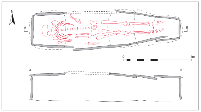
The cist is described as coffin-shaped and was aligned west/east. Internally, it measured 2.04m long by 0.47m wide (at centre) by 0.4m high (Fig. 4.10). It was constructed of mica schist flagstones set on edge, some overlapping slightly on the south side. Four slabs formed the south side and three the north (one of which had collapsed prior to Prendergast’s excavation), and the cist ends were each closed by a single slab. The side slabs were regular in outline,measuring a maximum of 0.05m thick and 0.96m long. The cist was covered with five lintels, four of which were still in place. These overlapped and would have completely covered the grave. There was no evidence of packing stones around the cist, and the pit dug to receive the cist was not located. The floor of the cist was formed of compacted clay.
The grave contained an extended inhumation of an adult male and some additional disarticulated human bone (1957:350). No associated artefacts were recovered. When excavated, the cist was filled with loose soil and compost that had fallen between the slabs. The articulated skeleton, an adult male, lay fully extended in a supine position with the head at the west end and the feet at the east. The forearms were slightly extended towards the pelvis. Most of the bones of this individual were present and his age was estimated as 25 years. Some bones other than those of the main burial were found. These consisted of the os coxae, left humerus and ulna and almost complete radius of a female.
Grave 2 (1991)
The area had been severely disturbed owing to the circumstances surrounding the discovery of the remains, and it was therefore not possible to trace the outline of the grave-cut. It is probable that the grave was a simple unprotected pit, as there was no evidence for grave-slabs. The pit was aligned west/east.
The grave contained an extended inhumation lying west/east (1991:40). The remains were analysed by Buckley and found to be those of an adult male. As the burial had been exposed with the underside of the lower half of the skeleton visible and the upper half still incorporated in the rootball with the vertebrae exposed in side view, it was not possible to record in plan the positions of the bones (Pl. 76). It appeared to consist of an extended inhumation, with the arms bent and crossed, the left over the right. It was not possible to recover the lower foot bones as these extended under the rootball, although animal bone was noticed in the vicinity of the feet. The remains of a second articulated skeleton were noticed close to the right elbow of this skeleton, but this was too firmly rooted to be excavated. A probable third burial, evidenced by the presence of a long bone, was visible under the root of another tree, some 1.5m east of the burial excavated here. A cut antler burr was also found in the vicinity of the second tree.
Comment
It is likely that the two graves discussed here are associated with the large early medieval cemetery and associated enclosures excavated by Malachy Conway in 1998 (Conway 2000).
HUMAN REMAINS
Grave 1 (1957:350)
J.F. KEENAN
The skeletal remains are human and are those of an adult male. Most of the bones were present and are partly fragmented. The skull is in fairly good condition, except for the right half and a small portion of a medial side of the left face. The mandible has right ramus and left condyle missing. The teeth were all present at death and show a good deal of wear. The skull, which appears large, and the mandible show strong male characteristics; age about 25 years.
Table 4.1—Summary of skull measurements, grave 1.

The upper and lower limb bones show strong male characteristics. The humeri and the right femur give a stature of about 5ft 4in. (162.6cm).
Table 4.2—Summary of stature measurements, grave 1

In addition to the above, there is a left femur (partly fragmented), two fragments of left os coxae, the lower half of a left humerus, a left ulna (lower portion missing) and an almost complete radius. These additional bones show female characteristics.
Estimated stature: 61.4in.
Grave 2 (1991:40)
LAUREEN BUCKLEY
The skull was fragmented and consisted of a complete frontal bone, fragments of both parietal bones, the mastoid area of the right temporal bone and a small amount of occipital bone around the external occipital protuberance. The zygomatic bones were complete and the left greater wing of sphenoid was present. The mandible was almost complete, although the anterior part was broken, and there were fragments of maxilla present. All the cervical vertebrae remained from the vertebral column but only the arches of the upper two thoracic vertebrae remained, along with the three lower lumbar vertebrae.
There were seven ribs from the left side and twelve from the right side present. Only the medial half of the left clavicle remained but most of the right clavicle was present, apart from the lateral joint end. Most of the right scapula also survived but the left scapula was missing. The left humerus was also missing but most of the right humerus was present, although it was fragmented at the proximal end of the shaft. Both ulnae were almost complete but their distal joint ends were missing. The left radius was virtually complete and the distal half of the shaft of the right radius was present. All the left carpals and the left metacarpals were present and there were five proximal and three middle hand phalanges. Only the right triquetral and two proximal and two distal phalanges survived from the right hand.
The pelvis was almost complete, with the ilia present but fragmented and both ischia and pubes complete. The sacrum was also virtually complete.
Only the proximal end remained from the left femur, but the right femur was virtually complete. The left patella and the proximal end of the left tibia were also present, along with the proximal half of the left fibula.
Age and sex
The pelvis was virtually complete and all observable features, the ventral arcs, sub-pubic concavities, sub-pubic angles and sciatic notches, were of the male type. The right mastoid process of the skull, the supraorbital ridges and the orbital rims were also of the male type.
The surface of the pubic symphyses and the auricular surface of the ilium were consistent with the individual being a middle adult of 26–45 years at the time of death.
Stature
Based on the regression equations of Trotter and Gleser (1952; 1958) and the length of the femur, it is estimated that the living stature of this individual was 170cm.
Skeletal pathology
Os acromale was present in the right scapula. In this condition the epiphysis at the end of the acromion of the scapula does not fuse to the rest of the acromion at puberty. It remains as a separate bone during adult life. The false joint present can develop osteoarthritis but that had not happened in this case.
There was some evidence of degenerative joint disease in the vertebral column. In general there was mild marginal lipping of some of the posterior joint surfaces, including the left joint between the sixth and seventh cervical vertebrae, the left superior surface of the fifth thoracic vertebra, the right superior surface of the seventh thoracic vertebra and the right inferior surface of the tenth thoracic vertebra.
Osteophytosis of the vertebral bodies was present to a mild degree in the ninth thoracic vertebra and was moderate on the third and fourth lumbar vertebrae. The fifth lumbar had severe osteophytosis on the superior edge of the body and moderate osteophytosis on the inferior edge. There was also severe porosity on the inferior and superior surfaces of the body of this vertebra.
In addition to the osteophytosis and the porosity of the lumbar vertebrae, there were crescent-shaped areas of erosive lesions near the anterior edge of the bodies on the inferior surface of the third lumbar, superior and inferior surface of the fourth lumbar and the superior surface of the fifth lumbar. This is a feature of osteochondrosis.
Schmorl’s nodes were present on the inferior surface of the bodies of the sixth and seventh thoracic vertebrae, the superior and inferior surfaces of the eighth thoracic vertebra and the inferior surfaces of the ninth and eleventh thoracic vertebrae.
There was a considerable amount of post-mortem damage to the sixth thoracic vertebra but the bone appeared to be wedge-shaped, which would indicate a crush fracture. The insertion for the quadriceps femoris muscle on the left patella was very well developed.
Anomalies
There was a third trochanter present on the right femur.
Dental pathology
Anomalies: the upper left canine, 23, was rotated 45˚ distally.
Abrasion: there was a slight vertical abrasion in the middle of the buccal/occlusal edge of the upper right canine and first premolar and also on the left central incisor.
Attrition: there was a moderate degree of attrition on the central incisors, with more wear on the upper incisors. The first molars had a heavy degree of wear and there was light wear on the second molars with virtually no wear on the third molars. Most of the heavy wear on the first molars was confined to the buccal half of the teeth on the mandibular teeth and to the lingual half of the maxillary teeth.
Calculus: in the maxilla there were moderate deposits on the buccal surfaces of almost all teeth, although deposits were light on the right second and third molars. The lingual surfaces of most of the maxillary teeth had light deposits of calculus, although they were moderate on the lingual surfaces of the right first and second molars. The upper left first and second molars also had moderate deposits on their distal surfaces.
In the mandible deposits were moderate on the lingual surfaces of the left incisors, canine and premolars and were considerable on the lingual surfaces of the left first and second molars. There were moderate deposits on the buccal surfaces of the first and second molars and on the distal surface of the third molar. The right side of the mandible had light deposits on the lingual surfaces of the incisors, canines and molars and moderate deposits on the lingual surfaces of the premolars. There were light deposits on the buccal surfaces of the premolars and first molar.
Periodontal disease: in the mandible there was a slight degree of alveolar recession around the roots of the right molars and left incisors, canine and first premolar. Recession was moderate around the roots of the left second premolar and the left molars. In the maxilla there was a slight degree of alveolar recession around all the teeth except the left second and third Molars.
Hypoplasia: linear enamel hypoplasia was noted on the upper right central, upper left lateral and lower lateral incisors. It was also present on the upper left canine and first premolar and the lower right canine.
Summary and conclusions
This was the skeleton of an adult male aged between 26 and 45 years at the time of death and with an estimated living stature of 176cm. He had an abnormality of the right shoulder, where the epiphysis had not united to the rest of the acromion. This usually occurs during puberty but non-union can occur where there is excessive use of the shoulder joint during a repetitive activity.
The individual had already suffered slight degeneration of some joints in the vertebral column, and there was evidence that he had carried out manual labour in the presence of Schmorl’s nodes in the lower half of the thoracic vertebrae. There may also have been more severe injury, with a crush fracture of the sixth thoracic vertebrae.
More severe degeneration was present in the lower lumbar vertebrae and there was osteochondrosis of the lower three lumbar vertebrae.
22. Parish of Killiney, barony of Rathdown. SMR DU026-119——. IGR 323728 224479.