1954:018 - SONNAGH DEMESNE, CO. WESTMEATH, Westmeath
County: Westmeath
Site name: SONNAGH DEMESNE, CO. WESTMEATH
Sites and Monuments Record No.: SMR WM018-013
Licence number: E1179
Author: BREANDÁN Ó RÍORDÁIN
Author/Organisation Address: —
Site type: Iron Age and early medieval graves, c. 300 BCc. AD 1200
Period/Dating: —
ITM: E 635141m, N 757158m
Latitude, Longitude (decimal degrees): 53.563157, -7.469552
Introduction
In August 1954 an inhumation in a stone-lined grave was discovered during quarrying operations at a sandpit near Mullingar, Co. Westmeath.124 A number of burials in unlined graves had apparently been discovered earlier but had been reburied elsewhere. The presence of stone lining alerted the workmen to the fact that the site may have been archaeological in nature, and the human remains were covered up pending an investigation. During this time, any bone discovered was placed on top of the burial. The NMI was not informed of the find until almost two weeks after the initial discovery. A two-day excavation carried out by Breandán Ó Ríordáin uncovered an extended burial, a short cist containing a cremation and a bowl, and an unprotected crouched inhumation (Pl. 90). Samples from all three burials have been dated and place the two inhumations in the early medieval period, while the cremation dates from the early Bronze Age. The human remains were initially examined by Mr D.M. Davies and have been re-examined by Laureen Buckley. Please refer to Vol. 1, Chapter 3, for a description of the Bronze Age grave (grave 2).
Location (Fig. 4.66)
The site was in the townland of Sonnagh Demesne, just 7km north-west of Mullingar town, Co. Westmeath.125 The sandpit was on a gradual incline at an altitude of 70–80m above sea level. Lough Owel lies 4km to the east, and the highest point in the area is Frewin Hill, approximately 3km north-east of Sonnagh Demesne, which stands 171m above sea level.
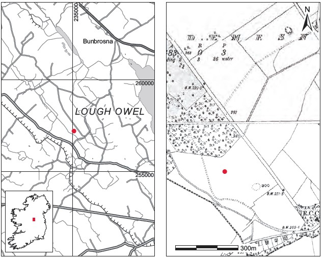
Fig. 4.66—Location map, Sonnagh Demesne, Co. Westmeath.
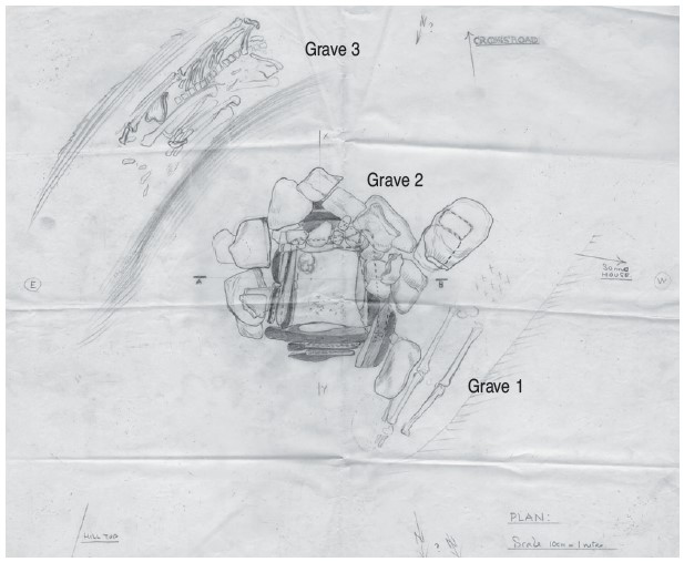
Fig. 4.67—Sketch of graves 1–3, Sonnagh Demesne, Co. Westmeath.
Description of site
Grave 1: early medieval stone-lined grave
The grave lay at a depth of 1.2m below ground level. It consisted of a rectangular pit, aligned north-east/south-west, lined with irregularly shaped stones (Fig. 4.67). The stone lining had apparently been visible on all sides of the pit but had fallen away at the west side by the time of Ó Ríordáin’s visit. The lining on the south-eastern side consisted of seven stones placed in a line approximately 1.7m long. The grave-pit measured 1.7m long by 0.5m wide by 0.2m deep. The maximum length of the stones was 0.45m and the maximum width 0.3m. The feet had apparently been covered by a stone measuring 0.63m by 0.43m but this had fallen down the side of the gravel pit. The floor of the pit does not seem to have been lined with stone. The grave contained an extended inhumation burial of an adult male (P1954:18) and no accompanying artefacts were found (Pl. 91). Only the legs and the top of the pelvic bones were in situ. The upper body bones had been badly disturbed and broken by the workmen, who had thrown sand and animal bones on the burial after its discovery, but the remains have been identified by Buckley as those of an older adult male, possibly aged over 60 years at death. The individual had suffered weapon wounds to the clavicle and femur. The body was lying north-east/south-west. The skull was not found. Fragments of animal bone were also found in the area of the burial but these may not have been contemporary with it, as the workmen stated that they had placed bone found around the site on top of this burial. The animal bone assemblage (P1954:20) has been identified as containing mostly ox bones and some pig bones, and apparently all had been ‘passed through fire’. A sample of the human remains was submitted for radiocarbon dating and yielded a date of 1545±40 BP, which corresponds approximately to the historical period 424–598.126
Grave 2: early Bronze Age cist grave
Vol. 1, Chapter 3, pp 552–9.
Grave 3: early medieval unlined grave
This burial consisted of an unprotected crouched inhumation located approximately 0.7m south-east of the edge of grave 2, the early Bronze Age cist (Pl. 92). The grave was a simple pit without stone lining, except for a few small stones placed over the pelvic region of the interred (Fig. 4.67).127 It lay on a level with or slightly above the capstone of grave 2, and was aligned south-west/north-east. No other features were noted.
The grave contained a crouched inhumation burial (P1954:19) and a piece of worked dog tibia (P1954:16). The body, that of an adult male, was crouched, apparently lying on its back with the legs flexed and placed to the right side of the body. As with burial 1, the head of this individual was not located, and Buckley found significant evidence for weapon wounds on the skeleton (see below). According to Ó Ríordáin, the bones were very well preserved and there did not appear to have been any previous disturbance in the region of the upper limbs or where the skull might reasonably be expected to be. The burial lay south-west/north-east. The exact location of the worked bone in relation to the interment is not known. This was part of the proximal end of a dog tibia, 11cm long, and is pointed, and possibly polished, at one end. Approximately ten animal bones (P1954:20)128 were also found with this burial, including some cow bones and some pig teeth. According to Davies, all of the bone had been ‘passed through fire’. A sample of the human remains was submitted for radiocarbon dating and yielded a date of 1520±40 BP, which corresponds roughly to the period 430–617.129
Bone object P1954:16 (Fig. 4.68)
Part of the proximal end of a dog tibia, 11cm long; cut and pointed, and polished at one end. The extreme tip is broken off. Some scratches are visible on surface.
Dimensions: L 11.1cm; max. width 2.35cm; width at point 2.35mm.
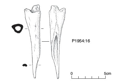
Fig. 4.68—Worked bone object, Sonnagh Demesne, Co. Westmeath.
Comment
This burial site was presumed to be prehistoric until radiocarbon dating indicated that the two inhumation burials were early medieval in date. The occurrence of a crouched burial is unusual in an early medieval context in Ireland. One crouched inhumation was excavated at a cemetery containing extended west/east inhumations at Knockea, Co. Limerick. O’Kelly (1967, 83) suggested a broad early medieval date for the site, based on the absence of gravegoods.
The burials were in close proximity to, although at a higher level than, an early Bronze Age grave. The reuse of prehistoric sites for burial in later periods is not unknown in Ireland and Britain (Thomas 1971, 53; O’Brien 2003, 67) and can also be seen at Lehinch, Co. Offaly (Vol. 1, pp 386–9; this volume p. 139ff). It is also worth noting that the extended burial, grave 1, was lying with the head to the north-east rather than towards the west, as would be expected in early medieval Christian burial contexts.
HUMAN REMAINS
LAUREEN BUCKLEY
Grave 1, 1954:18: older adult male, 178cm
This was an extended inhumation in a stone-lined grave, aligned north-east/south-west with the head to the north-east. Although most skeletal elements apart from the skull were present, the skeleton was in a poor state of preservation, especially the upper half, which had been disturbed and fragmented during discovery. There was no skull present; according to the workmen at the scene, there had been no skull found in the grave. The upper three cervical vertebrae were also missing, but the lower four cervical vertebrae were complete. All of the thoracic and lumbar vertebrae were present, although the lower half of the vertebral column was fragmented and incomplete. Twelve ribs from each side remained and there also was part of the manubrium and body of the sternum.
The clavicles were complete and the scapulae were virtually complete. Both humeri were present, although the left bone was fragmented at the proximal end of the shaft. The radii and right ulna were complete but the distal end was missing from the left ulna. Only the left scaphoid survived from the left hand. The surviving carpals of the right hand consisted of the scaphoid, lunate, trapezium and capitate. The metacarpals were very incomplete and, although the fourth was missing, the shaft of an unidentifiable metacarpal was present. There were also two proximal and two middle hand phalanges present.
The pelvis consisted of most of both ilia, the right ischium and both pubic bones. The sacrum was also present but was very fragmented. Both the femurs, tibiae and the right fibula were complete and only the proximal end was missing from the left fibula. All the tarsals except for the third cuneiforms survived from the foot bones, all the metatarsals were complete, and there were three proximal, one middle phalanx and one distal foot phalanx.
Age and sex
As the pelvis was virtually complete, it was relatively easy to sex this skeleton. All the features of the pubic bone were of the male type and the sciatic notch was narrow. The diameters of the femoral heads and the head of the humerus as well as the femoral bicondylar width also definitely indicated that this was a male individual.
The degeneration of the auricular surface of the ilium and the surface of the pubic bone indicated that this was an older individual more than 45 years of age and probably more than 60 years.
Pathology
Non-vertebral joint disease: there was degeneration of several joints in this individual. Severe osteoarthritis was present on the manubrium, with enlargement of the joint surface for the sternal ends of the clavicles on both sides. There was also cist formation on most of the joint surfaces. The first costal cartilages were also ossified and fused to the manubrium. There was, however, only mild porosity of the sternal end of the left clavicle and moderate porosity of the sternal end of the right clavicle. The head of the left humerus had mild marginal lipping and mild porosity of the joint surface.
There was moderate marginal lipping around the distal joint surface of the left radius.
On the right shoulder joint there was mild marginal lipping and porosity of the head of the right humerus, as well as a small surface osteophyte and mild porosity of the lateral end of the right clavicle. There was also mild lipping around the proximal joint surface of the right ulna, and mild lipping and porosity of the distal right ulna.
There was some degeneration of the left knee joint, with moderate lipping all around the distal femur surface and moderate lipping around the medial edge of the lateral condyle and the medial edge of the medial condyle of the proximal tibia surface. The right femur also had moderate marginal lipping around the distal joint surface.
Vertebral joint disease: several joints on the vertebral column were affected by degeneration. The fourth cervical vertebra had osteoarthritis with severe lipping and eburnation of the superior articular surfaces and severe lipping of the inferior articular surfaces. The fifth cervical vertebra had moderate lipping on all the posterior articular surfaces and also severe osteophytes on the inferior edge of the body as well as severe porosity of the inferior surface of the body. There was severe osteophytosis on the superior edge of the body of C6, moderate osteophytosis of the inferior edge, and severe porosity of the superior and inferior surfaces. The posterior articular surfaces had mild osteophytes. In C7 there was mild osteophytosis but severe porosity of the superior surface of the centrum. There were also moderate osteophytes on the inferior posterior articular surfaces.
The upper two thoracic vertebrae had mild degenerative changes on the posterior articular surfaces but there was severe osteophytic lipping around the facets for the head of the ribs. The third thoracic vertebra had mild marginal lipping and porosity of the superior articular surfaces but severe lipping and porosity of the inferior articular surfaces. There was also severe lipping and porosity of all the posterior articular surfaces of T4 and of the superior articular surfaces of T5. The inferior surfaces of T5 and the posterior articular surfaces of the sixth thoracic had mild degenerative changes. There were also mild changes on the superior articular surfaces of the tenth thoracic vertebra. The costal facets on the eleventh and twelfth thoracic vertebrae had moderate osteophytes and porosity on the right side but were only mildly affected on the left side. There was only a mild degree of marginal lipping on the posterior articular surfaces of the lumbar vertebrae and on the sacrum.
Fractures: the right clavicle was shorter than the left, suggesting that it may have been broken when the individual was very young. There was no callus present, however, so any break must have occurred early in life and the bone had a chance to remodel the repair. A slight callus was noted on the distal third of the right radius, suggesting a well-healed Colles fracture. The only deformity was a slight loss of the natural concavity of the bone in the distal third. There was also a slight callus and a lateral curve in the distal third of the right ulna. Since both these fractures are well healed with little deformity, it is probable that they occurred during childhood, when the bone is relatively soft and bends under pressure rather than breaks. The Colles fracture, the fracture of the ulna and a clavicle fracture can all occur as a result of a fall on an outstretched hand. It is highly probable that all these fracturesoccurred in the one incident during childhood, when the hand was outstretched in an attempt to break a fall.
Weapon wounds: there was a cut on the superior-posterior edge of the right clavicle. The wound was broad and ended in a sharp point, suggesting that the blade of a weapon had gone sharply into the bone, although the medial edge of the cut is more ragged, as if the weapon had then been roughly pulled up out of the bone. There was also a possible cut, very narrow and slight, in the medial side of the distal end of the right femur just above the condyle. Enthesophytes: there were strong attachment areas for the tibial-fibular ligament on the right tibia and fibula.
Grave 3, 1954:19: middle adult male, 181cm
This burial was found in a crouched position, lying on its back with the knees tightly drawn up. It was in a simple grave-cut with no stone lining, and was aligned south-west/north-east with the head to the south-west. The skull and upper three cervical vertebrae were missing, but the rest of the skeleton was complete and in excellent condition. The lower four cervical vertebrae were complete. All of the thoracic and lumbar vertebrae were present and most were complete. Twelve ribs from each side were present, as were the manubrium and part of the body of the sternum. Both clavicles were complete and both scapulae were virtually complete. Both humeri were present, although the left bone was fragmented at the proximal end of the shaft, and the radii and ulnae were complete. All the carpals and metacarpals from each hand were present and there were also ten proximal, eight middle and six distal hand phalanges. The pelvis consisted of most of both ilia, the right ischium and right pubic bone. The sacrum was also present and almost complete. The femurs, tibiae, fibulae and patellae from the leg bones were complete. All the tarsals and metatarsals were present, and there were ten proximal foot phalanges as well as two sesamoid bones.
Age and sex
The only feature available to examine from the pelvis for determining the sex was the sciatic notch. This was narrow on both sides, suggesting a male. All the metrics, such as the diameters of the femoral heads and the head of the humerus as well as the femoral bicondylar width, also fell into the male range. The degeneration of the auricular surface of the ilium and the surface of the pubic bone indicated that this was a middle adult individual not more than 44 years of age and probably more than 35 years at the time of death.
Pathology
The most remarkable feature of this skeleton was the number of weapon wounds on it. The individual had obviously been involved in a fight and had a number of defence wounds on the lower left arm and wrist. He had clearly lost his life in a battle, and had several sword cuts through the ribs as well as a stab wound to the side. There were almost too many cuts to describe fully, but the main wounds are listed below bone by bone.
Left arm: there was a very small cut on the medial surface in the proximal third of the humerus. The cut had a triangular profile but barely penetrated the outer surface of the bone, although the weapon must have cut through a lot of flesh and muscle before reaching the bone. This is a typical defence wound that occurs when the arm is raised to ward off a right-handed attacker. There was a cut in the distal third of the left ulna on the radial border. The cut was an oblique nick in the bone, going from the distal end upwards, and had a triangular profile such as would have occurred if it had been slashed with a sword held at an angle. The bone was broken adjacent to the cut, but this was a post-mortem break occurring because the weapon blow had weakened the bone.
There were several cut-marks in the distal third of the radius. There was a smooth cut obliquely through the medial edge of the bone at the junction of the proximal two-thirds with the distal third. Another smooth cut was present at the lateral edge of the bone, as well as a series of small cuts with V-shaped profile. The posterior surface of the radius had a series of small V-shaped cuts, and a chunk had been taken out of the bone in the distal third. There were also very shallow cuts, no more than scratches, on the posterior surface near the distal end. These also appear to be defence wounds to the lower arms, with the radius being broken by a combination of cuts and force.
Left hand: there was a small, straight cut across the base of the hamate bone, just on one corner.
Right arm: there were a few cuts to the lateral surface of the right humerus in the proximal third of the bone. The largest cut was only 1cm long and consisted of a broad chunk out of the bone, with some bone below being roughly broken away. A few ‘scratches’ were superimposed on this cut and must have occurred later in the combat. Below the broad cut there were a few small linear incisions that barely scratched the surface of the bone. The right radius had a sharp cut across the distal joint surface.
Right hand: there was a smooth cut across the base of the right scaphoid, and there were small scratches across the dorsal surface of the first metacarpal, near the proximal end.
Left ribs: there were cuts on several ribs on the left side. The sixth rib had three small cuts on the internal surface. One was a very small nick on the inferior surface at the level of the transverse articular surface. There was another cut 2.5cm lateral to this on the internal surface. The cut was 0.5cm long and had entered at a very slight angle to the left. The third cut was 2cm lateral to the second cut and was a small nick 4mm long that had barely penetrated the surface of the bone. The seventh rib had three small cuts in the superior margin. One was about 12cm along the shaft near the angle and was 3mm in length. The second cut was 6mm lateral to the first at an oblique angle towards the middle of the body and only penetrated the outer surface of the bone. It was 5mm in length. The third cut was 4mm lateral to the second, also at an oblique angle to the bone, and was 6mm in length.
The eighth rib had a large cut on the internal surface, which had virtually cut the head off. The wound extended obliquely towards the head from the level of the transverse articular surface. It was 2cm in length and the mark of the tip of the blade can be seen in the bone. The wound must have resulted from a heavy blow, as the bone around the cut was crushed and compressed and contained micro-fractures. There is also a slice across the superior margin of the same rib at the angle. The cut seems to go from right to left and ends with the bone roughly broken. At the medial edge of this cut there is a sharp incised cut that barely scratches the surface of the bone. There is also another shallow oblique cut to the left at the lateral edge of the slice. Post-mortem damage to the bone means that the full extent of the cut cannot be seen. The rib is broken in two, partly by the force of blows and partly by post-mortem pressures acting on bone that was probably weakened by secondary fractures.
The ninth rib had a rough fracture between the transverse articular surface and the head. Part of the bone is missing, but there is an oblique cut on the internal surface near the angle and another horizontal cut along the inferior edge. These cuts are rough and not incised like most of the other wounds. There was a sharp incised cut 3cm lateral to the lateral edge of the horizontal cut that cut the inferior surface of the bone. Just above this incised cut was another shallow cut at a slight angle on the internal surface of the bone. The twelfth rib had been roughly broken near the head.
Right ribs: only two of the lower right ribs had been affected by cuts. The eleventh rib had one sharp cut on the internal surface near the head. The wound was oblique, inclined to the right and was 0.5cm in length. The twelfth rib had been severed near the head. The cut was sharpest on the internal surface and there was a secondary fracture on the external surface.
Vertebrae: the ninth thoracic vertebra had a cut across the left side of the vertebral body. The cut barely penetrated the bone and must have been the very end of a wound that possibly cut through ribs before it reached the vertebra. The cut is very sharp and is 4.5cm in length. It extends from almost the anterior edge of the body to just above the inferior articular surface for the tenth rib, and there is also a small nick in the neural arch lateral to the inferior left articular surface. The second lumbar vertebra had a weapon wound on the left side of the body. The wound was 13mm in length and was a linear cut with a diamond-shaped profile in the centre. It was 8mm wide in the centre. It was not possible to say how far the wound penetrated into the vertebral body.
Scapulae: the left scapula had an incised sharp wound on the acromion just at the angle. The cut was 1.5cm in length and did not penetrate deeply into the bone. The right scapula also had a sharp cut in the acromion, near the angle. The blade had entered at an oblique angle.
Other trauma
There were two smooth areas of erosion on the head of the left humerus; one was circular, 2mm in diameter, and the other was oval, 1cm in length. These were probably lesions of osteochondritis dessicans caused by trauma to the joint.
Vertebral joint disease
As this individual was younger than burial 1, it is not surprising that there was less evidence of degenerative joint disease. In the cervical spine the fifth cervical vertebra seemed to be resting on the spine of the sixth cervical vertebra. There was compression of the intervertebral space, with severe osteophytosis on the inferior edge of the body and moderate porosity of the inferior surface. There was also moderate osteophytosis and porosity of the superior surface of the body of the sixth cervical vertebra. Mild marginal lipping of the posterior joints affected the seventh cervical and second and third thoracic vertebrae. The middle thoracic vertebrae T6–T8 had mild osteophytosis on the superior and inferior edges of the vertebral bodies.
The ninth thoracic vertebra had moderate osteophytes on the right side of the centrum at the inferior edge and severe osteophytes on the left side. There was also a crescent-shaped area of porosity near the anterior border of the inferior surface. There were mild osteophytes on the superior and inferior edges of the lower two thoracic vertebrae and the second and third lumbar vertebrae. There were moderate osteophytes on the superior edge of the centrum of the fourth lumbar. The anterior surface of the centrum of the fifth lumbar vertebra had new bone forming which had almost turned sclerotic. It was combining with the osteophytes at the edges of the vertebral body. Slight degeneration of the costal joints was present in only the two lower thoracic vertebrae. Schmorl’s nodes were present in the lower three thoracic vertebrae but were not very deep.
Summary and conclusions
The burials from this site consisted of three males; one was cremated and the other two were inhumations. The cremation, Bronze Age in date, consisted of 860g of efficiently cremated bone that had not been deliberately crushed (Vol. 1, pp 556–9). It appeared to represent only one individual, although it was mainly the larger obvious fragments of skull and long bones that had been collected. Neither of the inhumations had their skulls remaining, although there was no evidence of decapitation.
One of the inhumations was that of an older adult male, 178cm in height. He had some degenerative joint disease and osteoarthritis in the spine and other joints that are typically found in older individuals of this period. During his earlier life, possibly even in childhood, he had suffered a fall and broken his clavicle and wrist. These bones had healed, however, with the breaks being barely discernible at the time of death. There were some small, sharp weapon wounds on this person, on the superior edge of the right clavicle and possibly at the side of the right knee. As these are unhealed they must have occurred around the time of death. The second inhumation was also an adult male, although less than 45 years of age at the time of death. He suffered from some mild to moderate osteophytosis and degeneration of the spinal joints that would be typical of his age group after a strenuous lifestyle. The most remarkable feature of this skeleton was the number of sharp, unhealed weapon wounds to the arms and torso. These must have occurred at the time of death. The position of the wounds indicates that he received them in a fight or battle. There were cuts to the shoulder areas on both scapulae and proximal humeri that might have been caused by slashing movements with a sword or knife, although some of the cuts on the upper left arm appear to be defence cuts that occurred when he had his arm raised to ward off blows. There were also several defence cuts to the lower third of the left radius and ulna. Some of these blows were applied with such force that the radius had broken. As there were none of these defence wounds to the right forearm, it can be presumed that this man was able to defend himself with a weapon in his right hand for some part of the battle at least. There were also several cuts on the ribs on the left side, with only a few cuts to the lower right ribs. Most of the cuts on the left side occurred to the lower half of the rib cage. Some were made with slicing movements and had cut the superior and inferior margins of the ribs. Some of the wounds had come from the front but were on the internal surfaces of the back of the ribs, indicating that a weapon had been pushed through from the front. To get to the back of the ribs and vertebrae, the weapon has to go through several organs in the abdomen and the individual could not have survived so many wounds, accompanied by severe blood loss. It is possible that the weapon was pushed through while he was already on the ground. The second lumbar vertebra also had a sharp cut to the left side that may have been caused by a stabbing movement with a weapon. The external surface appeared to have a diamond-shaped profile in the middle, suggesting that it was caused by a spearhead. This might be an optical illusion, however, and the only way to be certain of the shape of the weapon is to determine an internal profile of the wound. In conclusion, these two individuals had certainly received blade injuries before they died, but the younger man seems to have taken part in a battle or fight and received many defence injuries to his arms before he finally succumbed.
124. Two extended skeletons had also been discovered two months previously, but these were deemed by the workmen to be of no importance and were reinterred.
125. Parish of Templeoran, barony of Moygoish. SMR WM018-013——. IGR 235198 257137. This townland is spelled Sonna Demesne on the Ordnance Survey maps, but the spelling given in the General alphabetical index to the townlands and towns, parishes and baronies of Ireland, based on the Census of Ireland for the year 1851 (Dublin, 1861) is used here.
126. GrA-24310.
127. It is probable that the extent of the pit was not revealed, owing to the disturbed nature of the site.
128. The same number was used to register all the animal bone from the site.
129. GrA-24313.