1953:010 - BELTRA, CO. MAYO, Mayo
County: Mayo
Site name: BELTRA, CO. MAYO
Sites and Monuments Record No.: SMR MA008-00201
Licence number: E1118
Author: MARY CAHILL
Author/Organisation Address: —
Site type: Graves of indeterminate date
Period/Dating: —
ITM: E 518437m, N 838107m
Latitude, Longitude (decimal degrees): 54.285234, -9.252618
Introduction
In August 1953 human remains and some possible grave-slabs were discovered following the erosion of a cliff face on a beach at Beltra, near Ballycastle, Co. Mayo. The site is known locally as the Green Hill. The bones were noticed by locals, who reported on the find in the Irish Independent newspaper on 7 August 1953. The site was visited by Ellen Prendergast. This report is based on Prendergast’s site notes and sketches. In June 1998 a zoomorphic penannular brooch (1998:41) of sixth/seventh-century date was found after further erosion of the Green Hill. The site was investigated by Mary Cahill. The finder of the brooch indicated that she had observed human and animal bones exposed in the area from time to time. A number of possible cist features were noted in the eroding face of the mound and bone was visible along the entire exposed section. All visible bone was collected by Cahill at the time of her visit and brought to the Museum.
Location (Fig. 6.32)
The site was in the townland of Beltra, north-east Co. Mayo.55 It was located on Lackan strand at an altitude of 0–10m above sea level.
Description of site
The burials lay in a mound known locally as the Green Hill, which was approximately 69m long by 5m high. The human remains occurred chiefly in a layer of sand approximately 0.3m thick that underlay the sod at the top of the mound. Underneath the sand was a layer of sandy clay about 0.9m thick. This lay on a thin layer of sea sand (0.05m thick), followed by charcoal debris (0.025m thick), followed by a 2.7m-thick layer of sea sand. Most of the human remains discovered appeared to have been deposited in unprotected pits in the mound, although some large slabs in a different area of the mound were thought by the excavator to be possible cist slabs. As the bones had been eroding out of the cliff for some time, it was not possible to estimate the number of burials; only one portion of a burial was excavated in situ and is therefore described here. The burial consisted of an extended inhumation, aligned east/west (1953:51). The pelvis, vertebrae and phalanges were present but had been previously collected by the Gardaí.

Fig. 6.32—Location map, Beltra, Co. Mayo.
Comment
Human remains have been eroding from the seaward side of the mound for many years. A visit at any time will almost certainly result in the observation of human and animal bone at the base of the cliff and in the exposed section. Elements of stone structures that may be interpreted as partially demolished cists of the lintel grave type have also been observed. This, however, would have to be clarified by excavation or survey. The site has been classified as a burial mound, a hearth and a midden by the Archaeological Survey of Ireland.56 These factors and the discovery of the zoomorphic penannular brooch at the site all strongly suggest that this site dates from the early medieval period. Nevertheless, because of the way human and animal bone has eroded from the site, as opposed to any remains having been excavated in situ, it was not thought worthwhile to radiocarbon-date samples. Further survey and excavation at this site would undoubtedly prove very worthwhile. Without some protective measures, however, it will continue to suffer loss by erosion as it occupies such an exposed position on the shore.
HUMAN REMAINS (1953)
LAUREEN BUCKLEY
Introduction
Although the excavation report states that there was one burial in situ as well as a number of disarticulated remains, all the bones were recorded under the one registration number (1953:51) and the articulated burial was not differentiated. For the purposes of this report all the bone is considered as disarticulated. A description of each bone found is given below, followed by a summary of the minimum numbers of skeletal elements present.
Adult bones
Left femur
(1) This consisted of the head and neck only of a left femur. The diameter of the head indicates that it is probably from a female individual (FeHd 40.9mm).
(2) This was the distal half of a left femur. The epiphyseal width indicates that it is likely to be from a female individual (FeE1 72.4mm), but it cannot be determined whether it belongs to the same individual as (1) above.
(3) This was also the distal half of a left femur. The size of the bone and the epiphyseal width indicate that it is probably from a male individual (FeE1 79.5mm). There was mild marginal lipping all around the articular surface.
(4) This consisted of the distal half of a left femur from a female individual (FeE1 71.6mm). There was a slight defect in the area of insertion of the medial head of gastrocnemius, which may indicate some damage to the ligament.
(5) This was the distal end of a left femur with most of the distal surface and part of the medial side of the shaft present.
Right femur
(1) This consisted of the proximal half of a right femur. From the diameter of the head it is likely that this belonged to a female individual (FeHd 39.1mm; FeD1 20.1mm; FeD2 29.1mm).
(2) This consisted of the distal third of a right femur. It is probably from the same individual as (1) above but part of the shaft is missing. The width of the distal end indicates that it is probably from a female (FeE1 73.2mm).
(3) This was the distal half of a right femur probably from a female (FeE1 72.1mm). As this bone also has a slight defect in the insertion area of the medial head of gastrocnemius, it probably belongs to the same individual as the left femur (4) described above.
Tibia
The proximal two-thirds of a left tibia was present.
Left radius
There was one complete left radius present (RaL1 243mm; RaHd 24.1mm).
Right radius
There was one complete right radius present (RaL1 241mm; RaHd 23.6mm).
Ulna
One complete left ulna was present (UlL1 264mm).
Pelvis
One complete right innominate bone from a male individual was present. The epiphyseal line was still visible on the iliac crest and examination of the sacral articulation area and the pubic symphysis indicates that this was a young adult aged 20–24 years at the time of death.
Foot bones
A right calcaneum, a right first cuneiform, a left first metatarsal and two left third metatarsals were present from the foot bones.
Skull bones
Only most of a left temporal bone from a male survived from the adult skull.
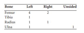
Summary of adult bones
The minimum number of adults based on the minimum number of left femora is four.
Adolescent bones
There were some bones present that were almost of adult proportions but the fact that all the epiphyses were unfused indicates that they were adolescent bones. These included a complete left and complete right femur, apart from the unfused epiphyses, which were missing, a matching left and right calcanea with posterior epiphyses unfused, a distal fibula epiphysis, a first metatarsal and a proximal foot phalanx.
Juvenile bones
The only accurate way to age juvenile skeletons is by the state of dental eruption. It is not possible to age single disarticulated juvenile bones, although the size, length and diameter can give an indication of whether the juvenile is an infant, younger child or older child. Where complete bones were found, their diaphyseal length was compared to the lengths of long bones from juveniles whose dental age was known in order to get an approximate age (Buckley 1991). The distal third of a right tibia shaft and the proximal third of a right femur that were present probably belonged to an older child (10–15 years). There were, however, a number of smaller bones that appeared to come from one younger individual. These included a complete left clavicle, complete left and right scapulae, complete left and right humeri and a complete set of cervical vertebrae. There were also four upper thoracic vertebrae and one sacral vertebra. The left ilium was also complete and the mid-shaft of a right femur was present. The left first rib was also present.
Epiphyseal fusion
All the epiphyses at the ends of the long bones were unfused. The vertebral arches were fused to the centra. The centrum of the second cervical vertebra was fused to the dens but the apex of the dens was unfused. This indicates an age of between four and twelve years (Scheuer and Black 2000).
The measurements of the bones compare well to other juveniles aged 6–8 years.
Juvenile skulls
In addition to the long bones, two juvenile skulls were found.
Skull 1
This was a complete skull in one piece, apart from the left temporal bone, which had become detached. The lambdoid and sagittal sutures and the right squamous temporal sutures had started to fuse ectocranially although they were not completely fused endocranially.
Age: the first molars were erupted and the second molars, although formed, were not erupted. Although the permanent incisors were missing, from the shape of the sockets they appear to have been fully erupted. This places the juvenile in the eight to ten years age range.
Dental pathology: there were moderate calculus deposits on the lingual surfaces of the lower molars and light deposits on the buccal surfaces of the upper teeth.
Skeletal pathology
There was a slight degree of cribra orbitalia in both orbits.
Skull 2
This skull consisted of an almost complete cranium. The occipital, both parietal, the frontal and right temporal bones were present. The left side of the maxilla was also present but all other bones of the skull were missing
Age: the first permanent molar was not erupted and this state of dental eruption indicates that the juvenile was aged between three and five years.
Summary and conclusions
The long bones indicate that an adolescent and at least two juveniles were present in this collection. There were also two juvenile skulls. Ageing of the juveniles from the long bone lengths is not as accurate as ageing from the state of dental eruption. Therefore, although the age estimation from the long bones does not exactly match the age from the skulls, it is not safe to assume that there were four juveniles present. It can only be stated that there were a minimum of two juveniles present, aged, from the state of dental eruption, as three to five years and eight to ten years. The minimum number of individuals in this collection is therefore seven, consisting of four adults, one adolescent and two juveniles.
HUMAN REMAINS (1998)
Introduction
As the material (2003:19) derives from an unknown number of graves, it was not possible to separate the bones into individual skeletons. The bones are therefore described individually below, with a summary of minimum numbers in order to determine the minimum number of individuals present.
Left femur
(1) The proximal half of a left femur with complete head and neck. The diameter of the femoral head (48.1mm) indicates that it was probably from a male individual. There were indications of early degenerative joint disease, with slight marginal lipping around the head (FeD1 26.0mm; FeD2 37.1mm).
(2) The proximal half of a shaft, from just below the lesser trochanter to the mid-shaft area. The bone is much smaller than the first left femur and may belong to a female individual (FeD1 23.1mm; FeD2 28.2mm).
(3) The distal epiphysis only. The bone was slightly damaged but was small, with the epiphyseal breadth measuring 73mm. This indicates that it was probably female and may in fact belong to the same individual as the proximal shaft (2), described above.
Right femur
(1) This consisted of two parts: the proximal third (complete) and the distal half. Although there was a small section in mid-shaft missing, it is highly probable that these two partial femora were from the one bone. The measurements from the head of the femur and the distal epiphysis were consistent with the remains being from a male individual (FeHd 50.5mm; FeD1 25.8mm; FeD2 36.2mm; FeE1 85.4mm). (2) This consisted of a very small fragment near the proximal end of the shaft from just below the lesser trochanter. It was severely weathered.
Tibia
Only two left tibiae were present.
(1) This consisted of the distal half of a left tibia. A lateral squatting facet was present.
(2) This consisted of most of the shaft of a left tibia. It appeared to be a larger bone than the
First.
Humerus
One complete right humerus was present. From the size of the bone and the diameter of the humerus head, it is probable that it came from a female individual (HuL1 299mm; HuHd 40.1mm; HuE1 56.3mm).
Radius
Two partial right radii were present.
(1) This consisted of the proximal half of the shaft of a right radius.
(2) This consisted of the distal third of the shaft of a right radius. From the size and appearance of the bones, it was concluded that the two radii fragments were from two different individuals.
Ulna
(1) This consisted of most of the shaft of a right ulna.
(2) This consisted of the distal third of the shaft of a right ulna as well as part of the distal joint end.
Fibula
Only one fragment of fibula was present; this was part of the proximal shaft, probably from the right side.
Pelvis
Only one fragment from the left ilium remained; it was from the most posterior part, with the sacral articulation area present.
Sacrum
(1) A complete sacrum was present.
(2) The right side of the first sacral vertebra from a second sacrum was present.
Vertebrae
There were three lower cervical vertebrae present, possible C3 or C4 and a C6 and C7. Three thoracic vertebrae were also present, a fragment of a T1, a complete T2 and a complete T10.
Pathology
There was some degeneration of the joint surfaces of the cervical vertebrae. The highest cervical vertebra, possibly a C3, had severe porosity of the surfaces of the centrum as well as mild osteophytosis at the superior and inferior margins. There were also some degenerative changes of the posterior joint surfaces, with mild lipping around the superior right articular surface and severe lipping and porosity of the superior left articular surface. The inferior right surface had definite indications of osteoarthritis, with enlargement of the joint surface, porosity and a slight degree of eburnation. The sixth cervical vertebra had severe porosity on the superior surface of the centrum as well as mild osteophytosis. There was also mild marginal lipping on the posterior articular surfaces of the right side. The first thoracic vertebra had mild porosity of the articulation area for the head of the first rib. The tenth thoracic vertebra had Schmorl’s nodes present on the superior and inferior surfaces of the centrum. The epiphyseal line was still visible on the inferior of the centrum. It was also still visible on the second thoracic vertebra. Although these vertebrae represent a minimum of one individual, it is probable that they are from two individuals. One was a relatively young adult with the epiphyses of the vertebrae just fused, and the other was probably an older adult with severe degeneration of the cervical Vertebrae.
Scapula
Part of the acromion of a right scapula only.
Ribs
Two left ribs and one right rib were present.
Tarsals and metatarsals:
There were two left calcanea present, one being bigger than the other. Two second right metatarsals were also present.
Skull
A left temporal bone was present and from the size of the mastoid process and shape of the zygomatic arch it appears to be from a male individual.
A section of frontal bone with the glabella and medial parts of the left and right orbits as well as part of the squamous area was present. This appears to be from a female. There were three fragments of occipital bone and most of the left and right parietal bones from the one individual present. The sagittal suture was relatively unfused. It is not certain whether this belonged to the same individual as the frontal bone above, as the coronal suture of the frontal bone was not present.
Juvenile bones
There were five fragments of parietal bone that were very thin and were assumed to be from a juvenile. Also present was a very decayed section of a left humerus shaft from a juvenile.
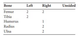
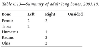
Summary
The minimum numbers of adult long bones present in this collection are summarised in Table 6.13. There were also two sacra and a minimum of two skulls present. The minimum number of adults present was therefore two; based on the characteristics of the skull and measurements of the heads of the femur and humerus, it appears that at least one was female and one was male. One of the individuals was a young adult and one was probably an older adult, as severe degeneration of the cervical vertebrae had occurred.
55. Parish of Lackan, barony of Tirawley. SMR MA008-00201-. IGR 118469 338102.
56. www.archaeology.ie; accessed 28/01/2009.