1950:015 - NURE OR LILLIPUT, CO. WESTMEATH, Westmeath
County: Westmeath
Site name: NURE OR LILLIPUT, CO. WESTMEATH
Sites and Monuments Record No.: SMR WM032-112
Licence number: E1174
Author: P.J. HARTNETT
Author/Organisation Address: —
Site type: Graves of indeterminate date
Period/Dating: —
ITM: E 637406m, N 744497m
Latitude, Longitude (decimal degrees): 53.449223, -7.436868
Introduction
In May 1950 an inhumation burial was discovered during quarrying operations near Castletown Geoghegan, south Co. Westmeath. The bones were discovered at a depth of approximately 0.2m below ground level and were disturbed, some of them having fallen out of the section. The site was reported to the NMI by Gardaí at Castletown Geoghegan. A rescue excavation was undertaken by P.J. Hartnett. The human remains were analysed by Laureen Buckley.
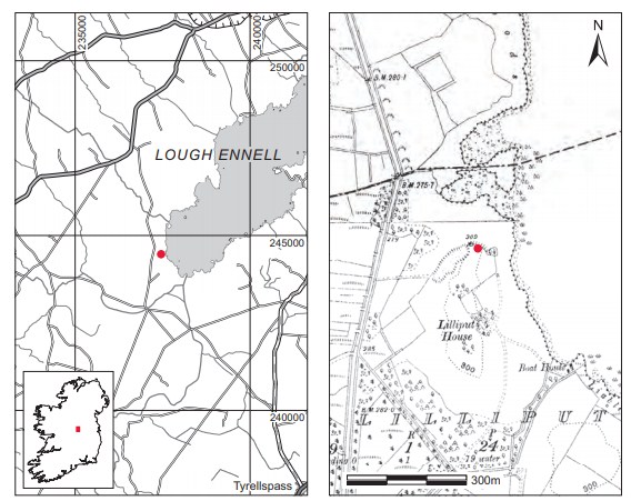
Location (Fig. 6.55)
The site was in the townland of Nure or Lilliput, just 3km east of Castletown Geoghegan, south Co. Westmeath.92 It was located at 80–90m above sea level overlooking the western shore of Lough Ennell, less than 1km west of the boundary between the baronies of Fartullagh and Moycashel. Immediately east of the site was an oval clump of trees surrounded by a low flat bank with an outer fosse. According to Hartnett, this oval enclosing bank and ditch were not ancient, and perhaps no older than the end of the nineteenth century.
Description of site
The site had been much disturbed by the time of Hartnett’s visit but it was possible to see that the grave was narrow and shallow, with the long axis running west/east. It measured approximately 1.7m in length by 0.3m in depth and was just wide enough to accommodate the body. There was no evidence for any stone or wood protection in the grave. The grave contained an extended inhumation of a middle-aged adult male (P1950:22), aligned west/east with the head to the west. The skull and upper bones on the right side had been disturbed, but it was clear that the body had lain fully extended, in a supine position with the left hand resting on the left hip. The bones on the left side were in position, except for the radius and the ulna. The bone was in poor condition but was collected and brought to the Museum for examination.
Comment
In the absence of associated finds or other evidence this burial is regarded as undated.
HUMAN REMAINS
LAUREEN BUCKLEY
Description of burial (P1950:22)
The skull had been disturbed and was fragmented but the frontal bone was almost complete and most of both parietal bones were also present. There were a few fragments of occipital bone and the mastoid areas of both temporal bones. The right wing of sphenoid and the right zygomatic bone were also present. The maxilla and mandible were present but were incomplete. Very little remained of the vertebral column; the first and second cervical vertebrae were present and there were four incomplete upper thoracic vertebrae. There were also two lower thoracic vertebrae and tiny fragments of lumbar bodies. Seven ribs from each side survived.
Both scapulae and clavicles were present and almost complete. The left humerus was complete apart from the proximal end, the distal end was missing from the left ulna and the middle third was missing from the left radius. The right humerus was almost complete but the proximal end was fragmented, the top of the olecranon was missing from the right ulna and the distal two-thirds of the right radius was present. Only the left scaphoid and capitate, second and third metacarpals and four proximal phalanges remained from the left hand, while the right hand consisted of the second and fourth metacarpals only. Both ilia and ischia remained from the pelvis but they were incomplete, and only the first sacral vertebra survived from the sacrum. The left femur was almost complete, with only part of the head and neck missing, but only the shaft and the head of the right femur survived. The left tibia was complete although the distal end was broken and decayed, and the shaft of the left fibula was present. The left patella was complete. Only the left talus remained from the foot bones.
Age and sex
All the features of the skull, the external occipital protuberance, the mastoid processes, the supraorbital ridges, orbital rims, posterior zygomatic arches and mental eminence, were of the male type. The sciatic notch was not very narrow but was not wide enough to suggest a female either. The diameter of the femoral heads and the clavicle length were also in the male range. The auricular surface of the ilium suggested an age of 35–39 years and the sutures of the skull were partially fused but not obliterated. This suggests that the individual was a middle adult, probably a late middle adult.
Skeletal pathology
There was a healed Colles fracture of the distal left radius (Pl. 104). There was only very slight displacement of the bone above the fracture, with the bone appearing slightly more convex than normal, and the bone below the fracture was more concave than normal.
Associated with this fracture was osteoarthritis of the left wrist. There was a moderate degree of marginal lipping all around the distal articular surface of the left radius and eburnation on the facet for the left ulna. Unfortunately the corresponding area of the left ulna was missing. There was also a moderate degree of lipping on the left scaphoid but most of the other left wrist bones were missing.
In the vertebral column there was a mild degree of lipping on the articular surfaces at the dens between C1 and C2. There were also moderate degenerative changes on the costal facets of the second thoracic vertebra and the inferior articular facets of the third thoracic vertebra. Since most of the vertebrae were so incomplete the extent of degenerative joint disease in the vertebral column could not be determined. There was severe porosity of the superior surface of the sacrum. Schmorl’s nodes were noted in the eleventh and twelfth thoracic vertebrae.
In the left tibia there was some healed periostitis on the lateral surface near the distal end. The right tibia also had healed periostitis on the lateral surface. The surface was porotic with some sclerotic new bone. There also appeared to be healed periostitis on the left fibula shaft but the right fibula was too decayed to be certain.
Dentition
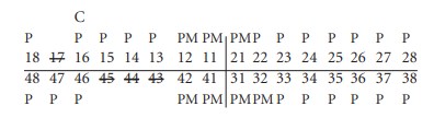
Attrition: there was moderate wear on the upper incisor, canines and premolars and the lower canine and premolars. Attrition was heavy on the molars and very heavy on the upper first molars (Pl. 103).
Calculus: there were light deposits on the lingual surface of the upper second premolar and upper first molars. Deposits were moderate on the buccal surfaces of the upper right canine, left first molar and both upper third molars. There were also moderate deposits on the lingual surfaces of most of the lower teeth except the left canine and right third molar, where deposits were heavy.
Caries: there was a moderate cavity on the distal surface of the crown of the upper right first molar at the cervical margin.
Periodontal disease: there was a slight degree of alveolar recession around the roots of the lower left canine and premolars. There was moderate recession around the upper premolars and first molars and the left upper second and third molars, as well as the lower incisors and lower right first and second molars. Recession was considerable around the roots of the upper incisors and left canine and around the lower left molars.
Hypoplasia: linear enamel hypoplasia was noted on the upper left canine and premolars.
Summary
This was the disturbed skeleton of a late middle adult male. The individual had suffered a fracture of the wrist some time during his life, probably as a result of a fall on an outstretched hand. This fracture had eventually led to the development of osteoarthritis in the wrist bones. There was some evidence of degenerative joint disease in the vertebral column, but the full extent could not be determined as the vertebrae were incomplete. The disturbance of the bones some months before they were reported to the Museum meant that there were no complete long bones to enable the living stature of the individual to be estimated. The teeth showed a considerable amount of wear, and there were also heavy calculus deposits that probably contributed to the development of periodontal disease in the alveolus around the teeth sockets. Exposure of the tooth roots had led to the development of caries at the cervical margin of one of the molars. The degree of wear and calculus deposits and the general low rate of caries are consistent with prehistoric and early medieval populations.
92. Parish of Dysart, barony of Moycashel. SMR WM032-112—-. IGR 237464 244473.