1950:017 - MARTINSTOWN, CO. MEATH, Meath
County: Meath
Site name: MARTINSTOWN, CO. MEATH
Sites and Monuments Record No.: SMR ME037-026
Licence number: E1139
Author: ELLEN PRENDERGAST AND A.T. LUCAS
Author/Organisation Address: —
Site type: Early Bronze Age graves
Period/Dating: —
ITM: E 687751m, N 753402m
Latitude, Longitude (decimal degrees): 53.523251, -6.676625
Introduction
In June 1950, sherds of a vase urn246 were discovered on top of screenings on the floor of a sandpit near Kiltale, Co. Meath. The sherds were noticed by Mr G.F. Mitchell of Trinity College, Dublin, who was on a geological tour of the area. Near the top of the pit he also noticed a depression containing burnt clay and charcoal. Mr Mitchell reported the find and brought the sherds to the NMI. The site was investigated on 10 June 1950 by P.J. Hartnett, who conducted a trial excavation in two areas of the sandpit where charcoal deposits were visible. Neither of the excavations revealed evidence of a burial and it seems likely that the grave had been completely destroyed, but some further sherds of a vase urn were recovered.247 In 1952 another grave was discovered when the sandpit at Martinstown was extended.248 This was also investigated by P.J.

Hartnett. In April 1953, two further early Bronze Age burials were reported from the site,249 and in June of the same year two more were excavated by Ellen Prendergast and A.T. Lucas (Pl. 51).250 This report is based on their excavation notes. Previously, in 1949, P.J. Hartnett (1951) had investigated the discovery of a burial of Neolithic date at the same quarry.
Location (Fig. 3.137)
The site was in the townland of Martinstown, south-east Co. Meath.251 The burial found in 1952 (grave 1, below) was located some 250m north-east of the Neolithic burial found in 1949 and on the same gravel ridge, all within the 300ft contour. The 1953 burials (graves 2–5, below) were found within approximately 8m of each other at the northern side of the sandpit. Their distance from those found in previous years is not shown. The site lay at an altitude of c. 90m above sea level. According to the landowner, he had frequently come across pockets of cremation and charcoal on this part of the ridge. Over 100 years ago a decorated bronze dagger and fragments of a bowl were found in a mound known as Croghan Erin, traces of which still remain, in the townland of Kiltale, just adjacent to this site.
Note that the registration numbers attached to the artefacts and human remains from graves 2–5 may seem not to be in the correct sequence, as outlined below. This is because the objects were numbered in a sequence that does not follow the actual calendrical sequence of discovery.
Grave 1
An area of 2m2 was cleared and brought to a level surface, enabling the outline of the burial pit to be seen. The pit was oblong in shape, with its long axis aligned north/south. It measured 0.8m by 0.5m and was scooped out of a yellow clay subsoil at an estimated depth of 1m below the turf level. There was no evidence for cist stones in the area of the burial.
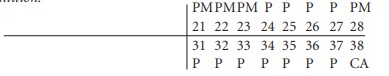
The grave contained an inhumation of an adult male (P1952:2.1), accompanied by a sherd of a bowl or vase (P1952:2.2; not illustrated) and a white stone pebble (P1952:2.3; Fig. 3.138). The body lay on its left side with the head to the north-east, the right hand resting under the chin and the left hand resting on the pelvic region between the legs. The knees were pulled up to the chest and the right foot crossed over the left. It would appear that the head, instead of lying on its left side, was tilted upwards, as only fragments of the base of the skull remained in position, and fragments of the front of the skull and part of the upper jaw were found at the southern end of the grave, having been disturbed by the scoop. The bones were articulated, and it was noted that the soil around the bones was darker than elsewhere. No traces of charcoal were found. One of the pottery sherds found in the north-east corner of the grave showed a fresh break. It is likely that the rest of the vessel was broken and removed by the digger. The spoilheap was searched but no more sherds were recovered. The stone pebble was resting on the mid-portion of the body and measured 0.04m by 0.03m.
Pottery (Fig. 3.138)
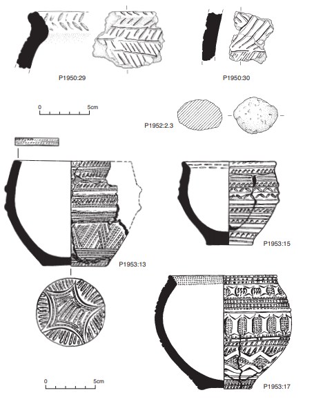
A single small sherd of undifferentiated vase or bowl was found (P1952:2.2). The outer surface is very worn, has lost some of its original outer surface and is decorated with a shallow groove.
Dimensions: max. L 2.52cm; max. W 2.26cm; max. T 1cm.
Grave 2
This burial had also collapsed by the time of Prendergast’s visit on 28 April 1953. The grave had been located at the top edge of the face of the pit. On investigation it was found that a trench had been dug into the upper layers of the sand, as if for a grave. The pit was loosely filled with soil and broken stones and traces of bones. There was no evidence for any stones forming a grave. The trench was fully excavated but no further remains were discovered. In three other places on the surface of the sandpit similar grave-pits were noticed and examined. Loose scatters of bone around these areas led to the assumption that the bones had been removed by the bulldozer as it skimmed the surface of the pit. The contents of this grave were collected at the base of the gravel pit where they had fallen. The remains were found to represent three individuals: an adult male, an adolescent and an infant (P1953:12). No accompanying artefacts were found with the remains.
Grave 3
This pit grave was discovered on 28 May 1953 and was investigated by E. Prendergast and A.T. Lucas that afternoon. Unfortunately the grave was completely destroyed at the time of its discovery. The grave had contained three inhumation burials, an adolescent, an adult male and an adult female (P1953:18, 19; registered as two individuals), accompanied by a bowl. The bowl had been removed for safekeeping and was complete but cracked when found.
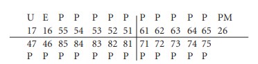
Ribbed bowl, P1953:17 (Fig. 3.138) This bowl is very finely manufactured, decorated with comb impressions and false relief (Ó Ríordáin and Waddell 1993, 126). The rim bevel is decorated with very fine comb impressions. The neck and shoulder of the vessel are decorated with comb impressions alternated with false relief. Towards the base of the vessel a chevron motif is executed in false relief. The base is plain.
Dimensions: H 11.55cm; ext. D rim 11.75cm; D base 5.35cm; T wall 0.75cm.
Grave 4
This grave and grave 5 were investigated on 24 June 1953 and were the only graves that were intact at the time of excavation. The burial had been placed at the base of a pit approximately 0.61m deep. The grave was not stone-lined, but a small stone measuring 0.15m by 0.23m had been placed over the skull and bowl. It was not possible for the excavators to examine the structure of this or grave 5 more closely owing to the danger of collapse of the pit face.
The grave contained an inhumation of a young adult male (P1953:14) accompanied by a bowl. Part of the burial, including the jawbone and teeth, was found lying on the surface near the grave. The skull and bowl lay underneath a stone, as described above. The crushed bowl had been placed in an upright position slightly overlying the skull.
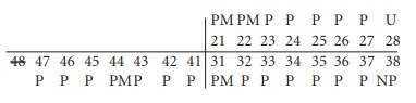
Tripartite bowl, P1953:13 (Fig. 3.138)
The bowl is very finely decorated with comb impressions and fingernail impressions (Ó Ríordáin and Waddell 1993, 125). The rim is decorated with three rows of comb impressions. The body of the vessel is decorated with rows of diagonal comb impressions alternated with horizontal lines of comb impressions and some fingernail impressions. The lower part of the vessel (below the shoulder) is dominated by a chevron motif executed by comb-impressed ornament. The base is decorated with a star shape executed by lunate impressions, and comb impressions. The vessel was fragmentary when excavated but has since been restored and is almost complete.
Dimensions: H 10.6cm; ext. D rim 12.1cm; D base 7cm; T wall 1cm.
Grave 5
This grave was approximately 8m west of grave 4 and consisted of an oval pit aligned north/south. The southern end of the pit had been cut away by quarry machinery. The grave contained a crouched inhumation of a juvenile (P1953:16) accompanied by a bowl, which had been placed upright close to the skull (Pl. 52). The skeleton was crouched and lay on its right side, with the arms slightly flexed and the knees drawn up. It lay in a north/south position with the head to the south, facing east.
Ribbed bowl, P1953:15 (Fig. 3.138)
The bowl has been restored and less than half of the rim survives (Ó Ríordáin and Waddell 1993, 126). The decoration is coarser than that of P1953:13, consisting of impressed decoration and some false relief. Bands of parallel incised lines alternate with either short impressed strokes or fingernail impressions to produce decoration in false relief. The base of the vessel is plain.
Dimensions: H 9.05cm; ext. D rim 9.5cm; D base 4.99cm; T wall 0.77cm.
Comment
The history of investigation at this site is complex, in that it was partially investigated on a number of occasions over a period of years from 1949 to 1953 by three officers of the NMI. Owing to the on-site circumstances, which are noted as being too dangerous to survey or to excavate the various pits properly, the records on the site are not as detailed as they might otherwise have been. It was also the case that there was a great deal of collapse, which meant that much of the archaeological evidence had fallen away. There is a sense from reading the file that reports to the NMI were not received as quickly as they might have been, given that it was known that discoveries were likely to be made.
The first report in 1949 was of a Neolithic burial from the western end of the gravel ridge. Sherds of a decorated necked bowl were found with a male skeleton (Hartnett 1951; Herity 1982, 300 and fig. 20:2). Nothing further of Neolithic date has been recorded. In 1950 sherds of vase urn were recovered but these were not in situ. The 1952 grave (grave 1 above) was from the eastern end of the ridge, while the 1953 graves (graves 2–5 above) were found towards the western end.
Only one burial has been dated. A sample of the remains from grave 4252 was submitted for AMS dating and yielded a date of 3670±40 BP, which calibrates to 2200–1920 BC.253 The ribbed bowl has been categorised as belonging to stage 2 of the development of pottery in the bowl tradition, which centres on a median date of 2030 cal. BC (Brindley 2007, 173, 246–7).
With the exception of the sherds of vase urn found in 1950 and the unidentified pottery, the remainder of the pottery found at Martinstown is of the bowl tradition, suggesting an early date for the commencement of the early Bronze Age phase of burial at the site. The ribbed bowl from grave 3 can also be assigned to Brindley’s stage 2 on the basis of its ornamental style, while the vessel from grave 5, with its more widely spaced, less complex patterns, probably belongs to stage 3, the latest date for which is c. 1930/20 BC. The presence of uncontexted vase urn sherds also suggests the continuation of burials at the site into the nineteenth century BC.
HUMAN REMAINS
LAUREEN BUCKLEY
Grave 1: early middle adult male, 169cm (P1952:2)
This skeleton was complete and in good condition, although the skull was crushed. It was lying on its left side in a crouched position in a simple pit aligned north/south. The skull was very fragmented, but most of the frontal, both parietal, the left temporal bone and part of the squamous occipital bone were present. The right side of the maxilla and part of the left side of the mandible were also present. All the vertebrae were present except the first cervical vertebra and, apart from the middle thoracic vertebrae, they were almost complete. Ten ribs from the left side and twelve from the right side remained.
The left scapula was virtually complete, and a fragment of acromion and the lateral border remained from the right scapula. Both clavicles were almost complete but their joint ends were missing. The distal halves of both humeri survived well, but the proximal halves were fragmented. The left radius and ulna were complete, and only parts of the proximal ends were missing from the right radius and ulna. The right scaphoid and trapezoid and both triquetral and pisiform bones were missing from the carpals, but all the metacarpals were present in a fragmentary state. There were also ten proximal phalanges and two middle hand phalanges present.
The left and right ilia, ischia and pubic bones from the pelvis were present and virtually complete, and the sacrum was also virtually complete. The left femur had a small piece of shaft missing and the right femur was fragmented at the proximal end. The distal half of the left tibia and distal two-thirds of the left fibula were present, and the right tibia and fibula were complete. All the tarsal bones and all the metatarsals, apart from the left fourth metatarsal, were complete and there were three of the proximal and one of the distal foot phalanges remaining.
Age and sex
All the observable features of the skull, external occipital protuberance, the mastoid process, supraorbital ridges and orbital rim, were of the male type. The sciatic notch and pubic bones the pelvis were also of the male type. The diameter of the heads of the humeri and the head of the radius were also within the male range. The surface of the pubic symphyses indicated an age at death of 19–34 years, and the auricular surface of the ilium indicated an age of 20–29 years. The sternal end of the ribs indicated an age of 30–34 years. Therefore, since all observable epiphyses were fused, the individual was probably an early middle adult aged 25–34 years at the time of death.
Based on the length of the fibula, stature was estimated at 169cm
Non-metric traits
Squatting facets were present on both tibiae and there was a Poirer’s facet on the right femur.
Skeletal pathology
There was slight compression of the body and a deep Schmorl’s node on the inferior surface of the eighth thoracic vertebra. Schmorl’s nodes were also present on the ninth to twelfth thoracic vertebrae and on the first, third and fourth lumbar vertebrae. In some cases they were very deep. The first lumbar vertebra was also slightly compressed on the left side. The only indication of degenerative joint disease in the vertebral column was the moderate osteophytosis on the posterior edge of the inferior side of the body of the fourth lumbar.
Osteochondritis was present in the middle facet of the left calcaneum. There was a very small lesion of osteochondritis on the middle facet of the right calcaneum.
Dentition

Abrasion: there were chips out of the occlusal edges of the upper incisor and canine and out of the occlusal/lingual corner of the lower first molar.
Attrition: there was moderate wear on the first molars and light wear on all the other teeth.
Calculus: there were light deposits on the buccal and lingual surfaces of most of the teeth, but there were moderate deposits on the buccal surface of the lower first molar and the lingual surface of the lower second molar.
Caries: there was a small cavity on the mesial surface of the crown of the lower third Molar.
Hypoplasia: linear enamel hypoplasia was present on the upper right lateral incisor and canine, and there were pits of hypoplasia on the upper right first premolar.
Grave 2 (P1953:12)
This burial was disturbed before it could be examined. It had been contained in a pit dug into the gravel and three other similar pits were found in the locality. All of the bone had been scooped away by bulldozer but the loose bone lying around was collected. Not surprisingly, it was found to contain more than one individual.
2a: adult male
The complete squamous occipital bone and posterior part of the right parietal bone were present. Only one lower thoracic vertebra was present and there were three right ribs. The acromion of the right scapula was present and the left humerus was almost complete, apart from the missing proximal end. The distal half of the right radius was also present.
Age and sex: the external occipital protuberance was very prominent and this suggests that the skull is from a male.
Skeletal pathology: Schmorl’s nodes were present on the superior and inferior surfaces of the vertebral body.
Dentition: there were no teeth remaining.
2b: adolescent (P1953:12)
Only a fragment of left femur shaft and the proximal two-thirds of the left tibia survived from the leg bones. The proximal articular surface was unfused.
2c: infant, 15–21 months (P1953:12)
This consisted of a skull only. Most of the frontal bone and most of the right parietal bone was present. There was also a fragment of mandible.
Skeletal pathology: severe cribra orbitalia was present in the left orbit. The lesions were extensive and extended over most of the orbital area. There was complete exposure of the diploe in the centre of the orbit.
Dentition:

The crown of the first permanent molar was one-third formed. From the stage of dental eruption the infant was aged 15–21 months.
Grave 3 (P1953:18–19)
This grave had also been completely destroyed on discovery, but it was thought to contain two inhumations as two skulls and some long bones were found. In fact, two adults and an adolescent were present among the remains.
Skull 3A: older adult male (P1953:18–19)
The cranium of this skull was almost complete. There were mineral deposits on the occipital bone, however, and the skull was decayed in part, both externally and internally. It had also been cracked post-mortem. The frontal bone was almost complete but the most lateral parts were missing. Both parietal bones were complete and most of the squamous occipital bone was present. The right temporal bone was almost complete and the left sides of the maxilla and mandible were present.
Age and sex: the mastoid process, supraorbital ridges, orbital rim and mental eminence were all of the male type. Virtually all the sutures of the skull were obliterated so it was probably an older adult.
Skeletal pathology: there were deep arachnoid pits in the internal surface of the left parietal and frontal bone. Some were almost worn through to the outer surface of the skull.
Dentition:
Attrition: there was very heavy wear on the first and second molars, with all the enamel virtually gone, and there was moderate wear on the remaining teeth.
Calculus: deposits were light on the lingual surfaces of all the teeth. They were moderate on the buccal surfaces of the lower molars and upper first molar and were heavy on the buccal surface of the upper second molar.
Periodontal disease: there was a slight degree of recession around the roots of the upper premolars and moderate recession around the roots of all other teeth.
Hypoplasia: linear enamel hypoplasia was present on the lower canine and premolars.
Skull 3B: older adult female (P1953:18–19)
This consisted of a virtually complete calvarium in one piece. The frontal bone and both parietal bones were complete and most of the squamous occipital bone was present, apart from the inferior part at the base of the skull. The left greater wing of sphenoid was also present.
Age and sex: the external occipital protuberance, supraorbital ridges and orbital rims were all of the female type. Almost all the sutures were obliterated so this is probably an older adult.
Skeletal pathology: mild cribra orbitalia was present in both orbits.
Dentition: there were no teeth present with this skeleton.
3C: adolescent (P1953:18–19)
The only part of the skull remaining was the left side of the maxilla and the mandible. Four complete cervical vertebrae remained from the vertebral column and there was one left rib. The lateral half of the left clavicle and an almost complete right humerus with only part of the proximal shaft missing remained from the upper half of the skeleton. The lower half consisted of a complete left femur, the proximal two-thirds of a right femur, a complete right tibia and the distal half of a left tibia.
Skeletal pathology: no pathology was noted on the bones.
Dentition:
The roots of the third molars were half-formed and therefore the individual is aged at 15–16 Years.
Anomalies: the upper second premolar, 25, is a very small, rudimentary, peg-shaped tooth. This seems to be an underdeveloped adult tooth rather than a retained deciduous tooth.
Attrition: there was light wear on the lower incisors and the first molars and virtually no wear on the other teeth.
Calculus: deposits were light in the lingual surfaces of the lower teeth and buccal surfaces of the lower lateral incisors. Deposits were moderate on the buccal surfaces of the upper first premolar and first molar.
Hypoplasia: linear enamel hypoplasia was noted on the lower incisors, all the canines and the lower premolars. There were also pits of hypoplasia on the upper second molar.
3D: adult, probably male (P1953:18–19)
These remains were from an adult and consisted of one right rib, the shaft of the left clavicle, the shaft of the left humerus, the shaft of the left tibia and an almost complete right femur with the proximal end missing. The clavicle was short but with strong muscular attachments, and the humerus also had well-developed insertions for the biceps muscle.
Age and sex: the thickness of the bones suggests that they were from an adult individual, and the strong muscular insertions suggest that this may have been a male.
Skeletal pathology: no pathology was observed on the bones.
Dentition: no teeth remained from this burial.
Grave 4 (P1953:14)
This burial lay at the base of a pit. The skeleton was in good condition and virtually complete, apart from the skull and cervical vertebrae. Only a fragment from the anterior of the left parietal bone, a small fragment from the posterior of the right parietal bone, a fragment of right squamous temporal bone and the right side of the maxilla remained from the skull. The cervical and first thoracic vertebrae were missing but the rest of the vertebral column was complete. There were nine ribs from the left side and eleven from the right, and the body of the sternum was almost complete.
The right scapula was virtually complete and the clavicle was complete. The proximal end was missing from the left humerus but the left radius and ulna were complete. The right humerus was virtually complete and the shafts of the right radius and ulna were present. The left hand consisted of all the carpals except for the trapezium and triquetral, all the metacarpals (although the first two were incomplete) and four decayed proximal phalanges. Only the right pisiform and distal half of the first metacarpal remained from the right hand.
The ilia, ischia and pubic bones from the pelvis were virtually complete and the sacrum was complete. Both femurs, the right patella, both tibiae and the right fibula were present and complete. The distal end of the left fibula remained. All the tarsal bones except for the left middle cuneiform were present, all the metacarpals were present and there were ten proximal, three middle and four distal foot phalanges. There was also one sesamoid bone present.
Age and sex
There was a ventral arc in the left pubic bone but, apart from that, all the other features of the pelvis, the sub-pubic concavity, sub-pubic angle, ischio-pubic ramus ridge and sciatic notch, were of the male type. The diameter of the humerus head was also consistent with a male individual. The medial end of the clavicle had just fused and the epiphyses at the heads of the ribs had just fused. The first sacral vertebra was just fused to the second sacral vertebra at the anterior surface. These indicate an age of 25–30 years, and the auricular surface of the ilium also indicated an age of 25–29 years. The stature was estimated, using the lengths of the femur and fibula, as 164cm.
Skeletal pathology
There were mild Schmorl’s nodes on the superior and inferior surfaces of the sixth thoracic vertebra, with deeper nodes on the remaining lower thoracic and first three lumbar vertebrae. In the eleventh and twelfth thoracic vertebrae the nodes were so deep that they had almost broken the vertebral body at the posterior side into the neural cavity. If they had caused the vertebral body to impinge on the spinal cord, this would have caused a great deal of pain and possibly some paralysis (Pl. 114).
Dentition

Attrition: there was moderate wear on the first molar, but only slight wear on the other teeth except the third molar, where there was no evidence of attrition.
Calculus: there were light deposits on the buccal surfaces of the canine, first premolar and second molar and on the lingual surfaces of the molars. There were moderate deposits on the buccal surfaces of the second premolar and first molar.
Grave 5 (P1953:16)
This burial was also found in a pit, which contained a crouched inhumation lying on its right side in a north/south direction with the head to the south. The skeleton was complete and in excellent condition. The skull was almost complete, with only small fragments of the frontal, parietal and squamous occipital bone missing. The temporal bones, sphenoid, zygomatic bone, maxilla and mandible were complete. The left side of the skull was more decayed than the right. The vertebral column consisted of the fused neural arches of all the cervical, all the thoracic and all the lumbar vertebrae, as well as the centrum of the fifth lumbar vertebra. There were twelve pairs of ribs, and the manubrium and body of the sternum were complete.
Both clavicles were complete and the scapulae were virtually complete. All the arm bones were complete apart from the distal end of the left ulna, which was missing. There were five metacarpals and six proximal hand phalanges present, but as they were decayed it is not known which hand they were from. The ilia were present from the pelvis but were incomplete. The first sacral vertebra was also present. The leg bones were all present and virtually complete apart from the distal ends of the tibiae and proximal ends of the right fibula, which were missing.
Epiphyseal fusion
The neural arches seem to have been fused to the centra of the vertebrae. The acetabulum of the pelvis was unfused and all the epiphyses of the long bones were unfused.
Dentition
Dental development: the crowns of the permanent central incisors, 31 and 41, were also visible and they were completely formed. The first maxillary permanent molar had just erupted and the root was one-quarter complete. The crown of the second permanent molar was not complete. From this stage of dental development the age of the juvenile can be estimated as 5–7 years.
Dental pathology: there were slight calculus deposits on the buccal and lingual surfaces of the left deciduous canine and molars.
Summary and conclusions
A total of nine burials were recovered from graves 1–5. They consisted of five adults, two adolescents, a young juvenile and an infant. Four of the adults were male and one was female. There were two early middle adult males (25–34 years), one older adult and one unaged adult male. The female was an older adult. The adolescents were probably both aged around 15–16 years, the juvenile was aged 5–7 years and the infant was 15–21 months. The infant and the adult female had both suffered from cribra orbitalia. This condition, characterised by pitting of the orbits, can be caused by iron-deficiency anaemia, particularly when the anaemia occurs during childhood.
The two early middle adult males had deep Schmorl’s nodes in the vertebral column. These depressions on the surface of the vertebral bodies occur as a result of herniation of the intervertebral disc when a heavy load is lifted. In one of the individuals the herniation was so severe that it had nearly caused the posterior surface of the vertebral body to be forced into the neural cavity, where it would have impacted on the spinal cord.
In the other individual two of the vertebrae were slightly compressed, and this was probably also caused by severe vertical pressure on the vertebral column such as occurs during heavy manual labour. This same individual had lesions of osteochondritis on the ankle bones and this is also caused by trauma.
There were teeth remaining from the juvenile, the adolescent, the two early middle adults and the older male. All the individuals, including the juvenile, had calculus deposits on their teeth, indicating poor oral hygiene. The deposits were generally light on the younger individuals, increasing with age, with the older adult having heavy deposits on several teeth. Attrition also tended to get heavier with age, with the younger individuals having very light wear and the older adult having heavy wear, with most of the enamel being worn away on the first and second molars. The older male also had periodontal disease, which was probably exacerbated by the heavy calculus deposits. Only one individual had evidence for dental caries: the early middle adult male in grave 1 had a small cavity on the mesial side of the crown of the lower third molar. This individual also had chips out of the occlusal edges of the upper front teeth and the lower first molar, suggesting that he had used his teeth as tools. Dental hypoplasia, which is indicative of childhood infection or nutritional deficiency, was present in burial 1, where the episodes occurred at 2–4 years, in the older adult, where the episodes occurred at 2–5 years, and in the adolescent, where the episodes occurred at 1–6 years. A rare dental anomaly was observed in burial 3C, which had a peg-shaped upper premolar.
APPENDIX I
TRANSCRIPT OF P.J. HARTNETT’S REPORT ON THE SITE STRATIGRAPHY ON FIRST VISIT TO SITE
Site stratigraphy/grave structure
The wall of the sandpit rose from 0 to 9m and was about 7.5m high from the floor at the point where the pit was sited. At 21m south-west of the spot a second such burnt clay/charcoal layer noted from grass surface: a third burnt layer (not containing charcoal) noted at the narrow south end of the pit.
Already 0.60m of earth had been removed during quarrying operations. This upper layer consisted of humus and hard-packed clay/sand. An area 1m x 0.50m in the section of the pit was removed to reveal the stratigraphy of the pit. The 0.20–25m of covering over the burnt layer was stony and tough and had to be broken with a pick. The charcoal occurred at a depth of 0.20–0.25m, some of it in small chunks and some pulverised. This rested on a gritty red layer of burnt soil, approximately 0.35m deep, which overlay a sticky [?] red soil layer of unknown depth. The red soil covered practically the entire area, and the charcoal layer thinned out to one end and continued into the quarry for an unknown depth. It reached a maximum thickness of 0.05m at land side of the cutting. It suggests intensive and prolonged fires at the spot since the soil under it, and for some distance beyond its periphery, is coloured a vivid red.
In the edge of the pit, on the same side, one metre to the south-west of layer 1 a second burnt layer was noted. This was also investigated. The soil above it had not been removed. The uppermost 0.40m consisted of a humic layer, changing to sandy loam gradient to stony gravel. The soil was looser than in layer 1. Here also was some charcoal in a thin uneven layer resting on a narrow band of burnt soil. The bottom of the red soil (5cm layer) was 0.60m below the turf.
At the south end of the pit a third layer was noted. There was no charcoal noticeable in this, but there was a band of burnt soil measuring 1m wide x 0.05m thick at a depth of 0.60m. This was not explored further due to pressure of time.
Grave contents
No bone was noted in the pit and no further sherds of pottery were recovered from the site.
246. 1950:29–30. Sherds of an unenlarged rim decorated with narrow bands of short oblique lines arranged in opposed directions, separated by deeply incised horizontal lines (Brindley 2007, 344).
247. See Appendix I for description of Hartnett’s excavation. Vase urn 1950:29–30 (Fig. 3.138).
248. Hereafter referred to as grave 1.
249. These will be referred to as graves 2 and 3.
250. These will be referred to as graves 4 and 5.
251. Parish of Galtrim, barony of Deece Lower. SMR ME037-026——. IGR 287820 253380.
252. P1953:14.
253. GrA-24155.