1944:003 - BUNDUFF, CO. SLIGO, Sligo
County: Sligo
Site name: BUNDUFF, CO. SLIGO
Sites and Monuments Record No.: SMR SL003-002
Licence number: —
Author: MAEVE SIKORA
Author/Organisation Address: —
Site type: Iron Age and early medieval graves, c. 300 BCc. AD 1200
Period/Dating: —
ITM: E 572557m, N 856390m
Latitude, Longitude (decimal degrees): 54.455285, -8.423195
Introduction
In early August 1944 a long cist containing an inhumation was discovered during military operations near Cliffoney, Co. Sligo. The cist was found approximately 0.75m below ground level and some of the slabs were broken in places during the discovery. The site was reported to the Garda Síochána at Sligo, who informed the NMI. The cist was not investigated by Museum personnel but the bones were sent to Dublin and detailed sketches of the cist were made by Sergeant T. Halligan, who was based at Bunduff.
Location(Figs 4.56–8)
The site was in the townland of Bunduff, north-west Co. Sligo, just 1.5km east of Mullaghmore.108 The grave was found only 8m south of the edge of the cliff, at an altitude of less than 10m above sea level, at the east side of a sandy beach, Bunduff strand. Many monuments are recorded in the townland, including an enclosure, a ringfort and a number of megalithic tombs.

Fig. 4.56—Location map, Bunduff, Co. Sligo.
Description of site
The grave was rectangular in plan, with its long axis running approximately east/west. Internally it measured 1.75m by 0.33m by 0.31m high (Figs 4.59–60). The cist was composed of six main slabs set on edge, with two each forming the northern and southern walls and one
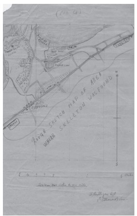
Fig. 4.57—Sketchmap, Bunduff, Co. Sligo.
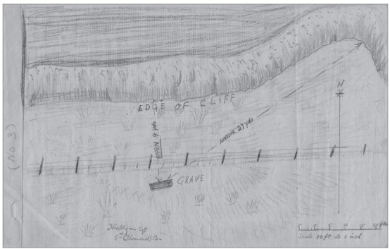
Fig. 4.58—Sketch showing location of grave, Bunduff, Co. Sligo.
at each end. The slabs seem to have been relatively thin, and at both the northern and southern sides the walls were chiefly made up of a very long slab with a shorter one added at each end. There was no evidence for capstones or lintels on the cist and its floor does not seem to have been paved. The grave contained an inhumation of an adult female (2010:91) and no accompanying artefacts were found. No details were recorded as to the disposition of the skeleton but it is probable, given the form of the grave, that the body lay in an extended position.
Comment
This burial is assumed to be early medieval in date, based on its form. No NMI investigation took place, and it is not known whether this was an isolated burial or whether others existed in the vicinity. It does not appear to be associated with an ecclesiastical site.
HUMAN REMAINS
LAUREEN BUCKLEY
Description of skeleton (2010:91)
The skull was complete and the long bones were present but decayed. Very little of the axial skeleton or small bones of the hands and feet survived.
Apart from some slight damage to the nasal area, the cranium was complete and in one piece, and the mandible was also complete. Only the first cervical, part of the second and third cervical and six middle thoracic vertebrae remained from the vertebral column. There were six ribs from the left side and two from the right side remaining, and the manubrium and most of the body of the sternum were present.
Only a fragment of the left scapula and the lateral half of the shaft of the left clavicle survived from the shoulder bones. The left arm consisted of the fragmented shaft of the left humerus and the complete shafts of the left radius and ulna. Only the shaft of the right
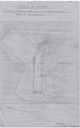
Fig. 4.59—Sketch-plan of grave, Bunduff, Co. Sligo.
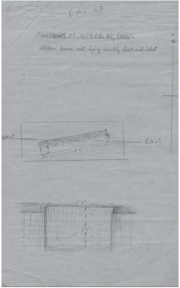
Fig. 4.60—Sketch-plan and section of grave, Bunduff, Co. Sligo.
humerus survived from the right arm. There were no joint ends present on any of the long bones of the arms. No hand bones remained. Fragments of both ilia and of the left ischium remained from the pelvic bones, along with fragments of the sacrum. The shaft of the left femur was complete and part of the proximal end was also present. The right femur was almost complete but slightly damaged at the joint ends. Both tibiae were present but their proximal ends were missing or badly damaged. The shaft of the left fibula was present and only the proximal end was missing from the right fibula.
Both calcanea, the right navicular and cuboid, the shaft of the left first metatarsal and incomplete first, second, third and fifth metatarsals were all that remained of the foot bones. The joint surface of the metatarsals and their shafts were very decayed, making them difficult to identify.
Age, sex and stature
All the morphological features of the skull—the mastoid processes, the supraorbital ridges, the orbital rim and external occipital protuberance—were of the female type. The only feature remaining from the pelvis was the left sciatic notch and this was also of the female type. The diameters of the femoral heads were also consistent with the individual’s being a female. Examination of the auricular surface of the left ilium indicated that the individual was aged 35–39 years at the time of death. As there were so few features remaining from the rest of the skeleton, the degree of suture closure in the skull was examined; this was consistent with the individual being an adult aged 28–44 years. Dental attrition was not heavy so it is probably safe to assume that this was a middle adult aged 26–45 years at the time of death. The living stature was estimated, using the length of the right femur and tibia, as 156cm.
Skeletal pathology
There were very deep Schmorl’s nodes on the inferior surfaces of the bodies of all the thoracic vertebrae that were present. In two of the vertebrae the nodes were so deep and large that they had broken the posterior surface of the body. Since the posterior surface, which lines the spinal canal, was damaged, it is more likely that the trauma that caused the nodes impinged on the spinal cord, causing a great deal of pain and possibly other neurological symptoms.
Attrition: there was a moderate degree of wear on the upper first right molar and on the lower first molar but very light wear on all other remaining teeth.
Calculus: no calculus deposits were noted.
Caries: there were very small pin-pricks of caries on the buccal surfaces of the second and third upper molars, with the distal surface of the third molar also affected; there were also small cavities on the buccal surfaces of both lower third molars.
Hypoplasia: linear enamel hypoplasia was noted on all the first molars and on the upper left second premolar.
Non-metric traits
In the skull, ossicles were noted in both sides of the lambdoid suture and there was a torus on the left side of the mandible. Squatting facets were present in both tibiae.
Summary and conclusions
This was the incomplete skeleton of an adult female with a living stature of 156cm. Apart from the skull, which was intact, most of the bones were in a very decayed state. There was very little pathology on the bones, apart from some deep Schmorl’s nodes on the vertebral column that must have caused considerable pain at the time they occurred. The teeth were in very good condition, although there were some very small cavities beginning to form on the molars.
108. Parish of Ahamlish, barony of Carbury. SMR SL003-002——. IGR 172600 356390.