1944:002 - CURRAGH, CO. LAOIS, Laois
County: Laois
Site name: CURRAGH, CO. LAOIS
Sites and Monuments Record No.: N/A
Licence number: E1106
Author: ELLEN PRENDERGAST
Author/Organisation Address: —
Site type: Iron Age and early medieval graves, c. 300 BCc. AD 1200
Period/Dating: —
ITM: E 668624m, N 678398m
Latitude, Longitude (decimal degrees): 52.852134, -6.981126
Introduction
In early June 1944 a number of extended skeletons were discovered during digging in a sandhill near Killeshin, Co. Laois. Mr Ovington, the landowner, immediately informed the Garda Síochána at Carlow, who in turn alerted the NMI to the find. The sandhill site had not been dug in living memory, but there was a definite area of depression where earlier disturbance had taken place. It consisted of a natural sand mound measuring approximately 60m across. The site was investigated by Ellen Prendergast and Joseph Raftery. At least four skeletons had been removed by workmen but the total number of burials excavated was five. This report is based on a manuscript on file written by Ellen Prendergast and on the site drawings and photographs. The human remains were analysed by Dr Amoroso-Centeno on behalf of Professor Keenan and were subsequently re-examined by Laureen Buckley.
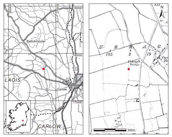
Location (Fig. 4.29)
The site was in the townland of Curragh, south-east Co. Laois, close to the border with Carlow and Kildare.64 It was a large, circular sand mound in a flat area. Cultivation ridges were evident at various angles to the mound in the surrounding field, and the old course of a stream was visible at the western edge of the mound.
Description of site
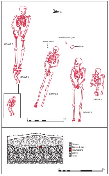
All the burials were concentrated at the same level and confined to a well-defined area in the centre of the mound. The burials seemed to follow the contour of the mound and lay under gravel and clay over purely sandy strata (Fig. 4.30) at a depth of 0.6m below the surface of the mound. The mound appears to be a naturally occurring feature. There was no evidence of a grave structure or slabs near any of the burials, but a darkening of the soil could be detected where the skeletal remains were in immediate contact with the soil. The graves all contained inhumation burials and no associated artefacts were found with any of the burials.
Grave 1
This contained the remains of an adult male and several disarticulated bones of a second individual (1944:243). The skeleton lay fully extended and supine, aligned west/east with the head to the west and the skull turned on its right side, looking south. The arms were placed parallel to the sides with the hands crossed over the pelvis. The lower leg bones and one femur had been uncovered prior to the excavation but the excavator was unable to establish their original position.
Grave 2
The remains have been identified as those of an adult female. The burial was approximately 0.2m north of grave 1, lying on a parallel axis. The lower limbs of the body were missing and were thought to have been removed by visitors to the site, as the side of the sandpit had been very disturbed in this area.65 The remaining portion lay in a supine position, also with the head to the west. The right arm was flexed over the ribs and the head rested on its right side, looking south. The left arm lay by the side. A 1986 study of skeletal material from Ireland found that this individual suffered from spina bifida (Saluja 1986).66 A sample of bone from this burial was submitted for radiocarbon dating and yielded a result of 1370±40, which calibrates to AD 598–767.67
Grave 3
This grave contained the remains of an adult male. It was approximately 0.4m south of grave 1 and was complete when excavated. The body lay fully extended, supine, and aligned approximately west/east. It measured 1.7m from the tip of the skull to the toes. Unlike the burials in graves 1 and 2, the skull was not turned sideways but lay facing forward. The left arm was flexed at the elbow and the hand lay over the pelvic region. The right arm lay parallel to the side, with the lower arm slightly flexed over the pelvic region. The legs were extended.The right patella and left toe bones were missing.
Grave 468
The remains have been identified as those of an unborn child aged between full-term and three months. This was located approximately 0.6m south of grave 3 and underneath the feet of the interment in grave 5. It lay in a supine position, on a parallel axis to the other burials, with the head to the west. The arms lay by the sides and the legs appear to have been slightly flexed. Owing to its young age, the skeleton could not be sexed.
Grave 5
This grave contained an adult male. It was located to the south of grave 3 and partly overlying grave 4. The body lay fully extended and supine, with the skull turned slightly to the left (north) and the arms parallel to the sides, with the hands resting over the hip joints. As mentioned above, the feet overlay the upper part of grave 4.
Other skeletal material
Various other human remains representing nine individuals were collected from the site: an isolated skull with the jaw underneath was found 0.3m west of grave 1, and a horse tooth and a few small teeth in a jaw were discovered approximately 0.2m west of graves 1 and 3 (Fig. 4.30).
Comment
This was clearly a cemetery of substantial size, and on the basis of the radiocarbon date is early medieval. The complete assemblage recovered during Prendergast and Raftery’s investigation represented at least fourteen skeletons. The analysis of the human remains indicates that the interred represent a normal graveyard population, consisting of male and female adults and juveniles. The cemetery does not appear to be associated with a church site.
HUMAN REMAINS
LAUREEN BUCKLEY
The number 1944:243 was applied to all the human remains from this site.
Skeleton 1 (grave 1): early middle adult male, 159cm
This skeleton was virtually complete and in excellent condition. The skull was complete and the cranium was in one piece, although it had been reconstructed and glued together rather than having been preserved intact. All the vertebrae were present and complete and there were thirteen ribs from the left side and twelve from the right side. The manubrium and body of the sternum were complete.
The left scapula was virtually complete and the right scapula and the clavicles were complete. All the arm bones were present and complete. Only the left triquetral and pisiform were missing from the carpal bones and all the metacarpals were present and complete. There were also ten proximal phalanges, eight middle and eight distal hand phalanges present.
The left and right ilia, ischia and pubic bones from the pelvis were present and complete and the sacrum was also complete. All the leg bones were complete, apart from the left patella and the right patella, which were missing. All the tarsal bones were present, apart from the left talus, cuboid, navicular and middle cuneiform. The metatarsals apart from the left fourth metatarsal were all present and complete and there was one proximal foot phalanx.
Age and sex
All the observable features of the skull, the sciatic notch and pubic bones of the pelvis and the measurements of the heads of the femur and humerus were of the male type.
The surface of the pubic symphyses indicated an age at death of 19–34 years, and the auricular surface of the ilium indicated an age of 25–29 years. The sternal end of the ribs also indicated an age of 25–30 years. Therefore this individual was definitely a young adult, aged 25–34 years at the time of death, and was most likely in the younger part of this age group. Stature based on the lengths of the femur and fibula was estimated at 159cm. This is very small for a male, but although the long bones were short in length they were very thick and strong, with well-defined muscular markings.
Skeletal pathology
The frontal bone and anterior parts of both parietal bones were very porotic, although as the skull was intact it could not be determined whether hyperostosis was present or not.
The proximal articular surfaces of a few of the middle hand phalanges seemed to be more cup-shaped than they should be, and one had evidence of slight chondral defects. There was a large, smooth, circular lytic defect on the superior surface of the right lunate in the prearticular area.
There were several problems with the vertebral column in this individual. The anterior surfaces of the cervical vertebrae had a very flattened appearance. The lower thoracic vertebral bodies have a squared-off appearance at the front; this, however, does not seem to be pathological but rather a developmental defect. There was a distinct scoliosis with convexity to the left in the lower thoracic region and there was also some compression of the intervertebral disc space, with the inferior articular surfaces of the ninth thoracic vertebra pressed onto the superior surfaces of the tenth thoracic with slight extension of the articular surfaces on to the transverse processes. The effect was more marked between the tenth and eleventh thoracic vertebrae but occurs only on the right side between the eleventh and twelfth thoracic vertebrae. There was some porosity of the right superior articular process of T12 and considerable extension of the articular surface onto the rudimentary transverse process.
The first lumbar vertebra had articular processes for the head of a lumbar rib on each side. Only one lumbar rib was found among the remains and it was small and rudimentary. Lumbar ribs are rare but they do occur. The body of the first lumbar vertebra was also slightly compressed on the left side and the anterior surface was slightly square. The second lumbar was compressed on the left side, with a localised area of osteophytes on the inferior surface on the left side. There was convexity to the right in the area of the second and third lumbar vertebrae. The third lumbar was also compressed on the left side, with moderate osteophytosis on the right side of the superior margin. The fourth lumbar vertebra was slightly compressed on the left side and there were moderate osteophytes on the inferior edge on the right and mild osteophytes on the right superior edge. The fifth lumbar had mild osteophytes all around the inferior edge. The body of the first sacral vertebra was severely compressed on the right side and is slightly tilted, possibly attempting to fuse with L5.
The scoliosis seems to originate with the abnormal development of the first sacral vertebra, with the compression on the left side of the lumbar vertebrae representing a possible attempt by the spine to straighten itself. There was a tighter curve to the ribs on the right side as a result of the scoliosis.
In addition to the scoliosis, there were very slight Schmorl’s nodes on the inferior surface of the body of the ninth thoracic vertebra, the superior surface of the tenth, the inferior surface of the eleventh and both superior and inferior surfaces of the twelfth thoracic vertebra. The superior surface of the first lumbar vertebra also had a slight Schmorl’s node.
Osteochondritis dessicans was present on the posterior surfaces of both calcanea (Pl. 109). The right navicular also had a well-developed tubercle. Both calcanea have welldeveloped peroneal tubercles, almost like an additional facet. The left bone was even more developed than the right and there was slight degeneration on the inferior part of the facet. The peroneal tubercle varies in size and is often not present. There are grooves above and below the tubercle for the peroneus longus and peroneus brevis tendon. The tubercle itself provides attachment for the middle third of the tendo calcaneus (Achilles’ tendon) and the anterior part of the tubercle articulates with the cuboid bone. Its presence here could be just a developmental trait of no particular significance or it could have overdeveloped as a result of strain on the leg muscles due to the marked scoliosis of the spine.
Dentition
Abrasion: the occlusal edges of the upper central incisors were chipped and broken and the left upper lateral incisor was also chipped to a slight degree. This may indicate that the teeth were used as tools.
Attrition: there was heavy wear on the upper and lower first molars on the left side. Attrition was moderate on the other molars, premolars and incisors.
Calculus: there were light deposits on the buccal and lingual surfaces of most of the teeth, with moderate deposits on the buccal surfaces of the upper left lateral incisor, canine and first premolar and on the buccal surface of the right lower incisors and canine. There were also moderate deposits on the lingual surfaces of the lower right incisors and lower left molars.
Caries: there was a small cavity on the distal surface of the root of the upper right second premolar, at the cervical margin. The upper left second molar had a moderate cavity on the distal surface at the cervical margin and there was a large cavity on the mesial surface of the crown of the adjacent third molar, also at the cervical margin. In the mandible there was a small cavity on the distal surface of the right second premolar at the cervical margin and the crown of the adjacent first molar was completely destroyed by caries, with only the roots remaining.
Periodontal disease: there was a slight degree of alveolar recession around the roots of most of the mandibular teeth, with recession being moderate around the lower right premolars and first molars. In the maxilla there was moderate recession around all the molars and premolars and right canine.
Hypoplasia: linear enamel hypoplasia was present on the central incisors, lower right canine and lower left first premolar
Skeleton 2 (grave 2): middle adult female, 158cm
This consisted of the upper half of a skeleton only; the lower half was disturbed before the burials were discovered. The skull was in a fragmented state; although it had been reconstructed in the past, some of the glue has now become unstuck. Apart from the orbital and facial area it was virtually complete. The cervical and upper half of the thoracic vertebrae were present and complete but the lower thoracic and first three lumbar that were present were fragmentary. There were twelve ribs from the left side and eight from the right and part of the manubrium was present.
Both clavicles were complete and most of both scapulae were present. The humeri were virtually complete and only parts of the proximal ends were missing from the right radius and ulna. The proximal thirds of the left radius and ulna were also present. The scaphoid, trapezium, capitate and hamate remained from the right hand, along with all the metacarpals and five proximal and two middle phalanges. No bones of the pelvis and legs were found in situ but some additional disarticulated fragments were with this skeleton and they are described below.
Age and sex
All the features of the skull, supraorbital ridges, mastoid processes, orbital rims, the external occipital protuberance and mental eminence were of the female type. The metric data were also consistent with the individual’s being a female. The only method of ageing available was the sutures of the skull and they indicated an age of 27–44 years. The stature was estimated, using the length of the ulna, as 158cm.
Skeletal pathology
Some degenerative joint disease was present in the vertebral column. It was generally mild with some lipping of the posterior articular processes, although there was severe lipping in the second and fifth cervical vertebrae and first thoracic vertebra. Mild lipping was also present in the middle thoracic with moderate lipping in the sixth thoracic vertebra. There was mild osteophytosis on T4–T6 only. Schmorl’s nodes were present on the inferior surfaces only of the lower six thoracic vertebrae.
There was mild degenerative joint disease on the left distal humerus, with mild lipping on the posterior and lateral edges of the trochlea. The costo-vertebral joints were also affected, with degenerative joint disease of four left ribs at the head and two right ribs at the transverse articular surfaces. Osteochondritis was present on the left superior articular surface of the second cervical vertebra.
Dentition
Attrition: there was moderate wear on the upper premolars and all the first and second molars. Attrition was light on the remaining teeth.
Calculus: there were light deposits on the lingual surfaces of the upper left third molar, upper right first premolar, lower left premolars and on the buccal surfaces of the lower right molars. Deposits were moderate on the lingual surfaces of the lower right incisors, premolars and molars and the buccal surfaces of the lower left second and third molars. There were heavy deposits on the buccal surfaces of the lower left first molar and the lingual surfaces of the lower left first molar and lower right canine.
Periodontal disease: there was a slight degree of alveolar recession around the roots of most teeth, with moderate recession around the roots of the upper left canine, premolars and molars and upper right premolars and first molar. There was considerable alveolar recession around the roots of the lower left first molar, with moderate recession around the roots of the second and third left molars.
Skeleton 3 (grave 2): young adult male, 164cm
This skeleton was virtually complete and in excellent condition. The skull was very fragmented but most of the bones were complete. The cranium had been reconstructed in the past although it was now falling apart. All the vertebrae were present and complete and there were twelve ribs from the left side and eleven from the right side. The manubrium and body of the sternum were complete.
The left scapula was virtually complete and the right scapula was complete. Both clavicles were complete. All the arm bones were present and complete. Only the left lunate and triquetral and both pisiforms were missing from the carpal bones, and all the metacarpals were present and complete. There were also eight proximal phalanges, four middle and two distal hand phalanges present.
The left and right ilia, ischia and pubic bones from the pelvis were present and complete and the sacrum was also complete. All the leg bones were complete, apart from the left patella and the right patella, which were missing. All the tarsal bones were present, apart from the left navicular and cuneiforms. The right metatarsals were complete but only the proximal end of the left third metatarsal remained. There were three proximal phalanges and one distal phalanx from the right foot.
Age and sex
All the observable features of the skull, the mastoid processes, supraorbital ridges, orbital rim and mental eminence were of the male type. The sciatic notch and pubic bones of the pelvis were also of the male type. Most of the metrical data were consistent with the individual’s being male, although the length of the clavicles and diameter of the heads of the humeri were in the female range. As the pelvis is the most reliable indicator of sex, it can be stated for certain that this was a male skeleton.
The surface of the pubic symphyses indicated an age at death of 19–34 years, and the auricular surface of the ilium indicated an age of 20–29 years. The sternal end of the ribs indicated an age of 20–23 years. The skull sutures were open and the first sacral was not fully fused to the second sacral vertebra, therefore the individual was probably a young adult aged 20–25 years at the time of death. Based on the lengths of the femur and fibula, stature was estimated at 164cm.
Non-metric traits
There was an ossicle at lambda and an ossicle in the left lambdoid suture. Lateral squatting facets were present on both tibiae.
Skeletal pathology
There were thirteen thoracic vertebrae instead of the usual twelve. The frontal bone was very porotic but the diploe was not expanded. The palmar surface of the right first metacarpal had a small exostosis near the distal end that probably represents some ligament damage. There was also some destruction of the distal end of the proximal phalanx of the little finger of the right hand that may have been caused by an injury.
In the vertebral column there was some slight compression of the joint spaces on the right side between T2 and T3, between T3 and T4 and between T4 and T5. There was also mild marginal lipping on the articular surfaces on the right side of the third, fourth and sixth thoracic vertebrae and also on the lower thoracic vertebrae.
Schmorl’s nodes were present on the inferior surface of the eighth thoracic vertebra, and on the superior and inferior surfaces of the ninth, tenth, eleventh, twelfth and thirteenth thoracic vertebrae and the upper two lumbar vertebrae. There were also Schmorl’s nodes on the superior surfaces of the third and fourth lumbar vertebrae. The nodes were very deep in the mid-thoracic region and mild in the lower thoracic and lumbar region.
Osteochondritis was present on the posterior surface of the right calcaneum and there was a chondral defect on the proximal articular surface of the first proximal phalanx.
Dentition
Attrition: there was moderate wear on the first molar and light wear on all the other teeth.
Calculus: there were light deposits on the lingual surfaces of the upper right incisors and molars and lower incisors, canines and premolars. There were moderate deposits on the buccal surfaces of the upper right incisors, upper right first molar and upper left second premolar. There were also moderate deposits on the buccal surfaces of the lower left second premolar and first molar and lower right third molar as well as the lingual surfaces of the lower right molars. Deposits were heavy on the buccal surfaces of the lower left incisors, canine and first premolar, the buccal surface of the lower right lateral incisor and canine and the lingual surface of the lower left first molar.
Caries: there were small cavities on the occlusal surfaces of the upper right and both lower third molars and a moderate-sized cavity on the occlusal surface of the lower left second molar. There was also a small cavity on the distal surface of the upper left second premolar at the cervical margin, and a large cavity on the mesial surface extending onto the occlusal surface of the upper right first molar.
Periodontal disease: there was a slight degree of alveolar recession around the roots of the upper right canine, premolars and first and second molars.
Hypoplasia: linear enamel hypoplasia was present on the upper right incisors and canine, all the upper molars and the lower canines, first premolars and second molars. There were also pits of hypoplasia on the lower right second molar.
Skeleton 4 (grave 4): infant, 0–6 months
This skeleton lay partially under the feet of skeleton 5. It was virtually complete and in excellent condition. The skull was totally fragmented but most of the bones were present, including the maxilla and mandible. The right halves of the neural arches of three lower cervical vertebrae were present; there were five centra, nine right and ten left halves of the neural arches of the thoracic vertebrae, and five centra and four arches remained from the lumbar vertebrae. There were eleven ribs from the left side and nine from the right side. The manubrium of the sternum was complete.
The scapulae and clavicles were complete and all the arm bones were present and complete. Only one metacarpal and one phalanx remained from the hand bones.
The left and right ilia and ischia and right pubic bone from the pelvis were present and complete and the first two sacral vertebrae were present. All the leg bones were complete apart from the patellae, but only four unidentified metatarsals were present from the foot bones.
Skeletal pathology
The anterior surfaces of both femurs and the entire medial surfaces of both tibiae were covered in a layer of porotic bone, which could be periostitis. The internal surface of the occipital bone was also very porotic.
Dentition

The crowns of the central incisors were formed and the crowns of the lateral incisors were almost complete. The crowns of the first deciduous molars were incomplete and the occlusal surfaces of the second deciduous molars had just formed. With this state of dental development the infant was certainly less than six months of age and was probably aged 0–3 months.
Skeleton 5 (grave 5): early middle adult male, 170cm
This skeleton was almost complete and in excellent condition. The skull was virtually complete and the cranium was in one piece, although part of the right side of the maxilla was missing. All the vertebrae were present and complete and there were ten ribs from the left side and twelve from the right side. The manubrium and body of the sternum were complete. Both scapulae and both clavicles were complete. All the arm bones were present andcomplete except for the distal third of the left ulna, which was missing. Only the left triquetral and trapezium and both pisiform bones were missing from the carpal bones, and all the metacarpals were present and complete. There were also ten proximal phalanges, seven middle and six distal hand phalanges present.
The left and right ilia, ischia and right pubic bones from the pelvis were present and complete and the sacrum was also complete. All the leg bones were complete, including the patellae. All the tarsal and metatarsal bones were present and complete and there were ten proximal phalanges, one middle foot phalanx and one distal foot phalanx.
Age and sex
All the observable features of the skull, the sciatic notch and pubic bone of the pelvis and the metric data were of the male type.
The auricular surface of the ilium indicated an age of 25–34 years. He was probably closer to the younger end of this age group, as the epiphyseal line was still visible at the proximal femurs and vertebral bodies.
Based on the lengths of the femur and fibula, stature was estimated at 170cm.
Non-metric traits
There was a sternal foramen in the body of the sternum. There were squatting facets on both tibiae.
Skeletal pathology
In the middle of the frontal bone there was a small glancing blow to the skull. The lesion was very small, approximately 9mm in length, and seems to have been made with a sharp object. The wound would have caused extensive bleeding but would not have been fatal, and indeed it was partially healed, indicating that the individual lived for some time after receiving it. There was no sign of infection around the wound. Schmorl’s nodes were present on the surfaces of the bodies of the lower thoracic vertebrae, T8–T12, and on the upper three lumbar vertebrae.
There was a smooth chondral defect on the trochlea of the left humerus. Osteochondritis dessicans was present on both superior articular surfaces of the second cervical vertebra.
The articular surface for the navicular on the left talus had a very large lesion of osteochondritis dessicans, 17mm long. There was also flattening of the joint surface and an area of eburnation around the lesion of osteochondritis. In the non-articular area behind the anterior facet there was a raised area of bone that was highly porotic and might indicate a slight callus after a fracture. The talar surface of the navicular was similar, with lots of destruction, a flat surface instead of a concave surface, and an area of eburnation in the middle. There was some destruction of the bone on the superior, non-articular surface and also some bone destruction of the medial, non-articular surface of the first cuneiform. These changes may be due to a trauma-related injury.
Dentition
Unfortunately the teeth had been glued back into their sockets years ago and it was not possible to examine them properly.
Attrition: there was moderate wear on the upper and lower first molars and very little wear on the other teeth.
Calculus: there were light deposits on the buccal surfaces of the upper right lateral incisor and canine, upper left teeth, lower left canine and lower right premolars and second molar. There were also light deposits on the lingual surfaces of the upper left premolars and lower right first and second molars. The buccal and lingual surfaces of the upper right premolars and first molars, the lingual surfaces of the other upper molars, lower left premolars and molars and lower right third molars all had moderate deposits of calculus.
Periodontal disease: there was a slight degree of alveolar recession around the roots of mandibular molars.
Hypoplasia: linear enamel hypoplasia was present on the incisors, canines, lower left premolars and lower right first premolar.
Skeleton 6
These were disarticulated bones collected from the site and were labelled skeleton 6, but in fact, as they are bones from the lower half of a skeleton only, they could belong to skeleton 2. Part of the right ilium and ischium was present. The proximal halves of both femurs, the distal two-thirds of the left tibia and fibula and the distal end of the right tibia were also present. Both tali and calcanea were present from the foot bones.
Age and sex
The sciatic notch was wide and the diameters of the heads of the femurs were in the female range.
Skeletal pathology
There was severe marginal lipping of the superior part of the right acetabulum and moderate lipping of the inferior part. The head of the right femur had a moderate degree of surface osteophytes
Disarticulated bone
A number of disarticulated skull and long bones were found on the site.
Skull bones
The following minimum numbers of skull bones were present.
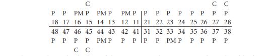
Table 4.6—Minimum numbers of skull bones.
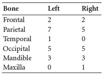
The minimum number of skulls present was seven. Three of the occipital bones were probably from male individuals.
Mandible 1:
There was moderate wear on the molar teeth and light wear on the premolars. The premolars had light calculus deposits on their lingual surfaces and the right molars had light deposits on their buccal surfaces, and there were moderate to considerable deposits on the lingual surfaces of the left molars. The crown of the right second premolar, 45, was almost completely destroyed by caries. There were grooves of hypoplasia on the right canine and linear enamel hypoplasia on the left first premolar.
Mandible 2:
Some of the teeth had been glued into their sockets. There was light wear on all teeth. The left lateral incisor had moderate calculus deposits on its buccal surface. There was linear enamel hypoplasia on the left incisors, canine, premolars and second molar.
Mandible 3:
The crown of the remaining molar was completely destroyed by caries and the sockets for the adjacent third molar that had been lost during life were completely healed. There was a large area of erosion around the root of the first molar, which may have been the remains of an abscess that had not healed. The carious second molar also had an associated external abscess.
Maxilla 1:

There was moderate attrition on the two remaining molars and light calculus deposits on their buccal and lingual surfaces.
Long bones
The following disarticulated long bones were present.
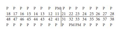
Table 4.7—Disarticulated long bones.
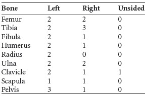
At least two of the pelves were from female individuals.
Juvenile bones
A fragment of humerus shaft from an older juvenile or adolescent was present. There was also one incomplete calcaneum and one sternaebrae. A juvenile and an infant rib were present. There was also another skull from an infant. It consisted of the right side of a frontal bone, left and right parietal bones and the squamous occipital bone. Also present were a fragment of rib and the proximal half of a right humerus. The infant was probably aged 0–6 months. Some burnt bone was also present. This included a thoracic vertebral body, a fragment of ulna and a well-cremated fragment of femur shaft, as well as a fragment of animal bone.
Isolated skull
This skull was labelled on the plan. It consisted of fragments of a juvenile skull. There was a tiny fragment from the middle of the frontal bone, with part of the crista frontalis visible and part of the frontal sinus. The basal, lateral and part of the squamous occipital bones were present and the lateral occipitals were fused to the basal occipital. Both temporal bones were present and the right temporal was almost complete. The sphenoid bone and fragments of the maxilla were also present.
The upper five cervical vertebrae were also present and virtually all complete. The arches were fused to the centra.
Dentition
The crown of the third molar, 48, was complete and the root was just starting to form. The individual was probably aged 12–13 years. The tips of the roots of the second molars were not fully closed. The left premolar, 34, was triple-rooted. Linear enamel hypoplasia was noted on the upper right first molar, lower right first and second molars and lower left premolars and first molar. There were also pits of hypoplasia on the lower right second molar.
Stray infant bones
Bones from another infant were present. These included three small fragments of skull, including a right orbit. Also present were six left neural arches and three right neural arches from the cervical vertebrae, and five left and four right neural arches of the thoracic vertebrae and a left first rib. The vertebral fragments could have belonged to the same individual as the disarticulated infant skull described above. A few fragments, including the lateral end of a right clavicle, two fragments of long bones and two teeth, from an older juvenile were also present.
Minimum number of individuals
The minimum number of individuals recovered from this site is therefore fourteen. This included four excavated adults, seven disarticulated adults, an older juvenile, one excavated infant and another disarticulated infant.
Summary and conclusions
Although only five individuals were excavated, fourteen individuals were found on the site. These consisted of eleven adults and three juveniles. The adults consisted of three males and one female from the in situ burials, with a further minimum of three males and two females from the disarticulated remains. Although the remains represent only a small sample of the buried population, there did not appear to be older individuals present. One of the excavated males was a young adult and two were in the early middle adult age range, with the female burial being a middle adult. The juveniles consisted of an older juvenile (6–12 years) or early adolescent and two infants less than six months old at the time of death. One of the infants may have suffered from an infection before death. All of the adults bore evidence of a vigorous lifestyle involving manual labour. They all had Schmorl’s nodes in the vertebral column and, despite their young ages, three had early indications of degenerative joint disease or compression of the intervertebral disc spaces caused by pressure on the spine, which might have gone on to develop into DJD if they had lived longer.
Skeleton 1, an early middle adult male, had a double scoliosis of the vertebral column, originating with the compression of the first sacral vertebra on the right side, causing a tilt in the vertebral column. The scoliosis is an adaption by the body in an attempt to straighten the spine. Other adaptive changes can also occur, such as the difference in the curvature of the ribs that was seen in this individual. It is also possible that the scoliosis affected this man’s gait, putting strain on the tendons of the foot.
The scoliosis can be seen as a developmental problem but there were other developmental anomalies seen in this population, although they may not have had any effect on the individuals concerned. Skeleton 1 also had a pair of lumbar ribs and skeleton 3, a young adult male, had thirteen thoracic vertebrae instead of the usual twelve.
Direct evidence for weapon trauma was seen in only one individual, skeleton 5, who had received a glancing blow with a sharp instrument to the front of the skull. It had not caused death and there was some evidence of healing. This same individual had also probably suffered a fracture of the talus in the ankle and possibly trauma to other ankle bones. One consequence of the trauma to the ankle may have been the large lesion of osteochondritis on the navicular surface of the talus, around which, despite the individual’s relatively young age, osteoarthritis had already developed. Osteochondritis dessicans is an area of bone death usually seen on a convex joint surface. It generally occurs in the ankle or knee but can also occur on other joints and is thought to be caused by trauma. Skeleton 5 had osteochondritis dessicans on both superior articular surfaces of the second cervical vertebra, and skeleton 2, a middle adult female, also had the lesions at the same location. Osteochondritis dessicans was also present in skeleton 1 in both calcanea and in the right calcaneum of skeleton 3. Therefore three of the four excavated adults had evidence of trauma to the ankle joints.
This may be aconsequence of walking unshod on rough ground or may be due to accidental falls. Skeleton 3, the young adult male, may also have suffered some trauma to his little finger and some ligament damage to his thumb. The teeth from the excavated remains, as well as the stray mandibles, suggest that attrition caused by coarsely ground food in the diet was high and dental hygiene was low, with moderate to severe calculus deposits on most of the dentition. There was also evidence of periodontal disease, although it tended to be slight owing to the relatively young age of the individuals. It was surprising that dental caries was found in two of the males and in two of the disarticulated mandibles. One of the males had four carious teeth and the other had six. Dental caries does not tend to be high in early medieval populations although, since only four adult individuals are represented here, it is not possible to determine the caries rate for the entire population. In conclusion, it can be said that the remains from this site probably indicate a normal cemetery, with males, females and juveniles present. The adults seemed to have had a vigorous lifestyle, suffering from stress on the spine, even at a young age, and most seemed to have had injuries to the ankle bones.
64. Parish of Killeshin, barony of Slievemargy. OS 6in. sheet 32. IGR 268689 178360.
65. The remains consisted of skull, vertebrae, arms, ribs and part of pelvis.
66. Although Saluja does not refer to the skeleton number, this was the only female skeleton from the site.
67. GrA-29054.
68. Apparently termed number 6 in the anatomist’s report.