1942:005 - ROSSNAREE, CO. MEATH, Meath
County: Meath
Site name: ROSSNAREE, CO. MEATH
Sites and Monuments Record No.: SMR ME019-059
Licence number: E1141
Author: JOSEPH RAFTERY
Author/Organisation Address: —
Site type: Iron Age and early medieval graves, c. 300 BCc. AD 1200
Period/Dating: —
ITM: E 699179m, N 772558m
Latitude, Longitude (decimal degrees): 53.693305, -6.498251
Introduction
In April 1942, during excavations for machine-gun posts, an inhumation was discovered by military engineers near Slane, Co. Meath. The bones were discovered approximately 0.3m under a pile of small stones. These stones were removed and a flexed burial was found. It was left intact by the military but was apparently disturbed overnight. The site was subsequently investigated by Dr Joseph Raftery. The human remains were initially analysed by R.G. Inkster, Trinity College, Dublin, but were re-examined by Laureen Buckley. As there are only some short notes on file, this report is based largely on Raftery’s plan and photographs of the grave.

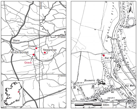
Location (Fig. 4.40)
The site was in the townland of Rossnaree, north-east Co. Meath85. It lay on the south bank of the River Boyne opposite Newgrange, at an altitude of approximately 15m above sea level.
Description of site
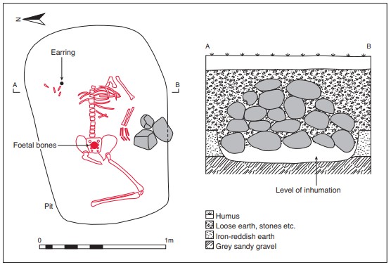
The grave consisted of a subrectangular pile of limestone stones averaging 0.3m by 0.3m by 0.15m. These had been removed by the time of Raftery’s visit, but according to his plan the burial lay under approximately 0.6m of stone, suggesting that a small cairn may have marked the grave (Fig. 4.41). The grave-pit appears to have been subrectangular in plan, with its long axis running east/west. There was no evidence for any stone lining around the grave and all of the stone appears to have been on top of the interment.
The grave contained the remains of three adult females and an infant (1942:1917.2), accompanied by a decorated silver earring (1942:1917.1). One of the adults—skeleton 1—lay with the head to the east and the feet to the west. Only the left leg remained, and this was flexed. The left arm was slightly flexed and lay beside the body. The right arm was missing and the skull also appears to have been missing. The rib bones were somewhat disturbed. From the plan and photographs, it appears that the burial may have been supine with the legs flexed to the left, but it is not possible to be certain about this as the burial was disturbed prior to Raftery’s inspection of the site. Raftery noticed what he thought were the bones of a dog or cat on and under the pelvis, but these were subsequently identified by R.G. Inkster as a foetus, and more recently Buckley (see below) found them to be the remains of an infant. The fingerring/earring was found to the north of the burial, close to where the right shoulder would

Fig. 4.41—Plan and section of grave, Rossnaree, Co. Meath.

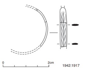
Fig. 4.42—Silver earring, Rossnaree, Co. Meath.
have been located. A sample of the human remains from skeleton 1 was submitted for AMS dating and yielded a date of 1660±40 BP, which calibrates to 257–533 at 95.4% probability.86
Silver earring, 1942:1917 (Fig. 4.42)
Two fragments of a silver earring were found on the right-hand side of the upper body of skeleton 1. As the skull, shoulder blade, arm and hand were not in situ at the time of the excavation, it is not clear whether or not the object, as marked on the plan, is in its primary position. As only two fragments survive, it may be that it was disturbed and broken during the interference with the site which was reported to have taken place. In the absence of any specific comment by the excavator, we cannot be certain that it was undisturbed.
It is a very finely made piece of silver metalwork. It consists of a narrow band of planoconvex section, decorated with a continuous panel of reserved/raised interlocking exaggerated trapezoidal shapes that superficially resembles a herringbone pattern. This is contained within an incised line just inside the edges of the object. Although incomplete, the smaller fragment has a broken tag at the narrow end of the band, which might be the start of the post that would have been inserted in the ear lobe. The fragments can be joined together and it is clear that the band narrows from its widest point in both directions.
Dimensions: combined L (both fragments) 17.78mm; max. W 3mm; T 0.8mm.
Comment
The object may be either a finger-ring or an earring. As the skeleton is female, if the custom of wearing earrings was derived from the Roman world through Britain, then the young woman should have had a pair of earrings. The Romans frowned on the idea of men wearing earrings, but those who did would have been more likely to wear a single earring rather than a pair (Allason-Jones 1989, 16–18). In the case of this burial, however, the remains are clearly those of a female.
The decorative pattern on the band is closely similar to those that are seen, for example, on the pins and pinhead rims of some disc-headed pins of the sixth to seventh centuries, such as the one from Craigywarren Bog, Skerry, Co. Antrim87 (Youngs 1989, 25). This motif may be derived from the stylised palm branch used in the Roman world to symbolise victory. It was also used to protect against evil forces and to assist the wearer to reach the afterlife. The palm branch motif was used on Roman finger-rings of gold, silver and bronze (Tyacke and Jackson 2008, 60, pls 107, 108). As used by the Christian church, the palm represented the victory of the faithful over the enemies of the soul.
In terms of comparative material from Ireland, the burial of a female at Rath, Co.88 Meath, is of some significance. This site was on the N2 Finglas–Ashbourne road scheme, about 0.5km north of Ashbourne (Schweitzer 2005) and about 25km south of Rossnaree. One of three ring-barrows produced evidence of burial over a period of time, including cremation deposits in the northern portion of the site. A recut of the backfilled ditch on the southern side of the barrow produced a crouched inhumation of a woman in her early to mid-twenties. Three copper-alloy rings were found on the toes of both feet. On the right foot, two toe-rings were found—the larger one encircling the big toe and the second toe, while the second ring was found around the fourth toe. This ring was decorated with a herringbone motif. The burial from Rath has not been dated owing to the poor condition of the bone, but Schweitzer (2005, 97–8) suggests that burials with toe-rings from Britain date from the late first century BC to the first half of the first century AD. Also found at Rossnaree in recent years was a bronze ring-headed pin (NMI 1998:80) of Raftery’s Type 1b, which he suggests may date from the third or second centuries BC to the first century AD (Raftery 1983, 152).
Given the result of the radiocarbon determination, the earring from Rossnaree fits, in an Irish context, well within the third- to sixth-century date bracket. The question arises as to whether this is a pagan or a Christian burial, and whether those buried at Rossnaree were Irish or, perhaps, British.
HUMAN REMAINS
LAUREEN BUCKLEY
The human remains were all registered as 1942:1917.2.
Skeleton 1: middle adult female, 165cm
Although this skeleton was disturbed, it was almost complete and the bones were in good condition. The skull was fragmented, but the occipital, both parietal, both temporal bones and the frontal bone were present, although only the left temporal was complete. The sphenoid, nasal bones and both zygomatic bones were also present. The left side of the maxilla was present and the mandible was almost complete. There was a lot of decay on the outer surface of the skull, with the outer cortex almost worn away in places.
The vertebral column was virtually complete, with only the second and fifth cervical vertebrae missing. There were ten ribs from the left side and eight from the right side present and fragments of the body of the sternum survived.
Both scapulae and clavicles were present but incomplete. Both humeri were virtually complete but their proximal ends were fragmented. The distal two-thirds of the left radius was present and the left ulna was almost complete but was missing part of the mid-shaft. The right radius was complete but the middle third of the right ulna was missing. All the carpals from the left hand were present, apart from the left trapezoid; all the metacarpals were present and there were five proximal, four middle and four distal phalanges. The right hand consisted of the scaphoid, capitate and hamate, all the metacarpals and two proximal phalanges and one middle phalanx.
Both ilia were present from the pelvis and were almost complete. The left ischium was present and the right ischium was complete. Only part of the left pubic bone survived. The sacrum was complete. The leg bones were present and complete. All the tarsals except for the left third cuneiform and right first and second cuneiforms were present and all the metatarsals were present.
Age and sex
There were a number of male and female features on this skeleton. The external occipital protuberance of the skull was pronounced, usually indicating a male, but the supraorbital ridges and the orbital rims were of the female type. The left mastoid process also seemed to be female but the right was more like a male. The mental eminence of the chin was of the female type. Only the sciatic notches were visible from the pelvis and these were wide, which normally indicates a female. The diameters of the heads of the femur and humerus and the width of the glenoid cavity were also in the female range.
Overall, it was felt that, despite the presence of some male features, most of the indicators of sex were of the female type and that this was a female skeleton.
The auricular surface of the pelvis and the sternal ends of the ribs indicated that this was a middle adult aged 26–45 years. The living stature—165cm—was estimated using the lengths of the humerus, femur and tibia.
Non-metric traits
Squatting facets were present on both tibiae.
Skeletal pathology
There was some evidence of slight degenerative change in some of the joints. In the left wrist there was mild marginal lipping around the distal surface of the left radius. At the right shoulder there was some porosity of the lateral end of the right clavicle. Mild degenerative joint disease was present in both hips, with moderate lipping of the lateral edge of the left acetabulum and mild lipping of the right acetabulum as well as mild lipping of the proximal ends of both femurs. At the knee there was mild lipping of the inferior edge of the left patella. The right tibia had a well-developed soleal ridge. The soleus muscle is used in walking and the insertion for it is well developed in individuals who do a lot of walking.
In the vertebral column there was moderate osteophyte formation at the dens of the first cervical vertebra, slight lipping of individual posterior articular surfaces in the sixth and seventh cervical vertebrae and in the eighth and ninth thoracic vertebrae. Only one lumbar vertebra, L3, had mild lipping of the right superior articular surface.
Osteophytosis was present to a very mild extent in the superior edges of the bodies of the third cervical and the eighth thoracic vertebrae and in the superior and inferior edges of the ninth thoracic body. There was also mild osteophytosis on the inferior edge of the body of the fourth lumbar and the superior edge of the body of the fifth lumbar. There was slight compression of the left side of the body of the fifth lumbar, possibly caused by lifting a heavy weight, and this would have contributed to the development of osteophytosis on the lumbar vertebrae. The twelfth thoracic vertebra was also compressed slightly on the left side of the body. A Schmorl’s node was present on the inferior surface of the second lumbar.
DJD was also present on some of the costo-vertebral joints, including the head of the right first rib and some middle ribs.

Abrasion: there was a vertical groove of abrasion on the buccal surface of 43.
Attrition: there was moderate wear on the first molars and light to moderate wear on most of the other teeth, although the lower central incisors had heavy attrition.
Calculus: there were light deposits on the buccal and lingual surfaces of the upper teeth except the right second molar, where deposits were moderate. There were light deposits on the buccal and lingual surfaces of the lower teeth except the lingual surfaces of the first molars and left second premolar, where deposits were moderate.
Periodontal disease: there was a slight degree of alveolar recession around the premolars and molars in the left side of the mandible.
Hypoplasia: linear enamel hypoplasia was present on the lower central incisors and lower left premolars. There were also pits of hypoplasia on the lower left first premolar, the lower right canine and upper right third molar.
Skeleton 1a: infant, 0–6 months
The bones from the infant were well preserved although very little from the upper half remained. The skull bones present included an almost complete occipital bone, fragments from the left and the right parietal bones, most of both temporal bones, the right greater wing of sphenoid and both zygomatic bones. There was also a small fragment of the right side of the mandible. Only three thoracic half-arches remained from the vertebral column and there were three ribs from the left side and six from the right side. No arm or hand bones were present. Only part of the right pubic bone remained from the pelvis. Both femurs and tibiae and the left fibula remained from the leg bones but there was only one metatarsal and one proximal phalanx from the left foot.
Age
The lengths of the long bones indicate that this infant is longer than the normal length of a fullterm infant and therefore it is not a neonate. There are no teeth or tooth sockets remaining to enable an accurate assessment of age at death, but from the length of the bones it was probably not much older than a neonate and would certainly have been less than six months.
Skeleton 2: young adult female
This consisted mainly of the lower half of a skeleton. Only the shaft of a left ulna remained from the upper half. The left ilium was present but was incomplete and there was one complete lumbar vertebra. The proximal half and the distal third of the left femur was present but the bone was very decayed. The left tibia consisted of the proximal joint surface and the distal half of the bone. Only the shaft remained from the right tibia.
Age and sex
The sciatic notch was wide and the bicondylar width of the femur was within the female range. It is therefore more than likely that this was a female skeleton.
The epiphyseal line of the posterior iliac crest was still visible and the epiphysis at the centrum of the lumbar vertebra was unfused. The individual was probably aged less than 25 years at the time of death.
Skeleton 3: probably female
This consisted of two femurs only. The left femur was almost complete but the distal third was missing. The shaft of the right femur was present. Although there were no features present to determine sex, the measurement of the head of the femur was in the female range.
Summary and conclusions
A total of three adults and one infant were recovered from this site. The most complete skeleton of an adult female, aged 26–45 years, was supine but the legs were flexed, and this has been reported in the past as a crouched burial. It is not the usual position for a crouched burial from the Bronze Age, however, and the radiocarbon date has confirmed this as it falls within the Iron Age/early medieval date range. Infant bones were found over the pelvic area but their exact position is not known, as locals disturbed the site before it could be investigated properly. The infant bones were not those of a neonate but of an infant not more than six months old, so the flexed position of the legs is not associated with the mother dying during labour.
The only pathology noted on the female was some mild degenerative changes in the shoulder and hip joints and in the vertebral column. There was some evidence that she had done a lot of walking during life and that she had lifted heavy weights, as there was some compression of the vertebrae. One of her tooth crowns was damaged, suggesting that she had used her teeth as tools. There was also evidence from the teeth that she had suffered periods of acute illness and nutritional deficiency during early childhood.
The second adult skeleton was also female and was a young adult less than 25 years of age. There were further leg bones from a third, probably female, individual. The bones of the second and third skeleton were not found in situ but were collected as disarticulated material from the site. It is unusual to find only females and an infant from one site. Even a small disturbance of a site normally provides a random sample of males, females and juveniles.
85. Parish of Knockcommon, barony of Duleek Lower. SMR ME019-059——. 299250 272540.
86. GrA-24354.
87. BM 1880.8-2.132.
88. 03E1214; excavated by Holger Schweitzer, CRDS Ltd.
