1940:006 - BALLYBRENNAN, CO. WESTMEATH, Westmeath
County: Westmeath
Site name: BALLYBRENNAN, CO. WESTMEATH
Sites and Monuments Record No.: SMR WM032-055
Licence number: E1162
Author: P.J. HARTNETT AND ELLEN PRENDERGAST
Author/Organisation Address: —
Site type: Early Bronze Age graves
Period/Dating: —
ITM: E 634338m, N 741361m
Latitude, Longitude (decimal degrees): 53.421248, -7.483400
Introduction
In August 1940 the first of several burials discovered in a sandpit at Ballybrennan, Co. Westmeath, was reported to the NMI. This grave was a small cist burial (referred to as cist 1 in the original report) that contained the remains of a child of about two years. In February 1945 another grave was reported. This was also a small cist (referred to as cist 2 in the original report), containing an inhumation of an adult female (mistakenly reported as an adolescent previously; see Buckley, below, grave 2). Animal bones representing an ox and a pony were found in the soil in the vicinity of the cists, but it is not clear whether these can be directly associated with either of the cists. Shells of a number of species of common snails were also found in cist 2, but their presence there may be fortuitous. There was no pottery found in either of these cists. It was reported locally that about twenty years previously an urn had been found but had been reburied. These sites, cists 1 and 2, were published by Ellen Prendergast (1945).
Subsequently, in January 1946, a third short cist (this report, grave 3) containing an inhumation and a bowl was discovered in the same sandpit on the lands of the Boyd-Rochfort family at Middleton Park, Castletown Geoghegan. 315
The NMI was informed of the find by the Garda Síochána at Castletown Geoghegan. The cist (grave 3) was found in the face of the sandpit at a depth of 0.4m below ground level, approximately 6.5m from where one of the cists (cist 2) was discovered the previous year. Work was suspended on the site pending a visit from the NMI. The site was excavated over two days by Dr Joseph Raftery and Ellen Prendergast. Two years later, in early March 1948, a fourth cist (this report, grave 4) was discovered at Ballybrennan after the collapse of one of the sides of the gravel pit and was investigated by P.J. Hartnett. The human remains from cists 1–2 and grave 3 were examined by Dr R.G. Inkster, Trinity College, Dublin, and the bones from grave 4 were examined by Professor E.J. Keenan, University College, Dublin. This report is based on Prendergast’s and Hartnett’s respective accounts of their excavations of graves 3 and 4. For the purposes of this report, the human remains from cist 2 and graves 3 and 4 were re-examined by Laureen Buckley.
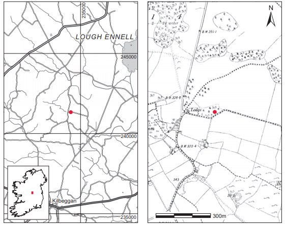
Location (Fig. 3.176)
The site was in the townland of Ballybrennan, south Co. Westmeath, just 2km south of Castletown Geoghegan.316 It is a small esker running east/west, the south side of which was being exploited for sand and gravel. The cists were found at an altitude of 90–100m above sea level. The area around Castletown Geoghegan has produced many early Bronze Age cist burials, such as Conranstown, north of the town, and Benalbit and Derryroe, a few kilometres south of Ballybrennan. In 1989 a number of inhumation burials were also discovered in Ballybrennan townland. An inventory report on these can be found in Vol. 2, pp 518–19.
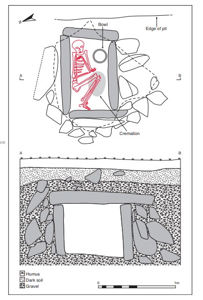
Grave 3 (Pl. 66)
The cist was rectangular in plan, with its long axis aligned north-west/south-east. It measured 0.96m long by 0.53m wide by 0.69m high (Fig. 3.177). The four cist walls were each formed of a single edge-set slab. The north-western end stone was flanked externally by a second edge-set slab. These two slabs were longer than that at the south-eastern end and extended out approximately 0.14m beyond that edge. All sides of the cist (apart from the south-eastern end at the face of the gravel pit) were supported by a packing of smaller stones. The cist was sealed by a large, irregularly shaped capstone which extended out approximately 0.5m on the south west side of the cist. It measured 1.42m long by 1.34m wide by 0.17m thick. The floor of the cist was not paved but consisted of sand. The pit dug to receive the cist does not appear to have been located.
The grave contained a crouched inhumation of an adult male (1946:13) and the cremated remains of an adult male (1946:14) accompanied by a bowl. The skeleton was crouched on its left side, with the head in the north-east corner and the feet at the north-western end. The cremation deposit had been placed carefully over the knees. The bowl was placed towards the south-eastern corner, close to the head, and had rested partly on its side. It was encrusted on the uppermost edge with limestone drip from the capstone of the cist.
Tripartite bowl, 1946:12 (Fig. 3.178)
This tripartite bowl is complete. The rim is slightly indented but undecorated. The vessel has two raised mouldings at the centre of the body, both of which are decorated with a row of vertical comb impressions. There is an undecorated gap on either side and between the mouldings. Above the first moulding the decoration consists of four panels, which from the
rim down consist of a panel of vertical comb impressions, a chevron in false relief, a row of slanted comb impressions and another chevron in false relief.
Below the lower moulding are three panels of ornament separated by undecorated panels. A double chevron row forming a series of lozenges is followed by a row of slanted comb impressions, which is followed by a row of vertical comb impressions. The base is plain.
Dimensions: H 12cm; ext. D mouth 16.3cm; int. D mouth 14.3cm; D base 7cm.
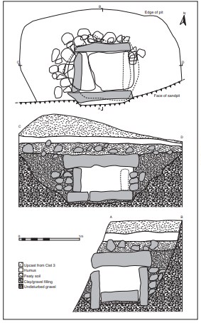
Grave 4 (Fig. 3.179)
The cist was almost square in plan, with its long axis aligned east/west, and lay 0.8–0.9m below ground level. Internally it measured 0.65m long by 0.6m wide by 0.3m high. It was formed of four main slabs set on edge, with one at each side. The southern and eastern slabs had been exposed by the collapse, and the eastern slab had been displaced. The western and apparently the eastern end slabs fitted between the northern and southern slabs. All sides of the cist appeared to have been supported externally by small packing stones. The cist was sealed by a large, subrectangular capstone, which measured 0.9m long by 0.8m wide by 0.2m thick. According to Hartnett, the capstone rested on the northern slab, the western slab and, at one point only, the southern slab, but did not touch the eastern slab.317 The floor of the cist was paved with two slabs, approximately 0.2m thick, and the interstices were filled with smaller angular spalls. The pit containing the cist was irregular in shape, measuring 2m long by 1.5m wide by 0.9m deep. It was dug through turf and humus into gravel.
The cist had contained a large deposit of soil when discovered, but had been emptied by the time of Hartnett’s excavation. The cremated bone appears to have been very disturbed and Hartnett refers to finding some fragments in the recently disturbed soil outside the cist, while some small deposits remained in situ. One piece of juvenile bone was also found.318 The deposit represented the remains of an adult male probably younger than 45 years. The vase was found lying on its side at the south-western corner of the cist, with the mouth to the east. Six flints, including two scrapers, a retouched flake, two flakes and a small nodule with cortex, together with a chert flake were found in the vicinity of the cist: two flint blades and one chert blade were located above the capstone of the cist (P1948:78, :80, :81), one flint flake was found west of the cist at a depth of 0.45m, one flint scraper was found in the packing behind the northern slab (P1948:77, :79), and a retouched flake (P1948:82) and a flint nodule (P1948:83) came from the western end of the cist floor.
Bipartite vase, P1948:76 (Fig. 3.178)
This bipartite vase is complete. The rim is flat and decorated with a single row of herringbone ornament. The body of the vessel has five imperforate lugs. The decorative scheme is simple in that the vessel is divided into narrow sections by grooves running around the body. Between these grooves, panels of short, slanted strokes form herringbone patterns above the lugs. Below the lugs are three panels of short vertical strokes. Panels of slanted strokes complete the decoration on the body. The base is plain.
Dimensions: H 12.5cm; ext. D rim 14.2cm; int. D rim 11cm; D base 7cm.
Lithics, P1948:77–83 (Fig. 3.178)
Six objects of flint and one of chert were found, as detailed above. Two flint flakes (:78, :80) and the chert flake (:81) are unretouched. The flint nodule (:83) appears to be a piece of natural flint. Of the remaining three flint objects, two are scrapers—one (:77) is a disc scraper and the other (:79) is a notched scraper—while the third (:82) is a retouched flake.
Comment
Over a period of time, burials were discovered as quarrying proceeded at this site. It appears to have been a flat cemetery, but owing to the partial nature of the investigations the full extent of the site is not known.
A sample of the cremation from grave 3 was submitted for AMS dating and yielded a date of 3660±40 BP, which calibrates to 2190–1926 BC.319 A sample from the inhumation was dated to 3650±40 BP, which calibrates to 2140–1914 BC.320 The dates indicate that the inhumation and cremation are roughly contemporary. Brindley (2007, 244, 246) places the tripartite bowl in stage 2 of the development of the bowl tradition pottery sequence, which is dated to the period 2080–1980 cal. BC.
The burial from grave 4 has not been dated. The bipartite vase is similar to those placed in stage 1 of the vase tradition by Brindley (2007, 184–5), which is dated to 2020/1990–1920 cal. BC, suggesting that the site continued in use for some time.
HUMAN REMAINS
LAUREEN BUCKLEY
Cist 2: middle adult female, 155cm (1945:24)
The burial consisted mainly of skull and long bones only. All the bones were in a poor state of decay, with most of the outer surface missing. Where some of the original cortex was present it stood proud of the remaining bone, indicating that the bones had lost width owing to decay. The skull consisted of most of the calvarium, with the frontal, both complete parietal bones and the occipital bone present. The left orbital part of the frontal bone was missing. There was no vertebral column and only one left and three right ribs remained. The glenoid fossa and acromial spines remained from both scapulae. The humeri were virtually complete but parts of their distal ends were decayed. Most of the shaft of the left ulna was present, as well as the shafts of the right radius and ulna. A left third metacarpal and one proximal and two distal hand phalanges were found with the cremated bone and probably belong to this inhumation. Only part of the right ilium and part of the right pubic bone remained from the pelvis. The shaft of the left femur was present and the right femur was complete. The shafts of both tibiae were present, as well as the distal end of the right tibia. Only fragments of the shafts of both fibulae remained.
Age and sex
The sciatic notch was wide, indicating that this was probably a female. The diameters of the femoral head and the humeral heads were also within the female range.
The only method of ageing was by examination of the skull sutures, and these indicated an age of 27–44 years. The stature was estimated as 155cm using the length of the humerus.
Skeletal pathology
Although there was a lot of decay of the skull bones, there was a small circular, smooth aperture in the right parietal bone, close to the sagittal suture near the bregma. The cause of this is unknown. There also appeared to be an enthesophyte or myositis ossificans on the posterior surface of the femur in the proximal third of the bone. Owing to the decay the lesion was smoothed over and it was difficult to assess its original extent.
Summary and conclusions
Grave 2 contained the burial of a middle adult female, aged 27–44 at the time of death, with an estimated living stature of 155cm. This is quite small even for a female and the bones also seemed very gracile, although they had been subject to extreme decay and most of the outer cortex had eroded away in some bones. At some stage in life the woman had suffered from a muscle or ligament injury to the back of the leg. Damage to ligaments may cause bleeding, which stimulates ossification of the muscle insertion point to produce a small exostosis known as an enthesophyte. If damage is severe, it can cause bleeding to extend into the muscle and produces a large area of ossification known as myositis ossificans. The lesion on the back of the femur is probably too large to be classified as an enthesophyte but its full extent cannot be determined as it has been reduced by bone decay. The area where the lesion occurs is near the insertion areas for the adductor brevis and magnus muscles and is a common site for myositis ossificans, although it could also be the insertion area for gluteus magnus. All these muscles are involved in lateral rotation of the hip, with the adductor muscles also used for adduction of the hip. Although painful at the time of occurrence, these soft tissue injuries do not cause long-term problems unless the bone produced in myositis ossificans is so irregular that it ends up pressing on a nerve.
Grave 3: late middle adult male, 161cm (1946:13)
This skeleton was found in a Bronze Age short cist, lying in a crouched position on its left side. A cremation of a male was found over the knee area. The bones were very decayed but most skeletal elements were present. There was more decay on the left side than the right and some of the outer cortex had been stripped away.
Description of skeleton
The skull had been fragmented when originally found but had since been glued together. The frontal bone was virtually complete and both parietal bones were complete. The squamous occipital bone was virtually complete and part of the basal occipital was present but very decayed. The temporal bones were complete and the sphenoid bone was almost complete. There were no zygomatic bones or maxilla remaining but the mandible was virtually complete. There were no cervical vertebrae remaining but the arches and most of the bodies of ten thoracic vertebrae were present. The tenth, eleventh and twelfth thoracic vertebrae were among those remaining. The left sides of the arches of the five lumbar vertebrae and the first sacral vertebra were also present. There were twelve ribs from the left side and the first costal cartilage was ossified.
The shafts of the clavicles were present. Part of the glenoid area, the coracoid, part of the acromial spine and the lateral border of the left scapula remained, but only a fragment of acromial spine remained from the right scapula. The left humerus was almost complete but was fragmented at the proximal end. Most of the left radius and ulna were present but their proximal thirds were missing. The right humerus was almost complete but the proximal joint surface was fragmented. The right radius and ulna were complete. The left hand consisted of the lunate, trapezium, trapezoid and pisiform, the first three metacarpals and four proximal and two middle hand phalanges. The right hand consisted of the capitate, scaphoid, trapezium and hamate, all five metacarpals and four proximal phalanges.
The left ilium was slightly decayed but in one piece, and part of the left ischium was also present. The left femur and patella were complete and most of the left tibia was present, but it was fragmented at the proximal joint surface. The left fibula was complete apart from the proximal end. The distal two-thirds of the right femur remained and the right patella was complete. The right tibia was complete but fragmented at the proximal end, and the shaft of the right fibula was present. All the tarsals and metatarsals were present, and there were four proximal phalanges from each foot as well as three middle phalanges and one distal phalanx from the left foot. A sesamoid bone was also present.
Sex and age
The sciatic notch of the pelvis was narrow, indicating a male individual. The features of the skull—the external occipital protuberance, left mastoid process, supraorbital ridges, orbital rims and posterior zygomatic arches—were also of the male type.
The auricular surface of the ilium was consistent with this being a late middle adult in the 35–44-year age range.
Non-metric traits
There was an ossicle at bregma and a foramen in the right parietal bone.
Degenerative joint disease
There was severe osteoarthritis and degenerative joint disease in this individual.
Mild DJD was found at the right shoulder, with moderate marginal lipping around the head of the humerus. At the left hip there was mild marginal lipping around the acetabulum and the inferior part of the head of the left femur. At the left knee there was also mild lipping all around the distal femur articular surface, mild marginal lipping around the patella and mild lipping around the medial edge of the medial condyle of the proximal articular surface of the tibia. There were much milder changes at the right knee, with very mild lipping around the distal right femur.
There was severe osteoarthritis at the right elbow. The capitulum of the right humerus had a large area of eburnation and there was a corresponding large area of eburnation on the head of the right radius. There was a very small area of eburnation on the proximal ulnar surface, where it was probably articulating with the edge of the capitulum. The trochlea of the humerus had moderate marginal lipping around the lateral and superior edges, with a significant build-up of bone on the lateral edge. The proximal joint surface of the ulna had severe lipping, with a considerable build-up of bone all around the joint surface. The inferior edge of this articular surface may have been broken and then healed, but there is so much bony build-up that it is not possible to be certain of this. There appeared to be destruction of the distal joint surface of the left fifth metatarsal, as well as destruction and surface osteophytes on the proximal articular surface of the fifth proximal phalanx. The distal end of the fifth proximal phalanx of the right foot had a large osteophyte which extended the joint surface inferiorly. From the angle of the joint it appeared that the individual might have been walking on this surface, possibly as a result of trauma to the other phalanges of this toe.
In the vertebral column there was mild degenerative disease in the upper thoracic vertebrae and in the upper two lumbar vertebrae. The third, fourth and fifth lumbar vertebrae had severe osteoarthritis, but the full extent could not be seen as only the left side of the arches of these vertebrae remained. The third lumbar vertebra had severe surface osteophytes on the superior and inferior articular surfaces. In the fourth lumbar vertebra the superior articular surface was enlarged and the surface osteophytes were so severe that they had almost covered the entire articular surface. The marginal osteophytes were so severe that they had grown backwards to form an articulation area for the severe osteophytes on the superior articulation surface of the fifth lumbar vertebra. There is also so much new bone formation that the superior and inferior articular surfaces of L4 are connected. The inferior articular surface of L4 was severely porotic and had a slight area of eburnation. The inferior edge of the surface had a large squared appearance owing to the formation of excessive osteophytes. The body of this vertebra is missing, but it seems likely that there may have been a crush fracture that compressed the vertebra so that the joint space was removed, and this may account for the build-up of bone to form a ‘buttressing’ effect.
The superior articular surface of L5 also had gross osteophytes and moderate porosity. The osteophytes had formed a shelf of bone to articulate with the squared-off inferior articulation surface of L4. The inferior articular surface of L5 was enlarged but there was only a moderate degree of marginal lipping and some surface osteophytes. The superior articular surface of the first sacral vertebra was also enlarged and had some moderate marginal lipping.
Enthesophytes
Enthesophytes are small areas of ossification where a muscle is inserted into the bone. They indicate either vigorous use of a muscle or injury to a muscle or ligament. In this individual there were enthesophytes at the greater tuberosity of the right humerus, where the muscles for stabilising the shoulder joint are inserted. There were also enthesophytes on the superior part of the linea aspera of the left femur and on the iliac crest, especially in the posterior half. These are both areas where the gluteus maximus muscle is inserted.
The ligamenta flava, ligaments that unite adjacent laminae and help maintain the curvature of the vertebral column, give support when in the flexed position and help to restore the column to the erect position, were considerably ossified in the upper thoracic vertebrae. This suggests considerable strain in maintaining the shape of the column and is probably due to the compression in the lumbar region.
Other pathology
The only other pathology noted in this individual was a possible osteochondroma at the end of the left fibula. An osteochondroma is a benign tumour of the cartilage that occurs during bone development but is converted to bone in the normal way so that the bone and the tumour are smoothly continuous. They are easily recognised by the backwardgrowing tip of the osteochondroma near a joint surface. Although there is some enlargement of the bone with an apparent backwardly projecting tip on the proximal end of the left fibula in this individual, unfortunately there was post-mortem damage to the bone and it could not be determined whether this was a tumour or a normal variant of the shape of the bone.
Dentition

Attrition: there was moderate attrition on the incisors and lower canines, with lighter wear on the premolars and molar teeth.
Calculus: deposits were light to moderate on the buccal surfaces of the maxillary teeth. In the mandible there were moderate deposits on the buccal surfaces of the right incisors and canine, light deposits on the buccal surfaces of the premolars and lingual surfaces of the incisors and canines, but heavy deposits on the lingual surface of the left second premolar and second molar and on the lingual and distal surfaces of the right third molar.
Periodontal disease: there was a slight degree of alveolar recession around the roots of the incisors and canines in the mandible, with moderate recession around the roots of the premolars and first molars and considerable recession around the roots of the third molars and right second molar.
Enamel hypoplasia: linear enamel hypoplasia was present on the lower right canine.
Summary and conclusions
The individual from this site was a late middle adult male, aged 35–44 years at the time of death, with an estimated living stature of 161cm. He had suffered mild degenerative joint disease in the spine, shoulders, left hip and knee, but it was very severe in the lower lumbar vertebrae and in the right elbow. The osteoarthritis present in the right elbow might have been secondary to a fracture but it was difficult to be certain, as there was so much extra bone around the joint. Fractures of the elbow are usually caused by falls. The severe osteophytosis and degenerative joint disease of the lower lumbar vertebrae may have been complicated by a crush fracture of the vertebrae. This may also have been caused by trauma or by lifting an excessive load, putting vertical stress on the spine. The vertebral column in this individual seems to have been under severe stress, as there was severe ossification of the stabilising ligament, ligamentum flavum, in several vertebrae.
Grave 3: cremation (1946:14)
This cremation consisted of 2,777 fragments of creamy-white, efficiently cremated bone weighing a total of 1,721g
Table 3.110—Fragmentation of bone, grave 3, cremation 1946:14.

The fragmentation of the sample is shown in Table 3.110, with the largest fragment being 110mm in length. It can be seen that nearly 60% of the sample consists of very large fragments, and that the large fragments more than 15mm in length make up nearly 90% of the sample. The sample must not have been much disturbed, or else only the larger fragments were collected.
Identifiable bone
As there was a high proportion of large fragments, a relatively high percentage of bone could be identified—a total of 872g (51% of the total bone). Table 3.111 shows the amount and proportion of the identified bone.
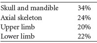
Table 3.112 summarises the main parts of the skeleton identified from this sample. It can be seen that while the proportion of skull is almost twice what is expected, the axial skeleton is almost exactly what it should be and the proportion of upper limb is what it should be. It seems that the skull has been collected at the expense of the lower limbs.
Skull
The supraorbital area and glabella and the right orbit from the frontal bone of a male were present. There was also a fragment from the left orbit and large fragments from the squamous frontal bone. There were large fragments of parietal bone and of the squamous occipital bone. The external occipital protuberance was prominent. The left mastoid area and squamous part of the left temporal bone from a male were present, as well as the anterior part of the temporal bone. There was also a small fragment of mastoid area from a right temporal bone and a left and a right petrous temporal bone.
Table 3.111—Proportion of identified bone, grave 3, cremation 1946:14.
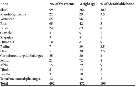
Table 3.112—Summary of identified bone, grave 3, cremation 1946:14.
Mandible, maxilla, teeth
Part of a left ramus with a condyle from an adult mandible were present, as well as another mandibular condyle. There was also a fragment from the body. The left side of a maxilla was also present.
Dentition
Sockets for the following teeth were present:
A lower molar and two upper molar roots were present. One of the molars had a large cavity on the distal side of the crown at the cervical margin. A premolar and five anterior teeth roots, including one canine, were also present.
Vertebrae
The first and second cervical vertebrae were almost complete and there were bodies from four lower cervical vertebrae. Ten thoracic bodies and eight partial arches remained, and there were five bodies and four arches from the lumbar vertebrae. One of the lumbar bodies had collapsed on the left side owing to a crush fracture. There was also some degeneration of the surface of the body and osteophytosis on a few fragments of thoracic and lumbar bodies.
Ribs
There were several fragments of shaft, and at least three ribs from the left and four ribs from the right side had their tubercles visible.
Pelvis
There were several fragments of ilium, including the posterior part and the auricular surface of the left ilium, and there was an almost complete left acetabulum. Part of the posterior right ilium with part of the auricular surface visible was present, and there was a fragment of another acetabulum and some iliac crest. A right ischium was present and there were the bodies of two sacral vertebrae.
Clavicle
The lateral end of the right clavicle and the shaft of a left clavicle were present.
Scapula
The left glenoid fossa and the base of two acromial spines were present.
Humerus
The distal third of the left humerus was present. There were also two proximal and two distal joint ends and several large fragments of shaft.
Radius
One very large fragment from the distal half of the shaft of a left bone was present. There were also other shaft fragments and two proximal and one distal joint ends.
Ulna
This consisted of distal and middle shaft fragments.
Carpals, metacarpals, phalanges
A partial capitate, hamate, lunate and scaphoid were present, and there were ten distal, ten proximal and three middle hand phalanges.
Femur
There were some large, thick fragments of shaft with the linea aspera present. Two large fragments of distal shaft, one neck and one fragment of femoral head were also present.
Tibia
Several fragments of shaft were present, including the posterior shaft near the proximal end with the nutrient foramina of two bones visible. There were fragments from the anterior surface of the bone, including the tubercle from the right bone.
Fibula
Fragments of shaft only were present.
Patella
A left and an almost complete right patella were present.
Tarsal, metatarsals, phalanges
Fragments of the right navicular and talus and a cuboid and a cuneiform were present, as well as a first metatarsal and one proximal and two distal first foot phalanges.
Minimum number of individuals
There was no repetition of skeletal elements so only one individual seemed to be represented. This was an adult male, possibly a middle or older adult, who had suffered trauma to the vertebrae and developed degenerative joint disease of the spine as a result.
Summary and conclusions
The cremation from cist 3 was that of an adult male, possibly a late middle adult or an older adult male. He had a vigorous lifestyle and had suffered a crush fracture of a lumbar vertebra. This had led to the development of osteophytosis of the spine.
This individual had a caries cavity on one molar tooth at the cervical margin. Although caries rates are generally low in early Bronze Age populations, they tend to occur at the cervical margins of the molar teeth, as carbohydrates can settle here and eventually turn into the sugars that encourage tooth decay. The rate of caries increases with age, so there is more likelihood of seeing caries cavities in an older individual from the Bronze Age than in a younger individual.
Grave 4 (P1948:76–84)
This sample consisted of a total of 730g of cremated bone. The bone was mostly creamy white, with numerous horizontal, concentric and vertical fissures of the bone. Some of the larger bone fragments had warped owing to the heat of the fire.
The fragmentation of the sample is shown in Table 3.113, with the largest fragment measuring 111mm in length. It can be seen that there was only a tiny number of small fragments, with nearly 90% of the sample consisting of large fragments more than 25mm in length. In fact, almost half the sample was made up of very large fragments more than 40mm in length. The sample had not been crushed as part of the cremation ritual. The total sample size was quite high, but was not the weight expected from a full adult cremation.
Since the sample was not highly crushed, identification was relatively straightforward.
It was possible to identify a total of 536.5g (73.5%) of the sample. The weight of bone recovered from each of the skeletal elements is shown in Table 3.114.
Table 3.113—Fragmentation of bone, grave 4, P1948:76–84.
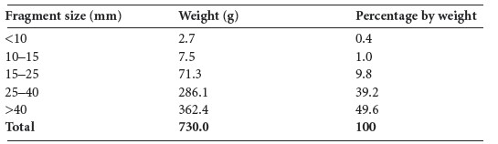
Table 3.114—Proportion of identified bone, grave 4, P1948:76–84.
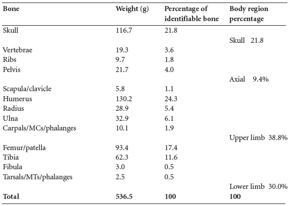
It can be seen that the percentage of skull recovered was similar to the 18.2% expected in a normal cremation, but that the amount of axial skeleton was much reduced from the 23.1% expected. There was a slightly greater amount of upper limb than the 20.6% expected, but the amount of lower limb was reduced from the normal 38.1%. The axial skeleton is the most fragile part of a cremation and breaks up easily. It is also possible that it may not have been collected fully from the site, as the large pieces of long bone would have been more obvious.
The identified elements are described below.
Skull
This included a fragment of squamous frontal bone from the right side with part of the fused coronal suture visible. There was also a large fragment from the left side of the frontal bone, with part of the orbital surface, the supraorbital ridge, part of the frontal sinuses and a large part of the squamous frontal bone present. From the appearance of the supraorbital ridge it appeared to be from a male skull. Other smaller fragments of squamous frontal bone were present.
There were also fragments of parietal bone with parts of the sagittal and lambdoid sutures visible, as well as fragments of the squamous occipital bone. The right greater wing of sphenoid was identified, and there was a small portion of a petrous temporal bone and most of a left petrous temporal bone. A mastoid process, probably from a male, was also present. A left and a right zygomatic bone were identified.
Most of the left side of the maxilla was present and there was a small amount of the right side, although the sockets on the right side were incomplete and could not be identified. A right mandibular condyle, one other mandibular condyle, a fragment of left coracoid and part of the body from the right side of a mandible were also identified.
Sockets present:

Teeth: the root of one canine, one premolar root and one upper molar root, possibly a second molar, were present.
Vertebrae
Cervical: the left side of the arch from a lower cervical vertebra, the left side of the arch from a C2 vertebra and a fragment of another cervical arch were present.
Thoracic: this included two thoracic spines, one with the inferior articular surfaces present, the left side of an arch from a lower thoracic vertebra, the right side of another thoracic arch, and most of another thoracic arch.
Lumbar: there were two vertebral bodies, one of which was lumbar and the other was probably lumbar, as well as parts of a lumbar arch including superior and inferior articular surfaces.
Ribs
There were a minimum of three right and three left ribs present, including one right transverse process. Most of the fragments were from the shaft, near the angle.
Pelvis
This consisted of fragments of ilium, including an area around a sciatic notch, part of an auricular surface and part of the acetabulum, as well as some of the iliac fossa.
Scapula
This included the acromion and part of the body of a left scapula.
Humerus
This consisted of several large fragments of shaft, including the distal half of a right bone. The bone was very thick and well developed. There was also a distal joint surface from a right bone with the trochlea and part of the capitulum present. There were several fragments of proximal and middle shaft and a fragment from a humeral head.
Radius
This consisted of the proximal end with part of the shaft of one bone, as well as several fragments from the mid-shaft and distal shaft areas of at least two bones.
Ulna
This consisted of part of the olecranon, and the proximal middle and distal areas of shaft of two bones.
Carpals/metacarpals/phalanges
This consisted of an almost complete second metacarpal, a left fifth metacarpal, three other almost complete metacarpals, one metacarpal shaft, one metacarpal head, two complete middle phalanges, the shaft of one other phalanx and the proximal end of a first phalanx.
Femur
This included the proximal shaft with the lesser trochanter and part of the neck area from a left and a right bone. Some of the gluteal tuberosity of a right bone was also present. There were fragments of anterior surface from the mid-shaft area and fragments of the distal shaft. Part of a proximal and a distal joint surface were also present.
Patella
Most of a left patella and part of one unsided patella were present.
Tibia
This included fragments from the proximal, middle and distal areas of shaft with the anterior, lateral and posterior surfaces present. One portion of posterior surface near the proximal end had a small exostosis, which probably represents an enthesophyte caused by trauma to the soleal muscle in the calf.
Fibula
This consisted of fragments from the mid-shaft only.
Metatarsals
This consisted of the head and most of the shaft of a first right metatarsal.
Juvenile bone (0.7g)
One fragment of infant or juvenile long bone with unfused epiphysis was present. It seemed to be an infant femur.
Animal bone
There was 31.8g of non-cremated animal bone.
Minimum number of individuals
There appeared to be one adult male and possibly one infant present.
Summary of grave 4
The remains from this cist consisted of 730g of cremated bone. The bone was creamy white in colour and had been fully cremated. Most of the bone recovered was in large fragments, indicating that it had not been crushed as part of the cremation ritual. The pieces probably fragmented owing to heat or cooling of the bones only. The proportion of the bone identified was quite high at 73.5% and this is a reflection of the large fragment size. Most skeletal elements were represented, although the amount of axial skeleton present was very small and the upper limb was overrepresented at the expense of the lower limb. The presence of a large piece of a well-developed humerus may have accounted for this.
There is sufficient evidence from the supraorbital ridge and mastoid process of the skull, as well as the size and development of the bone, to be certain that the one adult present was a male. There was one piece of juvenile bone, however, and this may be an infant femur.
The male could not be aged with certainty, but there was no evidence of degenerative joint disease and so he was probably less than 45 years of age. He enjoyed a strenuous lifestyle, as there was evidence for muscle strain on the bones.
315. The first cist discussed here is the third discovered at the site and is referred to as grave 3. The cist excavated by P.J. Hartnett in 1948 will be referred to as grave 4.
316. Parish of Castletown Kindalen, barony of Moycashel. SMR WM032-055——. IGR 234395 241336.
317. It is possible that a number of padstones had been laid flat on the side slab to act as an interface between it and the capstone.
318. Some of the cremated bone was found inside the vase also. Hartnett has ascribed different NMI registration numbers for the cremated bone found in different places: that found in the vessel is registered as P1948:76(2) and P1948:76(4); P1948:84(8) is cremated bone from the packing behind the northern stone of the cist; P1948:84(9) is cremated bone found in the dark peaty soil above the capstone; P1948:84(10) is cremated bone found in clean gravel underneath the western floor slab of the cist; P1948:84(11) is cremated bone found under the capstone on top of the packing behind the northern slab of the cist; P1948:84(12a) is cremated bone found scattered on the stone floor of the cist; P1948:84(13) is cremated bone collected from the sloping ramp of fresh trampled soil outside (and south of) the cist. Presumably these had been scooped out through the open eastern end of the cist when it was discovered.
319. GrA-24179.
320. GrA-24139.