1900:001 - LUG, CO. OFFALY, Offaly
County: Offaly
Site name: LUG, CO. OFFALY
Sites and Monuments Record No.: SMR OF009-036SMR OF009-039
Licence number: E704
Author: MICHAEL DUIGNAN
Author/Organisation Address: —
Site type: Early Bronze Age graves
Period/Dating: —
ITM: E 634525m, N 729270m
Latitude, Longitude (decimal degrees): 53.312585, -7.481908
LUG, CO. OFFALY, E704269
Introduction
In the early 1900s a number of short cists were discovered in a cairn at Lug, near Tullamore, Co. Offaly (Pl. 55). Owing to the fact that the site was to be put under cultivation for the first time, the cairn was in the process of being levelled by the landowner, Mr Michael Geoghegan. The site appears to have consisted of a large cairn of stones, as the owner describes removing over 100 cartloads of stone. During the removal of stones from the mound, the capstones of four cists were discovered. Two of the capstones were removed at this stage so as not to obstruct the plough.
Subsequently, in 1934, while preparing for the coming season’s crop, the owner discovered two more cists. These were later examined by Revd Professor William Moran of Maynooth College,270 who reported the site to Dr Adolf Mahr and to the members of the Harvard Expedition who had been excavating in Ireland at the time. A rescue excavation under the Minor Relief Scheme was undertaken by Michael V. Duignan, beginning in the spring of 1935 (Pl. 56). Work was temporarily halted to avoid the destruction of crops and resumed in October 1935.271 This report is based on Duignan’s account of the excavation.272 It is clear from the report that the disturbance that took place in the early 1900s had caused considerable damage to the contents of the cists from which the capstones had been removed at that time. In the course of the excavation seven cists (referred to by Duignan as cists I–VII) and two features (called ‘fire sites’) containing quantities of burnt bone, two post-holes and a charcoal spread were discovered.
In a summary account in the NMI archive Duignan says that ‘the site was so levelled in tillage operations as to be almost indiscernible. Little reliable information as to the original diameters and height of the cairn [survived]. It would appear to have been [a] flat cairn roughly circular, some 60 feet [8.2m] in diameter and some 4 feet [1.2m] high. It was composed of various sized stones, and before disturbance was covered with a layer of soil in which grew a number of whitethorn trees.’
In 1975 another cist (grave 10 in this report), some 400m south of the cairn site, containing a cremation and a vessel was reported to the NMI. This had apparently been found twenty years previously but had not been reported. The site was visited by Dr Joseph Raftery. As Dr Raftery’s report is not on file, this report is based on an account by the Offaly Historical Society.
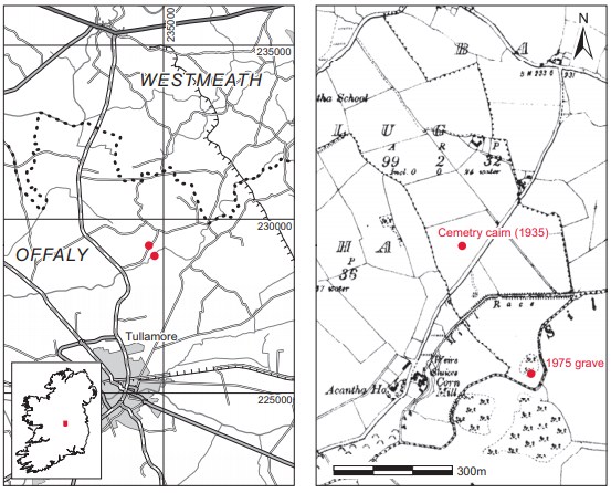
Location (Fig. 3.149)
The site was in the townland of Lug, north Co. Offaly, 2km south of the border with County Westmeath.273 At the time of Duignan’s investigation the site had the appearance of a flattopped mound approximately 18–20m in diameter. To the south and south-west of the site the field sloped away naturally to a wide hollow, making it difficult to see the limit of the cairn above ground. The site lay at an altitude of 60–70m above sea level.
Cairn
Owing to the disturbed nature of the site, it was not possible to undertake an excavation of the whole area. A trench 20.1m long by 1.2m wide (Fig. 3.150) was cut on an east/west axis across the mound at right angles to the nearest edge of the cultivated area. A north/south trench was also cut, dividing the site into four quadrants (I–IV), probably during the second season of excavation. The stratigraphy of the mound was found to be as follows (Fig. 3.150): the lowest layer of the cairn consisted of a substantial gravel layer (approximately 0.48m thick)274 overlain by yellow subsoil,275 which was in turn superseded by a layer of humus to the present ground level. A large post-hole measuring 0.31m in diameter by 0.46m deep was discovered at the north-eastern side of the cairn (Pl. 57). A second large post-hole, 0.2–0.22m in diameter and 0.15m deep, was found at the south-eastern edge of the cairn. Two ‘fire sites containing quantities of burnt bones’ were also noted in one of the documents. One of these was on the south-eastern side of the cairn south of grave 3. This grave was considered by Duignan to be the primary burial as it appeared to be centrally placed in the cairn.
No complete plan or drawing of the cairn, graves and other features has been located, and the positions and relationships of some features are not clear. One sketch-plan (which is not to scale and not illustrated here) shows the site divided into four quadrants, with grave 3 (the central burial) in the eastern part of the east/west trench and grave 2 0.6m to the north-east of grave 3. Graves 1 and 4 were in the western part of the trench, 6m and 5.4m (respectively) to the west of grave 3 (Fig. 3.150). Grave 5 was located south of grave 4 in the south-western quadrant (quadrant I), while grave 6 was immediately north of grave 4 in the north-western quadrant (quadrant III). Grave 7 was in the southern portion of the north/south trench, south-east of grave 4.
Grave 8 is not indicated on any sketch-plan and it may have been located outside the areas shown on the sketch-plans available. A large patch of charcoal was, however, noted close to the posthole in the north-eastern quadrant (quadrant II); as this is the only other feature noted on any plan, it may be one of the two ‘fire sites’, but this remains uncertain. It seems likely that the cremated remains noted on a label as ‘from fire site in Quadrant II’, the north-eastern quadrant, are from this feature (referred to here as grave 8).
Grave 9 is shown in the extreme south-east of the south-eastern quadrant (quadrant IV), towards the edge of the surviving cairn material. The location indicated as grave 9 on the sketchplan is described on another plan as an area of ‘charcoal and burnt bones 3˝ [7.5cm] deep resting on gravel and intermingled with stones of cairn’. This is the only feature marked in this quadrant and is noted as ‘IX’ on the first mentioned sketch-plan (referred to here as grave 9). Two original labels note, however, that a large fossil, a sherd of pottery (plain body sherd) and two fragments of cremated bone are from quadrant IV, i.e. the south-eastern quadrant, and this may be material from the charcoal patch or fire site noted as feature ‘IX’ on the sketch-plan.
Finally, Duignan also noted a ‘heap of bone fragments to the east of (a) [i.e. grave 3] but in the same strata’. This is not marked and the bone has not been located. According to Duignan’s notes, all the cists discovered in October were very badly disturbed.
Grave 1
This short cist lay approximately 0.1m below the surface of the mound, at the western side. It was almost square in plan, with its long axis aligned north-east/south-west (Fig. 3.150). Internally it measured 0.36m long by 0.34m wide by 0.47m high. The cist was composed of four main slabs, with one forming each wall. The side slabs did not meet the end slabs, there being a slight gap between them. Gaps were filled with superimposed flat stones. The western side stone was also considerably lower than the southern end stone and the gap in height was made up of wellcompacted earth and stones. There is no evidence in the plan or report for packing stones except for outside the north-eastern end, where a large limestone block had been placed. Between this block and the northern end stone earth and stones had apparently been packed closely together.
The cist was sealed by a large, irregularly shaped capstone measuring 0.71m long by 0.65m wide. The floor of the cist was roughly paved with jagged pieces of limestone and sloped sharply from the northern end to the centre. All of the cist stones were of limestone. The cist contained a bowl or vase (E704:1)276 and no human remains were found. When opened, it was full to the top: the upper, 0.23m-thick layer was loose in texture, as it had been cleared and then replaced by Professor Moran. Wheat had recently germinated deep in this layer and was growing out of the cist. Below this was a layer of gravel and sharp fragments of limestone. Some 0.08m below this the first pieces of the vessel were found. It was located near the northeastern corner of the cist and was badly crushed. A number of strong roots ran through the sherds and it was not possible to see whether the vessel had stood upright or lay on its side. It was also impossible to clear away the adjacent gravel without causing further damage to the vessel.277
Grave 2
This was located approximately 0.53m below the surface of the mound, about 0.6m north-east of burial 3.
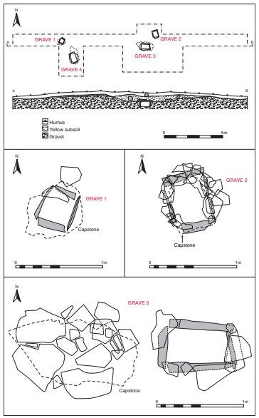
It consisted of a short cist, rectangular in plan, with its long axis running north/south. Internally it measured 0.53m long by 0.45m wide (Fig. 3.150). It was built of four main slabs set on edge, with one forming each wall. The slabs were irregular in shape, the two end slabs being much thicker than the two side slabs. As with the previous cist, the side and end slabs did not meet at the corners but were built up with smaller stones (Fig. 3.150). The gravel around the outside of the cist was filled with small packing stones, 0.23m wide on average. The capstone did not rest directly on the side slabs but on two layers of what Duignan describes as ‘corbels’ on the eastern, northern and western sides. These consisted of smaller stones placed flat on the side stones. The capstone was also irregular in shape and did not cover the extent of the cist, measuring 0.65m long by 0.51m wide. The floor of the cist was formed of very uneven limestone, which did not cover the whole cist but left a gap at the north-western corner. The cist contained a large quantity of cremated bone representing the remains of at least two adults and two infants (E704:2). No accompanying artefacts were found in the cist. The bone is described by Duignan as being ‘smashed up’ and apparently covered almost the whole floor of the cist (Pl. 58).
Grave 3
This appears to have been centrally placed in the cairn (Fig. 3.150). It lay approximately 0.64m below the surface of the mound. It was a short cist, rectangular in plan, with its long axis aligned approximately east/west. Internally it measured 0.76m long by 0.38m wide by 0.58m high. It was composed of four main slabs set on edge, with one forming each wall. The slabs appear to have been relatively regular in shape and Duignan describes the cist as being ‘extremely well made’. Again, the difference in height between the side slabs was compensated for by the positioning of flat stones on the southern and eastern slabs. There is no evidence in the plan for packing stones around the cist, and Duignan does not mention them in his report. The cist was covered with a large spread of slabs and the capstone was placed on top. The capstone was irregular in shape, measuring 1.5m by 0.77m. The floor of the cist was paved with small fragments of limestone embedded in the underlying gravel. The cist had been subject to flooding, which left a water mark 0.08m high on the northern side stone. The silt on the floor of the cist showed that the water had flowed from the south-east towards the north-west. The pit dug to receive the cist
measured approximately 1.2m long by 0.8m high.
The grave contained a crouched skeleton (E704:3) lying on its right side, with the skull in the south-western corner and the pelvis lying across the north-eastern corner (Pl. 59). The remains were those of an adult female and bore evidence for multiple myeloma—a form of bone marrow cancer (see Buckley, below). The burial was accompanied by a small ribbed bowl, which had been placed mouth upwards beside the skull. The skull lay almost face downwards against a fragment of ‘corroded blue stone’ which was roughly rectangular, measuring 13cm long by 10cm wide by 2cm thick. All of the bones above the silt material were well preserved, but those embedded in it were badly decayed.
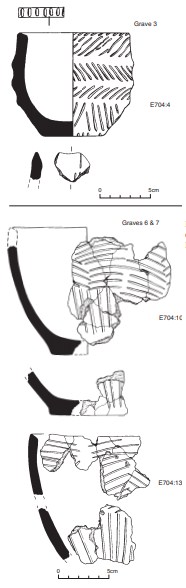
Ribbed bowl E704:4 (Fig. 3.152)
This is a small, complete ribbed bowl. The rim is slanted slightly inwards and is decorated with a single row of impressed broad strokes. The body of the pot is divided into four zones by the slightly raised ribs. The first three upper zones are decorated with incised, broad slanted strokes that form a herringbone pattern, with the ribs forming the central element of the design. The fourth panel is also decorated with slanting incised strokes, but these are grouped together to form broad V-shapes. The base is plain.
Dimensions: H 9.56cm; ext. D rim 11cm; int. D rim 9cm; D base 6cm.
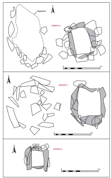
Grave 4
This was located approximately 5.4m west of burial 3, and lay approximately 0.15m below the surface of the mound (Fig. 3.151). It was a rectangular short cist with its long axis aligned approximately north/south and measured 0.78m long by 0.48m wide by 0.56m deep internally. It was composed of four main slabs set on edge, with one forming each wall. The slabs did not meet at the corners, but the spaces were filled with a packing of smaller stones. As the western side slab was slightly higher than the eastern and southern sides, flat stones were placed on the latter to level the cist walls. The pre-excavation plan of the cist shows a concentration of packing stones around the southern, eastern and western sides of the cist. The cist was sealed by a massive capstone of irregular shape, 1.62m long by 0.82m wide by 0.18m thick. The floor was paved with ‘fragments’ of limestone embedded in the underlying gravel. The pit dug to receive the cist does not seem to have survived (this cist had been previously opened by Dr Moran, and was therefore probably somewhat disturbed).
The cist contained the unburnt remains of a young adult female (E704:5) and there were no accompanying artefacts. The cist was filled to the top when opened (as stated above, it had been previously opened by Dr Moran). The upper layer consisted of loose surface soil with occasional inclusions of charcoal and fragments of ‘white bone’ (E704:6, 7) representing one adult. Below this layer was a very compact bed of gravel and small limestone fragments. The remains were in poor condition and had been badly disturbed, presumably as a result of the previous excavation. From the position of the skull, pelvis and leg bones, it was clear that the body had been in a contracted position, lying on the right side with the head in the south-eastern corner. There were no recognisable traces of the vertebrae. The bones of the face between the eye sockets and the lower jaw had completely disappeared. The teeth of the upper jaw were found loose in the soil some 0.1m from the southern end of the cist. The pelvis was badly damaged and lay towards the north-western corner. Two large pieces of limestone (one almost in the exact centre of the cist, and the other touching the northern end 0.18m inches from the north-western corner) lay upon the bones. It is not known whether these were deliberately placed, as the cist had been disturbed prior to Duignan’s excavation.
Graves 5–9
Five more graves were discovered in the second phase of excavation in the autumn of 1935— three cists (graves 5, 6 and 7) and two cremation deposits (graves 8 and 9). These are not well documented on file (other than Duignan’s remark that the contents of all were ‘violently disturbed’). The following is based on descriptions from Waddell’s 1990 corpus, together with some additions from original notes found in the Department of Archaeology at NUIG by Prof. John Waddell.278
Grave 5
Grave 5 was a small, slab-built cist, almost square in plan, measuring 0.61m long (Pl. 60). The plan shows that it was covered by a layer of stone slabs covering an area of 1.52m by 1.45m (Fig. 3.151). The floor of the cist was ‘roughly paved with large fieldstones’. This grave contained the remains of an adult, possibly female (E704:8), and no accompanying artefacts were found. The body was placed in the cist in a crouched position, with the skull in the south-eastern corner and the pelvis at the western side stone. The bones were described as being ‘all very rotten’.
Grave 6
Grave 6 was a small, almost square, slab-built cist, approximately 0.35m long (Fig. 3.151; Pls 61–2). It contained cremated human remains representing a young adult and a juvenile (E704:9). One rim sherd and a few fragments of pottery were apparently found.
Fragments of an anomalous vessel, E704:10 (Fig. 3.152)
Ó Ríordáin and Waddell (1993, 127) describe the surviving rim fragment (28mm by 33mm) as having ‘a simple bevel, and two slight vertical lines on its exterior are uncertain decoration’.
Grave 7
Grave 7 was a short cist measuring 0.46m long by 0.43m wide by 0.4m deep. It contained the cremated bones of an adult (E704:11, 12) and ‘numerous sherds of a small vessel with a simply bevelled rim and an exterior decorated with incised lines and shallow grooves’.
Fragments of an anomalous vessel, E704:13 (Fig. 3.152)
Ó Ríordáin and Waddell (1993, 127) describe ‘numerous sherds of a small vessel with a simply bevelled rim and an exterior decorated with incised lines and shallow grooves’.
Grave 8
Grave 8 was a small, unprotected pit at the base of the mound and may be the ‘fire site’ in the north-eastern quadrant (quadrant 2) mentioned in Duignan’s notes. The pit apparently contained charcoal and cremated human remains. A quantity of cremated human remains labelled ‘from fire site in Quadrant II’ may be from this feature (E704:14).
Grave 9
Grave 9 was a small, unprotected pit at the base of the mound in the south-eastern quadrant. It is possible that a sherd of pottery (E704:15), a fossil (E704:16) and two fragments of cremated bone (E704:17) are all that was found or retained from this feature. The pottery sherd (not illustrated) is very similar to the sherd from grave 6. It is plain except for three shallow grooves.
Grave 10
This was discovered by the landowner, Michael Geoghegan, some twenty years after Duignan’s excavations. It was located about 400m south of Duignan’s excavation.279 According to the account from the Offaly Historical Society, the cist measured approximately 0.46m wide by 0.3m long. It contained cremated human remains representing one adult female and possibly a juvenile as well (1975:265). A piece of animal bone was also found in the cremation. Apparently a vessel was discovered, but this was not recovered. No details are recorded as to the disposition of the cremation within the cist. As this site is some distance from the cairn excavated by Duignan, it may be considered as a discrete burial site, broadly contemporary, perhaps, with the cairn, but in the absence of the vessel said to have been found this cannot be confirmed.
Comment
As a result of the circumstances of discovery this important site was very disturbed. Although a period of about five weeks was spent investigating the site, which should have revealed much about its construction and use, the fact that many of the records relating to the second period of excavation in September/October 1935 are missing makes interpretation difficult. In all, seven cists and two deposits of cremated bone were discovered. The total number of individuals from the burials recovered from this site was fifteen, twelve adults and three infants, with at least five adults being female and two being male. The most remarkable pathological discovery was the multiple myeloma of the adult female in the central burial, grave 3.
The vessel discovered in grave 1 has not been located and is not recorded, but three other vessels were recovered. These are a ribbed bowl from grave 3 and sherds from two anomalous vessels from graves 6 and 7 and grave 9.
A sample of the human remains from grave 3 was submitted for AMS dating and yielded a
date of 3575±40 BP, which calibrates to 2032–1775 BC.280 The vessel belongs to Brindley’s stage 3 in the development of the bowl tradition, dated to 1980–1930/20 cal. BC. This single date does not provide sufficient evidence to date the period during which this site was being used for burial purposes, but it does give an indication.
Four of the seven cists produced some pottery but only the ribbed bowl from grave 3 can be categorised. Three cists produced no pottery or other grave-goods. Duignan remarked on the level of disturbance of some of the graves but it is not known whether this was due entirely to the recent agricultural activity at the site or to earlier damage. It may be that some of the graves had been robbed in the past. Nevertheless, as has been remarked above, a high percentage of early Bronze Age burials (42% in this corpus) were not accompanied by pottery vessels. It is not possible to argue that earlier disturbance or robbing is the sole cause, as in many cases the cists appear to have remained undisturbed until their accidental discovery.
The fossil noted above is a good specimen in two fragments of an orthoconic (i.e. straight cone shape) nautiloid c. 7cm in length (Matthew Parkes, Natural History Division, NMI, pers. comm.). Unfortunately it is not clear from the records whether this is a deliberate inclusion in the fire site in the south-eastern quadrant of the site or an accidental recovery. It is not burnt. Fossils have been noted at other early Bronze Age burial sites, such as Nevinstown, Co. Meath (this volume, pp 371–7).
HUMAN REMAINS
LAUREEN BUCKLEY
Grave 1
No human remains have been identified from this grave.
Grave 2 (E704:2)
This short cist contained a large quantity of cremated bone that covered the entire floor of the cist, but the depth of bone was not specified. A number of obviously infant bones were apparently identified on site and were stored separately from the main cremation. These will be dealt with separately.
The main body of the cremation consisted of 3,030 fragments of bone weighing a total of 2,664g. Although this could be within the weight of bone expected from a full adult male cremation, this is unlikely given the amount of bone normally found in early Bronze Age cremations, and it is more probable that it represents the remains of more than one adult.
The bone was white in colour and efficiently cremated, with numerous horizontal fissures and warping of the bone.
Table 3.77—Fragmentation of bone, grave 2, E704:2.
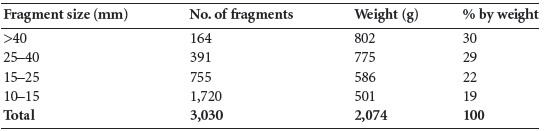
The fragmentation of the bone is shown in Table 3.77, with the largest fragment being 108mm in length. It can be seen that over half the sample consists of very large fragments and that almost all the sample is made up of large fragments more than 15mm in length. There was a small proportion of medium-sized fragments and no very small fragments. Although it is probable that the smaller fragments were not collected, the high proportion of larger fragments nevertheless indicates that the bone was not deliberately crushed after collection from the funeral pyre.
Identifiable bone
A total of 1,448g (54% of the total bone) was identified. This high percentage is due to the number of very large fragments.
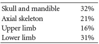
Table 3.78 shows the proportion of identified bone. Table 3.79 summarises the main parts of the skeleton identified from this sample. Compared to what would be expected from a normal adult cremation, the proportion of skullis nearly twice what it should be. This usually happens when more than one individual is present since skull is so easily identified, but it can be harder to separate fragmented long bones. The amount of axial skeleton is close to that expected from a normal cremation but the amount of upper and lower limb bones is slightly lower than expected.
Skull
Bones present included two right orbits, one from a male and the other possibly from a female, one incomplete left orbit, a partial glabella area of the frontal bone, also from a male, a fragment of another glabella and a small part of the supraorbital area from the right side, possibly from a female, and several large fragments of the squamous frontal bone. Two left and two right petrous temporal bones were present, as well as the left temporal fossa and anterior suture and the mastoid areas of a left and a right temporal bone from a female. The superior part of the left mastoid area of another temporal bone with part of the temporal fossa and superior margin of the external auditory meatus was present. The left and right zygomatic bones were present. There were several large fragments of parietal bone, including large fragments from the anterior part of the right bone and the posterior part of the left bone. At least two squamous occipital bones were also present, one with a more pronounced external occipital protuberance than the other.
Table 3.78—Proportion of bone identified, grave 2, E704:2.
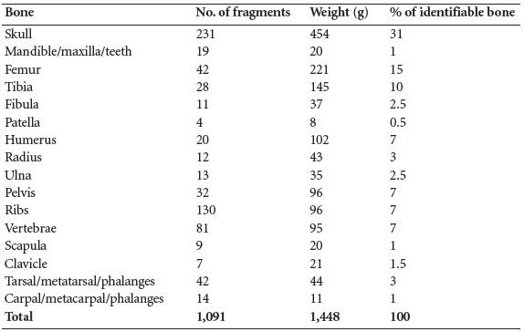
Table 3.79—Summary of bone identified, grave 2, E704:2.
Mandible and maxilla
There was a large section of the right side of the mandible with some sockets visible. Most of the right ramus and a fragment of the left ramus at the internal angle were present, and there were two mandibular condyles. There was also a fragment of the internal surface with the genial tubercles present. There were fragments from both sides of the maxilla.
Dentition
The following tooth sockets were present:

The roots of the mandibular first molar were still in the socket and there were roots of two other molars. The roots of the mandibular premolars were also present.
Vertebrae
The right side of the first cervical vertebra was present, as well as the body and right side of the second cervical vertebra. At least five bodies and disarticulated arches from the lower cervical vertebrae were present. Parts of the bodies of nine thoracic vertebrae were present, as well as some fragments of neural arches. Four of the thoracic vertebrae had Schmorl’s nodes. Five complete lumbar bodies survived, as well as a few articular surfaces from the neural arches. Schmorl’s nodes were visible on four of them.
Ribs
Most of the fragments were shaft fragments but there were at least two left and one right tubercle heads remaining.
Pelvis
Two left ischia with the inferior parts of the acetabula were present, and there was a fragment of right ischium. The right superior part of the acetabulum and part of the body of the ilium were also present. The accurate line of the right ilium was present, as well as part of a sciatic notch. The posterior auricular area of a left ilium was present and there was also an auricular area possibly from the same left bone. The bone was decayed so it was difficult to be certain. Various other fragments of ilium were present, as well as a decayed fragment of pubic symphysis from a young individual. The right ala of the sacrum also survived.
Clavicle
The lateral third of the shaft of a left and the medial half of a left clavicle were present. There was also a lateral half of a right clavicle shaft and a medial end of a right bone with fused sternal epiphysis. There was also a fragment from the middle of the shaft of one bone.
Scapulae
The right glenoid fossa and most of the lateral border was present, as was the right acromion. There was also a left acromial spine and a left lateral border.
Humerus
The proximal third of a right humerus with the biccipital groove, lesser tuberosity and part of the head was present. The proximal third of another right humerus with the same features was also present but it was from a larger bone. Most of the proximal articular surface of this bone was present and there were two other proximal articular surfaces. The mid-shaft area of a right humerus was present, as well as the distal shaft of a right bone. A fragment of the left distal shaft was also present, and there were fragments from at least four distal articular surfaces.
Radius
The proximal third of a radius shaft and one radius head were present (RaHd 20mm). The distal third of a left radius with part of the distal joint end was present, and there was also a distal end with most of the articular surface from another left bone. Fragments from the middle of the shaft were also present.
Ulna
There were several large fragments of shaft from the proximal and the distal part of the bone, and one fragment of distal shaft had the joint surface visible.
Carpals, metacarpals and phalanges
A left scaphoid and left and right capitates were present from the carpal bones. A first and a second metacarpal, a fifth right metacarpal and at least two other metacarpal shafts were present, as well as two fragments of metacarpal heads. One proximal phalanx and one complete middle phalanx were present.
Femur
Several large pieces of shaft from the proximal, middle and distal parts of the bone were present. There were a minimum of three proximal articular surfaces, as well as fragments of two distal articular surfaces. The neck areas from one left and one right bone were present, and there was also a lesser trochanter and posterior part of the shaft from a left bone as well as from a right bone.
Tibia
Several fragments of shaft with the anterior and interosseous borders visible were present. Two large fragments from the proximal posterior shaft with nutrient foramina from two bones were present, and there were also an anterior tubercle and an anterior shaft fragment from a right bone with part of the lateral surface visible, as well as the distal end of a right tibia.
Fibulae
Several large fragments of shaft and the distal end of a left bone were present.
Patellae
Two almost complete left patellae were present; one was smaller than the other. There was also a fragment of a right patella and the anterior surface of one other patella. Both of the left patellae had a vastus notch.
Tarsals, metatarsals, phalanges
One right talus and fragments from one other talus, two navicular and two calcanea were present from the tarsal bones. There were four first metatarsals, consisting of the heads, most of the shafts and two proximal articular surfaces. Eight other metatarsals consisting of shafts and some distal ends were present, and there were one first proximal phalanx, one other proximal phalanx and two first distal phalanges.
Juvenile bones
In addition to the two adults present in the main cremation there were some fragments of juvenile teeth and juvenile skull, including a left and a right petrous temporal bone. The teeth consisted of the almost complete crown of a deciduous canine, two deciduous upper first molars and one deciduous lower first molar. The partial crowns of two upper molars and a lower second deciduous molar were also present. These teeth could have come from an infant aged 7–11 months.
Infant bones
A total of 46g of infant bone was present. Most of this (65%) consisted of skull fragments, including a complete frontal bone with both orbits present and the metopic suture fused. This usually fuses during the first year of life. The left and right petrous temporal bones were present and there was also the right mastoid area from one temporal bone. There were large fragments of parietal bone and some occipital bone present.
Other bones present included most of the shaft from the left femur, the mid-shaft area of another femur and a virtually complete left tibia. These long bones seemed to be from the one infant aged around one year old. There were also two ribs present. It is possible that the teeth found in the main cremation belong to this infant. Since, however, there are two pairs of petrous temporal bones from juvenile skulls, it is likely that two infants were present.
Minimum number of individuals
There appear to be at least two adults, one male and one female, and two infants present, giving a minimum number of individuals of four. This is confirmed by the fact that there were four pairs of petrous temporal bones present, but there was also repetition of other skeletal elements.
Summary of grave 2
This was a very large sample of cremated bone from one cist, consisting of 2,664g. It all appeared to be very efficiently cremated. Most of the bone was in large fragments, with over half of the sample being in very large fragments, the largest being 108mm in length. It seems that the bones were kept in large pieces and were not deliberately fragmented as part of the cremation process. It was possible to identify 58% of the sample and, although all parts of the skeleton were represented, the proportion of skull was almost twice what it should have been. This tends to be an indication that more than one individual was present, and so it was in this case. The main part of the cremation consisted mainly of two adults, one male and one female. There was also part of a juvenile present. A further sample of infant bones had been kept apart from the main sample. It was estimated that two juveniles were present in the cist, one approximately one year of age and the other 7–11 months.
Grave 3: (E704:3)
This cist lay in the centre of the cairn. The burial consisted of a tightly crouched inhumation with the skull almost face down in the south-west corner and the pelvis in the north-east corner. A small ribbed bowl had been placed upright to the side of the skull in the north-west corner. The skeleton was virtually complete and was in a relatively good state of preservation, although it was very fragile owing to the pathological condition of the bones described below.
The skull, including the mandible and facial bones, was virtually complete. All of the vertebral column, seven cervical, twelve thoracic and five lumbar vertebrae were present and all were virtually complete. There were twelve ribs from the left side and ten from the right side remaining. The body of the sternum was complete.
Both clavicles and scapulae were present and were almost complete, but the joint ends of the clavicles were fragmented. The left humerus was complete but the right humerus was fragmented at the proximal joint end; both radii and ulnae were complete. The carpal bones consisted of the left and the right lunate, triquetral and hamate, the left trapezoid and the right scaphoid, pisiform and capitate. The left second, third, fourth and fifth metacarpals were present, as well as five proximal, four middle and four distal hand phalanges. In the right hand only the first and fourth metacarpals survived, and there were two proximal, four middle and three distal phalanges.
Both ilia, ischia and pubes were present from the pelvis and they were virtually complete. The sacrum was also present and almost complete. Both femurs were present but fragmented at the distal joint ends. The tibiae were almost complete but were fragmented at the proximal joint ends. The shafts of both fibulae were also present. Very little remained from the foot bones and the bones were very decayed. Both calcanea, tali and left navicular were present. A cuboid and an unidentified cuneiform were present but too incomplete to side properly. Only a right first metatarsal and both fifth metatarsal shafts as well as one proximal phalanx survived from the remainder of the foot bones.
Age and sex
All the observable features of the pelvis and most of the features of the skull were of the female type. The available bone measurements were all in the female range.
It was difficult to age this individual owing to the pathological lesions present on most bones. She was certainly over 25 years of age, and the wear on the mandibular teeth suggests an age of 25–40 years.
Skeletal pathology
Several bones were affected by various-sized lytic lesions that had destroyed the bone, but there was no indication of bony repair.
Left scapula: there was a very small lytic lesion just at the glenoid area on the pleural surface.
Left clavicle: the sternal end had been completely destroyed by ante-mortem lytic lesions. The shaft was also affected for a short distance (1cm) beyond the joint surface. There appeared to be a sclerotic edge to some of the lesions.
Left humerus: the area of the anatomical neck of this bone had several lytic lesions. On the
medial surface there was a large scooped lesion just below the head, and several smaller lytic
lesions with the internal structure of the bone visible. The anterior surface also had lytic
lesions just below the head. The posterior surface had a very large lesion. It was possible to see
right through the bone because of the extent of bone destruction.
Right scapula: just below the inferior margin of the glenoid there was a large lytic lesion that had destroyed some of the medial border, although there was also some post-mortem damage to this area. On the posterior surface close to the glenoid area there were smaller lytic lesions, and there was at least one destructive lesion in the body of the scapula. The end of the acromion was almost completely destroyed by destructive lesions, with no indication of bone repair. At the base of the coracoid process there was a small lytic lesion on the posterior surface and a much larger area of bone destruction on the pleural surface.
Right clavicle: there was destruction of most of the sternal end of the clavicle, with small lytic lesions on most of the articular surface and also some lysis of the shaft for 1cm beyond the joint surface. There also appeared to be a small area of porotic new fibre bone on the shaft.
Right humerus: the proximal end of this bone was fragmented, but it was apparent that it had been weakened by several lytic lesions. The inferior edge of a large destructive lesion was visible on the posterior surface of the remaining intact bone shaft. The edges of this lesion appeared to be slightly sclerotic. Other smaller lytic lesions on the lateral surface of the shaft near the greater tuberosity were visible on a bone fragment. The edges were also sclerotic. A fragment of humerus head had lytic lesions just below the surface in the inside of the bone.
Cervical vertebrae: the first cervical vertebra had some slight destructive lesions on the posterior surface of the arch and also on the anterior surface of the arch on the right side. Most of the anterior surface of the body of the second cervical vertebra had been destroyed. There was a scooped-out lesion on the left side of the body and a severe lytic lesion on the right side. Most of the right side of the neural arch, including part of the superior and inferior articular surfaces, was destroyed, with the destruction extending slightly on to the left side of the neural arch. On the third cervical vertebra there was some destruction of the tip of the arch and a small destructive lesion on the inferior surface of the body on the posterior surface. The right tip of the neural arch on the fourth cervical vertebra had completely resorbed. There was also slight destruction of the inferior edge of the body on the posterior surface. There was a small lytic lesion on the anterior surface of the body of the fifth cervical vertebra on the left side at the superior edge. The sixth cervical vertebra had a very slight degree of destruction on the left side of the neural arch, on the inferior surface of the pedicle. On the seventh cervical vertebra there was a large destructive lesion on the right side of the centrum,penetrating from the posterior to the anterior surface, although some post-mortem damage may have been superimposed on this.
Thoracic vertebrae: T1—neural arch was incomplete but there were severe lytic lesions in the right side of the body, mainly in the posterior half. The right transverse process was also severely affected and there appeared to be lysis on what remained of the neural arch.
T2—there were several small destructive lesions on the left side of the body towards the anterior half, and also to a lesser degree on the right side of the body. The transverse process on the right side was almost destroyed with lytic lesions.
T3—there were destructive lesions on the left side of the body in the anterior half but not as severe as in T1. There was also a very slight degree of destruction on the right side of the body and on both transverse processes. The posterior surface of the neural arch had a scooped-out lesion on the left side and a small lytic lesion on the right side. In addition to the destructive lesion, the body was compressed on the right side. This was probably a crush fracture caused by the weakness of the trabaculae in the vertebra. The inferior surface of the body on the left side was also caved in, similar to Schmorl’s nodes, but the collapse probably resulted from destruction of the internal structure of the vertebra.
T4—there was a slight degree of destruction of the left side of the body, but relatively large lytic lesions were present on the left side of the neural arch and on both transverse Processes.
T5—there were destructive lesions all through the body. Some small lesions were evident on the anterior surface of the body but there were a few large lytic lesions on the posterior side.
T6—only the right side of the body remained but there appeared to be a lot of destruction, with collapse of the vertebral body on the left side resulting from a crush fracture. There were also some lytic lesions on the transverse processes.
T7—there were large, scooped-out lytic lesions on the left side of the body, extending on to the inferior surface. The right side had smaller lytic lesions. On the left side of the neural arch there was a large lytic cavity that had gone from the outside to almost right through the bone.
T8—very little of this vertebra remained, but there appeared to be a slight amount of lysis forming on the arch and left transverse process.
T9—there was a large lytic lesion on the anterior surface of the body and smaller, scoopedout lesions on the left side of the body. There were also some lytic lesions on the right transverse process and the left side of the neural arch. In fact, this lesion had almost destroyed the inferior articular surface. Large lytic lesions were visible in the posterior half of the vertebral body.
T10—large lytic cavities were visible on the left side of the body and small areas of lysis on the right side. The right side of the body had suffered a crush fracture, however; the body was severely compressed on the right side and the superior surface of the body was broken and partially healed. There were some lytic lesions on the transverse process and one lesion on the right had gone from the outside, near the articular surface for the head of the rib, right through the pedicle of the neural arch. There were other small, destructive lesions on the posterior surface of the neural arch, and the spine seemed to have been destroyed.
T11—the body was broken and incomplete, so a large cavity in the centre was visible, as well as several smaller cavities on the right side. The transverse processes were also affected. The edges of large destructive lesions were visible on each side of the neural arch and it is possible that the split in this vertebra was caused not by post-mortem damage but by a destructive lesion that had completely destroyed the spine and centre of the neural arch.
T12—several destructive lesions were visible on the anterior surface of the body. There was a lesion beginning to break through on the inferior surface, and very slight lesions on the pedicles and posterior surface of the neural arch (Pl. 122).
Lumbar vertebrae: L1—several lesions were visible all around the anterior surface of the body, with two small lesions on the superior surface, possibly breaking through from the inside. The posterior part of the body had large lytic lesions visible. There were also lesions on the left side of the neural arch, including the pedicle and posterior part. One lesion was penetrating the left superior articular surface. The spine had been almost completely resorbed.
L2—there was a very large lytic cavity on the right side of the vertebral body, going right through the vertebral body and connecting with other large lesions going through to the anterior and posterior surface. So little of the internal structure remained that the left side of the body had collapsed, with a large circular depression on the inferior surface on the left side. Two smaller lesions were emerging on the superior surface.
L3—there were a number of small lytic lesions on the anterior surface of the body, with a large lesion on the left side extending to the superior articular surface. The posterior half of the centrum also had a number of lytic lesions visible, and there was a smaller lesion emerging from the middle of the body to the inferior articular surface. The left superior articular surface appeared to have been destroyed by lesions, and the right superior articular surface was partially destroyed. There was some post-mortem damage to the spine.
L4—there was a very large cavity in the middle of the superior surface of the body, extending into the centre of the bone and connecting with moderate-sized lesions on the anterior surface. There was a depression on the left side of the superior surface where the internal structure had collapsed. Lesions were also visible on the posterior half of the body, and the left side of the neural arch was missing. From the appearance of the lesions it is probable that the left arm of the arch had been destroyed.
L5—there were a few small lesions visible on the anterior surface and also beginning to emerge on the superior and inferior surfaces. The lesions were more destructive on the ala and on the neural arch, where the spine has been destroyed, and numerous other lesions were present on the remainder, so that only a small connecting piece of bone remained between the two halves.
Sacrum: the sacrum had been partially destroyed by lytic lesions. There was a large lesion on the anterior surface of the body of S1, which had partially destroyed the superior surface also. The lesion penetrated the bone, connecting with lesions in the posterior part. There were also lesions on both ala, and the first foramina were very enlarged. The lateral parts of the second and lower sacral vertebrae seemed to have been completely destroyed by lesions. The dorsal surfaces of the bodies of these vertebrae had also been affected by lesions. There was a sharp, 90º angle at the junction of the third and fourth sacral vertebrae, with the lower two vertebrae sitting perpendicular to the rest of the sacrum. This may have been caused by the effect of sitting on severely weakened bones (Pl. 123).
Sternum: the body of the sternum was present, with severe destruction of most of it. A large destructive lesion in the upper half penetrated through the anterior and posterior surfaces and only edges of the bone remained. The upper edge was partially destroyed. There were other smaller, destructive lesions in the lower half of the bone, although these were combined in one large lesion on the posterior surface. As both lesions were larger on the posterior surface, they may have originated on this side (Pl. 124).
Ribs: almost all the ribs on each side, especially the upper ribs, had a number of destructive lytic lesions. The first right rib in particular had a large lesion in the middle of the shaft and another near the sternal end, which had gone right through the bone. The lesions on the ribs varied from small cavities penetrating the bone to scooped-out lesions on the internal surface and scalloping of the inferior edge. The right side was slightly more affected than the left side. There was evidence of at least one unhealed pathological fracture on the right side.
Pelvis: apart from the skull, the pelvis seemed to be the most severely affected area of the skeleton. The left ilium had a large number of destructive lesions. The posterior part, including part of the articular surface, had so many large and medium-sized lesions penetrating the lateral and the medial surfaces that most of the bone was gone and only a framework remained. The iliac fossa and the top of the iliac crest had a number of lesions, some of which penetrated only one surface or the other and some which pierced the bone completely. A very large lesion just above the sciatic notch had destroyed most of the interior of the bone and part of the acetabulum.
The right ilium was similarly affected, with most of the posterior part and articular surface destroyed by lesions. On this side the iliac crest had fragmented, partly as a result of the lesions and partly as a result of post-mortem damage. Most of the anterior iliac crest had been destroyed by a large lesion (Pl. 123).
Both ischia were affected, with a number of large lesions on the internal surface behind the tuberosities. The right side was again affected slightly more than the left, with the ischiopubic ramus being almost eaten through at one point.
The pubic bones were severely affected, with the superior edge of the ramus partially destroyed in the left bone as well as large lytic lesions in the body of the pubis. In the right pubis the body was almost completely destroyed by a large lesion that had penetrated the anterior and posterior surfaces.
Left femur: the proximal end of the left femur had a number of large lytic lesions. The articular surface of most of the head was missing and, although there may have been some postmortem destruction, most of the destruction had occurred during life. The posterior surface of the neck was missing, but there was a large destructive lesion on the anterior surface and also on the proximal shaft. The destruction of the interior structure of the bone was clearly visible. No lesions were present in the distal two-thirds of the bone.
Right femur: the right femur was affected in a similar way to the left, although not as severely. There were two small lesions penetrating the articular surface of the head and there also seemed to be large cavities in the internal structure of the head. There were two large lesions on the neck, penetrating from the anterior surface through the internal part of the bone to the posterior surface. The posterior surface of the neck and the greater trochanter area seemed to be completely destroyed.
Tibia/fibula: there were no lesions on the tibiae and fibulae. The foot bones were very decayed, so it was not possible to state whether the post-mortem decay had removed traces of antemortem erosions.
Skull: there were a number of lesions on the skull. The largest was on the left side of the frontal bone, measuring roughly 3cm in diameter. The edges had a very rough, ‘moth-eaten’ appearance. The lesion seemed to be progressing from the inner surface of the bone to the outside, with the internal extent being larger than the external. There was an area of generally porotic bone on the outer surface of the frontal bone around the lesion. It seemed to be an aggressive lesion with no indication of bone repair. In the right parietal bone there was a small lesion, 1cm in diameter, close to the sagittal suture. Again this lesion appeared to be progressing from the inside out, with an area of porosity around it. In the posterior part of the right parietal bone, near the lambdoid suture, there was a very small aperture with an area of porosity around it. In the middle of the right parietal bone there was a very small destructive lesion, as well as some general porosity over the bone.
On the base of the occipital bone, immediately posterior to the foramen magnum, some destruction of the outer surface of the bone was visible but the lesions had not penetrated the bone. There were also destructive lesions on the basal part of the occipital bone, particularly on the left side. The left orbit was also affected, with the most lateral part of the orbit having evidence of destructive lesions.
A CT scan of the skull, carried out at the Advanced Radiology Centre, revealed the existence of several other lesions not visible to the naked eye. Most were in the internal structure of the skull bone, i.e. the diploe, although some had penetrated the inner table. There were several lesions present in the frontal, parietal and occipital bones. These kinds of lesions are typical of a form of bone marrow cancer known as multiple myeloma. This is a primary cancer of bone and gives the typical moth-eaten appearance seen in the skull, with many lesions progressing in the diploe of the skull for a long time before they are evident on the surface. It has a preference for the axial skeleton but typically affects many bones of the skeleton. In the long bones, scalloping of the edges of the lesion, giving a ‘punched-out’ appearance as seen in this case, is also typical. There is also no bone reaction or sclerosis of the edges of the lytic lesions. A differential diagnosis could be metastatic carcinoma from breast cancer, for example, but the extensive involvement of the skeleton indicates a diagnosis of multiple myeloma (Ortner 2003, 377). Also, the involvement of the clavicles and scapulae frequently occurs in multiple myeloma but not in metastatic carcinoma. The condition usually shows a greater prevalence in males than in females and is rare before 40 years of age, so it is very unusual to find it in archaeological samples. The fact that the burial was in a cist has protected the bone, which would have been weakened by the disease, from being broken and damaged after burial. It is remarkable that this skeleton with evidence for this disease has survived so well.
Other pathology
The left wrist was dislocated, with the left radius curved laterally so that the ulna articulated with the radius on a false joint just above the true ulnar notch of the radius. The curve may have occurred as a result of childhood trauma.
Dentition
Attrition: there was moderate wear on the incisors, light wear on the canines and premolars, and moderate to heavy wear on the first and second molars.
Calculus: there were light deposits on the buccal and lingual surfaces of the upper teeth and moderate deposits on most of the buccal and lingual surfaces of the lower teeth, but there were heavy deposits on the lingual surfaces of the lower left molars.
Periodontal disease: there was a slight degree of alveolar recession around the roots of the mandibular teeth and the maxillary incisors.
Grave 4: young adult female (E704:5)
This burial was found in a short cist that had been opened and disturbed prior to its full excavation, and this had hastened the decay of the bones. The skeleton had been disturbed but it appears to have been crouched, lying on its right side with the head in the south-east corner. Most skeletal elements were present but they were very decayed and fragmented, with virtually no joint ends remaining.
Most of the calvarium of the skull survived, the occipital, both parietal and occipital bones were complete, and both temporal bones were present although the mastoid areas were missing. The sphenoid bone was also present and the left zygomatic bone was complete. Most of the mandible was present.
The right side of the first cervical and dens and body of the second cervical vertebrae survived, as well as fragments of neural arches from a minimum of four thoracic vertebrae and some arches and partial bodies from four lumbar vertebrae. At least seven ribs from the right side were present and another three were probably from the left side.
The glenoid area and part of the acromion of the left scapula remained, and the glenoid and lateral border of the right scapula was present. Only the shaft of the right clavicle remained. The shafts of both of the humeri were present but the proximal half of the left bone was shattered. The shafts of both radii and ulnae were also present. The hand bones consisted of incomplete left lunate and trapezium, the shafts of both first, left third and right second metacarpals, and five proximal and two distal hand phalanges.
The pelvis consisted of most of the ilia, part of the left ischium, the ramus of the right pubic bone and five incomplete sacral vertebrae.
Both of the femurs were almost complete, but the joint ends were fragmented and incomplete. The shafts of both tibiae were present and there were also fragments of the joint ends in the right bone. Fragments from the shafts of both fibulae were present. The foot bones consisted of part of the right talus and right first cuneiform, two other fragments of tarsal bones, the shaft of the left first metatarsal and most of the fifth left metatarsal. One proximal phalanx was also present.
Age and sex
All the observable features of the skull, the external occipital protuberance, supraorbital ridges, orbital rims and mental eminence, were of the female type. The sciatic notches on the ilia were wide, also indicating a female.
The auricular surfaces of the ilia and the lack of wear on the teeth indicated that this was probably a young adult.
Skeletal pathology
Only the left side of the fifth lumbar vertebra was present, but there was spondylolysis of the neural arch.
Dentition
There was some brown staining on the right maxillary incisor and canine. Mineral deposits were present on some of the other teeth.
Attrition: there was light wear on most of the incisors, canines, premolars and first molars, and no wear on the third molars. The mesial side of the lower right canine seemed to be worn Obliquely.
Calculus: there were light deposits on the lingual surfaces of most of the maxillary teeth, on the lingual surfaces of most of the mandibular teeth and on the buccal surfaces of the lower right first molar, lower right lateral incisor and lower left second molar. The lower right and upper left third molars had slight deposits on their occlusal surfaces.
Hypoplasia: linear enamel hypoplasia was present on the lower right incisors and canine, lower left canine, upper left canine and upper right lateral incisor.
Grave 4: cremated bone (E704:6), sample 1
Nineteen fragments of cremated bone weighing a total of 13g were present with this inhumation. Most of the fragments were more than 15mm in length, the largest measuring 31mm. The bone was white in colour and efficiently cremated.
The fragments consisted mainly of human long bone, with the proximal end of a tibia and a fragment of thoracic vertebra also present.
Grave 4.V [cist IV.V] (E704:7), sample 2
[Editors’ note: This sample was labelled ‘Cist IV.V’, and in the absence of any record it is assumed to relate to the cremation described immediately above from grave 4. The excavator may have numbered the cremation from grave 4 (cist IV) as the fifth (V) burial from the site. Both samples represent the remains of a single individual. Grave 5, a cist burial, contained an inhumation in poor condition, as described below.]
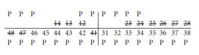
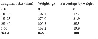
This sample consisted of 205.3g of cremated bone. The bone was very black with charcoal but the underlying colour was very white, indicating efficient cremation and the surface of the bone was cracked. The fragmentation of the sample is shown in Table 3.80.
Table 3.80—Fragmentation of bone, graves 4.V, E704:7; sample 2..
There were no very small fragments and almost 40% of the sample consisted of large or very large fragments more than 25mm in length. Despite the large size of the fragments, identification was difficult owing to the colour of the bone, and it was only possible to identify 42.7g (21%). Most of the identified bone (74%) consisted of skull fragments.
Identifiable fragments
Skull (31.8g): this consisted of fragments of calvarium only.
Ribs (0.5g): the medial end of a first rib was present.
Humerus (4.1g): this consisted of fragments from the mid-shaft area of the bone.
Femur (6.3g): this consisted of fragments from the posterior surface of the distal shaft.
Minimum number of individuals: one adult.
Grave 5: young adult male (E740:8)
This skeleton was also crouched in a short cist. The skull was very fragmentary but the complete frontal bone, fragments of the parietal bones, most of both temporal bones and fragments from the occipital bone were present. Both zygomatic bones were also present and the mandible was almost complete. No vertebral column or ribs remained.
The shafts only of both of the humeri, radii and ulnae were present, and a fragment of one clavicle remained. There were no hand bones.
Most of both ilia and the right ischium remained from the pelvis.
The shafts of both femurs were present, although both were fragmented in their distal thirds. The shafts of both tibiae and fibulae were also present. Only the left talus and calcaneum remained from the foot bones.
Age and sex
The supraorbital ridges and mental eminence of the mandible were of the male type. The sciatic notch of the pelvis was also of the male type.
Most of the metaphyses and epiphyses of the long bones were unobservable but the ischial tuberosity and iliac crest were unfused. The proximal tibia was unfused but the distal femur was partially fused. This would indicate an age of 16–20 years, but since the third molars were erupted the age of this individual is probably 18–20 years.
Dentition
Mineral deposits over some of the teeth made it difficult to determine the presence of calculus.
Attrition: there was moderate wear on the upper incisors and first molars. Most other teeth had little or no wear.
Calculus: there were light deposits on the lingual surfaces of most of the teeth in the mandible except the third molars, which had no calculus, and there were also light deposits on the buccal surfaces of the lower right incisors and upper first molars.
Hypoplasia: linear enamel hypoplasia was noted on the upper canines and lower left canine as well as the upper right lateral incisor. There were pits of hypoplasia on the upper left second premolar.
Grave 6 (E704:9)
This consisted of 10.1g of very decayed but non-cremated bone. It consisted of fragments of skull from a juvenile. The largest was a fragment of occipital bone that was very decayed, but the internal occipital protuberance was observable. There were also fragments of juvenile parietal and frontal bones and one deciduous incompletely formed tooth crown, the upper second molar, 65. At this stage of development the individual would have been aged between six months and one year of age.
Grave 7 (E704:11), sample 1
This sample consisted of 19.1g of cremated bone. The bone was cream in colour and was efficiently cremated. The largest fragment was 35mm in length. As the sample size is so small, the proportion of bone in the various categories is not given. The fragmentation of the sample is shown in Table 3.81.
Table 3.81—Fragmentation of bone, grave 7, E704:11; sample 1.
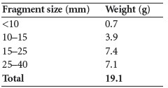
As most of the fragments were quite large it was possible to identify 9.5g (50%) of the sample. Most of this, 8.2g (86%), consisted of skull fragments; the only other bone identified
was part of a metacarpal, although there were some long bone fragments present. The
identifiable fragments are described below.
Skull: there was one small fragment of calvarium with open suture that may have been
from a juvenile.
Fragments of mandible included the anterior part of the body with sockets for the
following teeth:

There was another fragment of body but the sockets were too damaged to identify. Also
present was an isolated tooth socket and part of a mandibular condyle.
Teeth: roots of two lower molars and part of a premolar root.
Metacarpals: part of the head and shaft of a first metacarpal.
Minimum number of individuals: the remains represent one adult.
Grave 7 (E704:12), sample 2
This sample consisted of 846g of cremated bone. The bone was mainly white in colour,
although there was 40.6g (5%) that was not fully cremated and was partially blue or black.
These blue fragments were mainly confined to the left humerus and part of a femur. They may
have been lying in a position on the pyre where oxygen flow was restricted. The fragmentation
of the sample is shown in Table 3.82, with the largest fragment 72mm in length.
Table 3.82—Fragmentation of bone, grave 7, E704:12; sample 2.
Over half of the sample consisted of large or very large fragments more than 25mm in length. As there was only a small proportion of smaller fragments this made identification easier.
Identifiable bone
It was possible to identify 399.6g of bone (47% of the sample). The proportion of identified bone is shown in Table 3.83.
Table 3.84 summarises the main parts of the skeleton identified from this sample. It can be seen that although the proportion of skull is slightly higher and the proportion of axial skeleton is slightly reduced, the proportion of limb bones is similar to what would be expected in a normal cremation.
Table 3.83—Proportion of identified bone, grave 7, E704:12; sample 2.
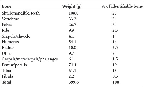
Table 3.84—Summary of identified bone, grave 7, E704:12; sample 2.
Description of identifiable features of the bones
The identified elements are described below.
Skull: adult; large fragments of squamous frontal, parietal and occipital bone, as well as numerous small fragments. There were several fragments of temporal bone, including the area just above the mastoid process of a left bone, a mandibular fossa, anterior suture and part of the zygomatic arch from a left bone, as well as fragments of squamous temporal bone. Other identifiable elements included a fragment of occipital bone including the posterior part of the foramen magnum, a left orbit, a fragment of supraorbital protuberance, a left zygomatic bone and a fragment of frontal bone with the internal crest visible.
Maxilla: most of the right side and a small fragment of the left side from what appears to
be the same bone were present. The following sockets were present:

Mandible: there was a fragment of the right side of the body, split through the alveolus, with sockets for the following teeth available:

There was another fragment with a few partial sockets present but they could not be identified.
Teeth: fragments of tooth roots included a maxillary first molar, maxillary second molar, two lower molars, two premolars and a few unidentified fragments.
Vertebrae: this included the odontoid process of a second cervical vertebra and the bodies of at least five thoracic and four lumbar vertebrae, as well as other smaller fragments of vertebral bodies and a few articular surfaces from thoracic and lumbar vertebrae.
Pelvis: this consisted of fragments of ilium, including some from the body, fragments of iliac crest, fragments of acetabulum and the auricular surface.
Ribs: this consisted of several fragments of shaft, with one rib head and one tubercle Present.
Scapula/clavicle: this consisted of the lateral half of the shaft of a left clavicle and part of the lateral border of a scapula.
Humerus: this included most of the proximal half of a left bone with deltoid tuberosity present. The deltoid tuberosity from a right bone was also present. There were fragments from the proximal articular surface and the tubercles. Also present were some smaller fragments from the mid-shaft.
Radius: this includes fragments from the mid-shaft and distal half of the bone.
Ulna: this consisted of some of the proximal shaft of a right bone with a small amount of articular surface. There were also fragments from the mid-shaft area.
Carpals/metacarpals and phalanges: there were fragments of scaphoid and hamate, one metacarpal head, and five proximal, three middle and two distal hand phalanges.
Femur/patella: this included fragments from the proximal, middle and distal shaft with the anterior and posterior surfaces present. The distal articular surface of a right bone was also present. One fragment of the medial half of a right patella was present.
Tibia: this consisted of several fragments, including the proximal end of a right bone, most of the lateral surface of the shaft of a right bone, some lateral and anterior surface from a left bone and several fragments of distal and proximal shaft, including one fragment with the nutrient foramen visible.
Fibula: this consisted of one fragment of proximal shaft.
Minimum number of individuals: the remains appear to represent one adult individual.
Sample 1 from this grave is probably part of this same cremation.
Grave 8, from ‘fire site in quadrant II’ (E704:14)
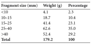
This sample consisted of 179.2g of cremated bone. The bone was heavily coated with charcoal even after washing and so appeared black, although the underlying colour was white. The bone was efficiently cremated, with cracking and fissuring of the surface.
The largest fragment measured 68mm and 29% of the fragments were more than 40mm in length, with a further 35% between 25mm and 40mm. Therefore nearly two thirds of the sample consisted of large or very large fragments. The fragmentation of the sample is shown in Table 3.85.
Despite the large size of the fragments, the colour of the bone made identification difficult; nevertheless, it was possible to identify 65.9g (37%) of the bone. As not all skeletal parts were represented, the proportions of the various parts of the skeleton are not given, but the weights and description of the identified bone are given below.
Skull (7.8g): small fragments of skull cap, parietal and occipital bone.
Clavicle (2.6g): lateral third of right clavicle.
Humerus, 18.3g): fragments of shaft, mostly from the proximal half of two bones.
Ulna (3g): part of the proximal shaft of a left bone.
Table 3.85—Fragmentation of bone, grave 8, E704:14.
Femur (28g): several fragments of shaft, including the distal half of the shaft posterior surface and some anterior mid-shaft, as well as part of the proximal joint surface.
Tibia (6.2g): a few fragments of shaft. Most of the bone consisted of long bone fragments and the remains represented one adult.
Grave 9 (E704:17)
Only two small fragments of cremated bone from this feature have been identified. Both appear to be skull fragments.
Grave 10 (1975:265)
This cist was located 400m south of the main mound and was excavated 40 years after the other burials. It consisted of 619 fragments of bone weighing a total of 631g. The bone was mainly white with a chalky texture, apart from a few fragments that were a blue/black colour. There were numerous horizontal fissures on the bone. The fragmentation of the bone is shown in Table 3.86, with the largest fragment being 68mm in length.
Table 3.86—Fragmentation of bone, grave 10, 1975:265.
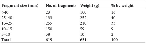
It can be seen that over half the sample consists of very large fragments and that almost all the sample is made up of large fragments more than 15mm in length. Most of the smaller fragments probably came from post-excavation fragmentation of the bone. It seems reasonable to assume that the bone was not deliberately crushed after collection from the funeral pyre. The total weight of bone recovered does not represent a full adult cremation, however. Some of the bone may have been lost in the twenty years since it was originally discovered.
Identifiable bone
A total of 372g (59% of the total bone) was identified. This is a high percentage of identifiable bone and is due to the number of very large fragments. The proportion of identified bone is shown in Table 3.87.
Table 3.87—Proportion of identified bone, grave 10, 1975:265.
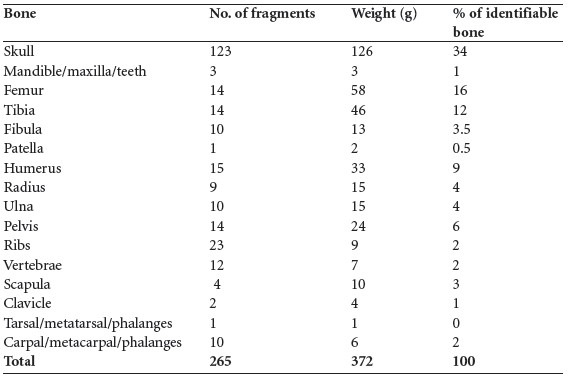
Table 3.88—Summary of identified bone, grave 10, 1975:265.
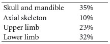
Table 3.88 summarises the main parts of the skeleton identified from this sample. The proportion of skull is almost twice what it should be, while the amount of axial skeleton is half what it should be. The skull fragments are easier to identify and therefore easier to collect from a disturbed site. The vertebrae and ribs nearly always get totally fragmented when a site is disturbed, and subsequently are overlooked or considered insignificant when cremated bone is collected. The amount of upper limb is almost exactly what it should be and there is only a slight reduction in the amount of lower limb expected.
Description of identifiable features of the bones
Skull: there were several fragments of frontal and parietal bone. One large fragment of occipital bone was present. The external occipital protuberance was not pronounced, so the individual was probably a female. One fragment of orbit with cribra orbitalia was present.
There was one temporal fossa and one partial petrous temporal bone.
Mandible and maxilla: most of the left ramus of the mandible and a segment of the body, possibly from a juvenile, were present. There seemed to be a socket for a deciduous tooth present, as well as an undeveloped tooth crown. The roots of two incisors and one molar were also present. A fragment from the nasal area of the maxilla was present.
Vertebrae: two partial bodies and two partial arches from the lower cervical vertebrae were present. There was one partial body and two partial arches of thoracic vertebrae, and a few articular surfaces from the lumbar vertebrae.
Ribs: most were fragments of shaft, although there were a few heads and transverse processes present. Some of the fragments were small and may be from a juvenile.
Pelvis: fragments of ilium with one small fragment of acetabulum were present. An almost complete right pubic bone and part of a left pubic bone from a female were also present. The symphyseal surface of the pubic bone indicated that it was probably from a young individual aged fifteen to nineteen years.
Clavicle: the mid-shaft area and lateral end of a left clavicle were present.
Scapulae: a fragment of left scapula from near the glenoid fossa with part of the acromial spine was present. There was also a fragment of right acromion and a fragment of the lateral border.
Humerus: all the fragments were shaft fragments and they were mostly from the proximal end of the bone.
Radius: all the fragments were of the shaft, including some from the middle and some from the distal end of the bone.
Ulna: there were fragments of shaft from the middle and distal parts of the bone. One fragment from the distal end had the distal joint surface attached; there were also fragments from the proximal shaft.
Carpals, metacarpals and phalanges: a few metacarpal shafts were present, and there was one almost complete middle phalanx and fragments of five others.
Femur: this all appeared to be from adult bone, as there were some very thick fragments of shaft, mostly from the proximal and middle parts of the bone. A few fragments had the linea aspera present.
Tibia: all the fragments were of the shaft, including some from the proximal and some from the distal ends. A fragment from the proximal posterior shaft with a nutrient foramen was present, and there were fragments with the interosseous border visible. At least two adult bones were present.
Fibula: this consisted of fragments of shaft only.
Patella: one incomplete patella, probably the left bone, was present.
Tarsals/metatarsals: one fragment of talus only was present.
Minimum number of individuals: there appears to be one individual present as there is no repetition of skeletal elements. It seems to be the remains of one young female, although there may also be a juvenile present.
In addition to the human bone, one large fragment of animal bone was present.
Summary
A total of four inhumations and six separate cremations were recovered from various cist graves from this site. The central burial (grave 3) was that of an adult female, and the other adult inhumations consisted of a younger female and a younger male. There was another infant inhumation in grave 6 which, owing to its colour, had been described on site as a cremation. The most startling pathology was the multiple myeloma that had probably resulted in the death of the female in grave 3. This is a very rare type of bone cancer, although it has been found from Neolithic sites in Europe. This woman had also suffered from osteoporosis, secondary to the bone cancer, and had suffered from a greenstick fracture or dislocation of the wrist in childhood.
Other pathology noted on the skeletons was spondylolysis in the young female. In this condition the neural arch of the fifth lumbar vertebra becomes detached from the body of the vertebra, probably as a result of trauma, and the fracture rarely heals. It will result in an aching back but it is usually a stable fracture. Two of the individuals had suffered from nutritional distress or acute infection during early childhood.
The larger cremations consisted mainly of large fragments of bone and were probably not crushed as part of the cremation ritual. This meant that identification was high, ranging from 21% to 59%, with the small identification rate being due to the bone being excessively coated in charcoal in two of the cremations. In all samples the bone was very efficiently cremated.
The total number of individuals found in the cremations was six adults and two infants. Most of the samples consisted of a minimum of one adult only, but grave 2 contained four individuals: two adults, one of whom was male and the other probably female, and two juveniles. One of the juveniles was an infant of around one year old and the other was probably aged seven to eleven months.
In two of the smaller cremations there was a fragment of skull that could have been from a juvenile, but this was not considered enough evidence for a juvenile individual and they were not included in the count of minimum numbers.
The total number of individuals from the burials recovered from this site was therefore twelve: nine adults and three infants, with at least four adults being female and two being male. The infants were one year or less in age.
269. For reasons which are not obvious from the file the objects and human remains recovered during this excavation were not registered. The fact that the finds were not registered in 1935 suggests that there may not have been an agreement with the landowner regarding the donation or purchase of the finds. At that time the acquisition of the finds by the NMI would have had to be negotiated with the landowner. In recent years the excavation has been assigned the excavation number E704.
270. Professor of Dogmatic Theology and first curator of Maynooth College Museum.
271. 24 April–5 May and 23 September–15 October 1935.
272. There is a disparity between the surviving records from the spring excavation report and that of October. No notes, plans etc. have been found, although there are short progress reports from Duignan to Dr Mahr on file.
273. Parish of Durrow, barony of Ballycowan. SMR OF009-036——. IGR 234582 229243. Grave 10, excavated by Raftery in 1975, was located further south: SMR OF009-039——; IGR 234750 228940.
274. The pit containing the cist of grave 3 appears to have cut this layer (Fig. 3.150).
275. This was also found in the fill of the pit for grave 3. 276. Other than some tiny sherds, the vessel from grave 1 has not been located in the NMI. It is possible that it was not acquired. As none of the finds from the excavation were registered at the time, however, it may be that it has been mislaid.
277. Waddell (1990, 130) states that the vessel had ‘lain on its side near the north-west corner, its mouth facing roughly west’. It is not clear from the file that this was the case, so it is possible that Waddell obtained this information from another source.
278. We are very grateful to Prof. John Waddell for providing us with an original file relating to the excavation which was located in the Dept of Archaeology in NUIG. Amongst these papers plans of graves 5 and 6 were found, but there were no plans of graves 7–10, nor any notes or accounts of them.
279. SMR OF009-039—. IGR 234750 228940.
280. GrA-24309.